
HEMATOLOGY-ONCOLOGY
PLATELET DISORDERS
THROMBOCYTOPENIA (PLT COUNT <150,000/µL)
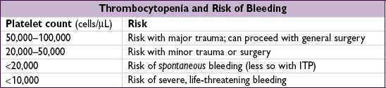
Etiologies
• ↓ production
hypocellular bone marrow: aplastic anemia (qv), rarely MDS, drugs (eg, thiazides, antibiotics), alcohol, cirrhosis
hypercellular bone marrow: MDS, leukemia, severe megaloblastic anemia
marrow replacement: myelofibrosis, hematologic and solid malignancies, granulomas
• ↑ destruction
immune-mediated (distinguish primary from secondary; Blood 2009;113:2386) Primary (idiopathic): immune thrombocytopenic purpura (ITP, see below)
Secondary: infxn (HIV, HCV, HSV), collagen vascular diseases (SLE), APS, lymphoproliferative (CLL, lymphoma), drugs (many, including heparin, abciximab, quinidine, sulfonamides, vancomycin), alloimmune (posttransfusion)
non–immune-mediated: MAHA (DIC, HUS, TTP), ticlopidine/clopidogrel, vasculitis, preeclampsia/HELLP, cardiopulm bypass, CVVH, IABP, cavernous hemangioma
• Abnormal distribution or pooling: splenic sequestration, dilutional, hypothermia
• Unknown: ehrlichiosis/anaplasmosis, babesiosis, RMSF
Diagnostic evaluation
• H&P: meds, infxns, underlying conditions, splenomegaly, lymph nodes, bleeding hx
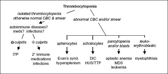
• CBC with differential: isolated thrombocytopenia vs. multilineage involvement
• Peripheral smear
↑ destruction → look for large plts, schistocytes (see “Peripheral Smear” inserts)
↓ production → rarely limited to platelets → look for blasts, hypersegmented PMNs, leukoerythroblastic Ds; can see inclusion bodies (anaplasma), parasites (babesia)
r/o pseudothrombocytopenia due to platelet clumping (✓ platelet count in non–EDTA-containing tube, eg, citrate-containing yellow top tube)
Figure 5-5 Approach to thrombocytopenia
• Additional laboratory evaluations as indicated (eg, viral titers, flow cytometry, ANA, APLA)
if anemia: ✓ reticulocyte count, LDH, haptoglobin, bilirubin to detect hemolysis
if hemolytic anemia: ✓ PT, PTT, fibrinogen, D-dimer, Coombs, ANA
BM bx for unexplained thrombocytopenia, esp. if associated with splenomegaly
Primary immune thrombocytopenic purpura (ITP) (Blood 2010;115:168)
• Primary ITP: isolated thrombocytopenia due to immune plt destruction & ↓ production (auto-Ab to megakaryocytes); (2° ITP a/w disease/drug exposure; Rx underlying disorder)
• Primary ITP is diagnosis of exclusion; no robust clinical or lab parameters, but typically:
CBC: isolated ↓ plt (<100,000/µL); 10% have ITP + AIHA = Evans syndrome
Peripheral smear: large platelets
BM bx: ↑ megakaryocytes; perform in adults >60 y to r/o myelodysplasia
R/o other etiologies: viral serologies (HIV, HCV, HBV, EBV), H. pylori Ab, ANA, pregnancy test, APLA, TSH, parvovirus, & CMV PCR. Anti-plt Ab tests not useful.
• Clinical manifestations: insidious onset of mucocutaneous bleeding;  :
: = 3:1
= 3:1
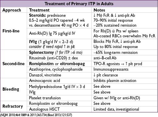
• Treatment: goals based on individual Pt rarely indicated if plt >50,000/µL unless bleeding, trauma/surgery, anticoag, comorbidities steroids, IVIg, & splenectomy mainstay of initial Rx; romiplostim/eltrombopag if refractory
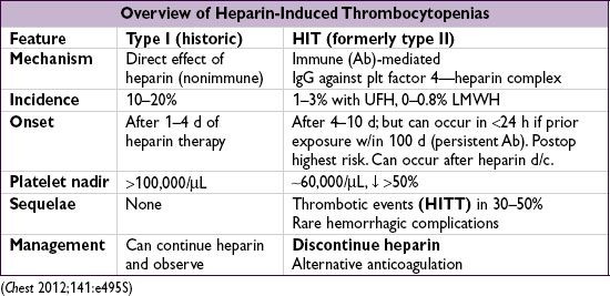
• Pathophysiology (type II): Ab binds heparin-PF4 → immune complex binds to plt → plt activation, further PF4 release → plt aggregates removed from circulation → thrombocytopenia; procoagulants released by plts and tissue factor released by endothelial cells damaged by HIT Abs → prothrombotic state
• Diagnosis (need clinical + pathologic)
Clinical: plt <100k or ↓ 50% from baseline; or venous (DVT/PE) or arterial (limb ischemia, CVA, MI) thrombosis (4:1 ratio); skin necrosis; ? ↑ heparin resistance
Pathologic:  HIT Ab using PF4-heparin ELISA (≥90% Se, IgG-specific ELISA Sp 94%), may confirm w/ functional plt aggregation (serotonin-release) assay (>90% Sp)
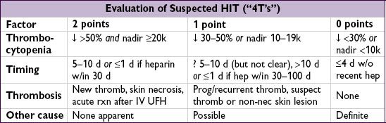
Pretest prob w/ “4T’s” criteria (Blood 2012;120:4160): ≤3 points → 99% NPV, investigate other causes; 4–5 points 22% PPV & 6–8 points 64% PPV, ✓ lab test and replace UFH
• Treatment of HIT (type II) (Chest 2012;141:e495S; Blood 2012;119:2209; NEJM 2013;368:737)
Discontinue heparin (including flushes, LMWH prophylaxis, heparin-impregnated lines)
Avoid plt transfusions if not actively bleeding (anecdotally linked w/ thrombotic events)
Nonheparin anticoag (argatroban, bivalirudin; NEJM 2013;368:737) regardless of thrombosis; start warfarin when plt >150k, overlap ≥5 d (✓ chromogenic Xa to titrate)
thrombosis (HITT): anticoagulate for ≥ 3–6 mo
 thrombosis (HIT): screen for DVT; unclear duration of subsequent anticoag (until plt count recovers, often ~2–3 mo if no clot); 25–50% thrombosis rate w/in 30 d
• Heparin use if h/o HIT: if PF4 Ab (typically >100 d after dx) → re-exposure to UFH reasonable (eg, for surgery); HIT recurrence low
Hemolytic-uremic syndrome (HUS) & thrombotic thrombocytopenic purpura (TTP)
• Definition: vascular occlusive disorders w/ systemic (TTP) or intrarenal (HUS) plt aggreg.
→ thrombocytopenia & mechanical injury to RBCs (MAHA) (NEJM 2002;347:589)
HUS triad = thrombocytopenia + MAHA + renal failure
TTP pentad (all 5 in only ~5%) = thrombocytopenia + MAHA (100%) ± Δ MS (65%) ± renal failure (50%) ± fever (25%)
• Pathophysiology: mechanism in most HUS cases is distinct from TTP (NEJM 1998;339:1578)
HUS: Shiga toxin binds & activates renal endothelial cells & plts → intrarenal thrombi
TTP: ↓ ADAMTS13 protease activity or inhibitor→ persistence of large vWF multimers on endothelial surface → adhesion and aggregation of passing platelets → thrombosis
• Clinical manifestations and associations
HUS: usually in children; prodrome of bloody diarrhea due to enterohemorrhagic E. coli
TTP: usually in adults; idiopathic, drugs (CsA, tacrolimus, gemcitabine, mitomycin-C, ticlopidine, clopidogrel, quinine), HIV, pregnancy, HSCT, autoimmune disease, familial
• Dx: unexplained thrombocytopenia (typically <20k) + MAHA → sufficient for dx schistocytes (>2–3/hpf), Coombs, normal PT/PTT & fibrinogen, ↓↓ ADAMTS13 ↑↑ LDH (tissue ischemia + hemolysis), ↑ indirect bili., ↓↓ haptoglobin, ↑ Cr (esp. in HUS)
Biopsy: arterioles filled with platelet hyaline thrombi
Ddx: DIC, vasculitis, malignant hypertension, preeclampsia/HELLP syndrome
• Treatment: urgent plasma exchange ± glucocorticoids if suspected; FFP if delay to plasma exchange (Blood 2010;116:4060); ? eculizumab in HUS (NEJM 2011;364:2561); plt transfusions contraindicated → ↑ microvascular thrombosis (NEJM 2006;354:1927)
Disseminated intravascular coagulation (DIC): see “Coagulopathies”
DISORDERS OF PLATELET FUNCTION
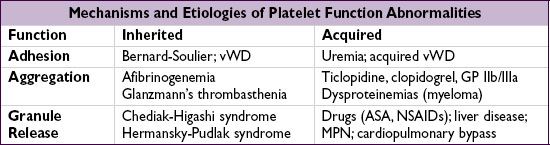
Tests of platelet function
• Bleeding time: global screen of platelet function; not reliable and rarely used
• Platelet aggregation tests: measure aggregation in response to agonists (eg, ADP)
von Willebrand’s disease (vWD) (NEJM 2004;351:683 & 2012;367:1954)
• von Willebrand’s factor (vWF) function = platelet glue & plasma carrier of factor VIII
• vWD most common inherited (usually auto dom) bleeding disorder; ~85% (type 1) have partial quantitative defic of vWF, ~15% (type 2) have qualitative defic in vWF
• Acquired vWD: a/w many disorders (malig, MPN w/ ↑ plt count; autoimmune; hypo-thyroidism; drugs) and caused by different mechanisms (anti-vWF Abs, ↑ clearance, ↓ synthesis); Heyde’s syndrome = vWF destruction by severe AS, a/w GI AVMs/bleed
• Diagnosis: ↓ vWF:Ag, ↓ vWF activity (measured by ristocetin cofactor assay), ↓ factor VIII, ± ↑ PTT, ± ↓ platelets; confirm with vWF multimer analysis
• Clinical condition, factor VIII levels and ristocetin cofactor assay useful to guide Rx decision
• Rx: desmopressin (dDAVP, IV/IN) → ↑ endothelial cell release of vWF; efficacy depends on type (avoid in Type 2), ∴ ✓ response before use w/ subseq. bleeding or procedures;
vWF replacement: cryoprecipitate, factor VIII concentrates rich in vWF, recomb. vWF
Uremic bleeding
• Uremia → platelet dysfunction including ↓ aggregation, impaired adhesiveness
• Treatment: dDAVP, cryoprecipitate, correct anemia (improves plt aggregation and
adhesion by increasing plt interactions with endothelium), consider holding anti-plt agents