
CARDIOLOGY
PA CATHETER AND TAILORED THERAPY
Rationale
• Cardiac output (CO) = SV × HR; SV depends on LV end-diastolic volume (LVEDV)
∴ manipulate LVEDV to optimize CO while minimizing pulmonary edema
• Balloon at tip of catheter inflated → floats into “wedge” position. Column of blood extends from tip of catheter, through pulmonary circulation, to a point just proximal to LA. Under conditions of no flow, PCWP  LA pressure LVEDP, which is proportional to LVEDV.
• Situations in which these basic assumptions fail:
(1) Catheter tip not in West lung zone 3 (and ∴ PCWP = alveolar pressure ≠ LA pressure); clues include lack of a & v waves and if PA diastolic pressure < PCWP
(2) PCWP > LA pressure (eg, mediastinal fibrosis, pulmonary VOD, PV stenosis)
(3) Mean LA pressure > LVEDP (eg, MR, MS)
(4) Δ LVEDP-LVEDV relationship (ie, abnl compliance, ∴ “nl” LVEDP may not be optimal)
Indications ( JACC 1998;32:840 & Circ 2009;119:e391)
• Diagnosis and evaluation
Ddx of shock (cardiogenic vs. distributive; esp. if trial of IVF failed or is high risk) and of pulmonary edema (cardiogenic vs. not; esp. if trial of diuretic failed or is high risk)
Evaluation of CO, intracardiac shunt, pulmonary HTN, MR, tamponade
Evaluation of unexplained dyspnea (PAC during provocation w/ exercise, vasodilator)
• Therapeutics (Circ 2006;113:1020)
Tailored therapy to optimize PCWP, SV, SvO2 in heart failure (incl end-stage) or shock
Guide to vasodilator therapy (eg, inhaled NO, nifedipine) in pulm HTN, RV infarction
Guide to perioperative management in some high-risk Pts, pretransplantation
• Contraindications
Absolute: right-sided endocarditis, thrombus/mass or mechanical valve; PE
Relative: coagulopathy (reverse), recent PPM or ICD (place under fluoroscopy), LBBB (~5% risk of RBBB → CHB, place under fluoro), bioprosthetic R-sided valve
Efficacy concerns (NEJM 2006;354:2213; JAMA 2005;294:1664)
• No benefit to routine PAC use in high-risk surgery, sepsis, ARDS
• No benefit in decompensated HF ( JAMA 2005;294:1625); untested in cardiogenic shock
• But: ~½ of CO & PCWP clinical estimates incorrect; CVP & PCWP not well correl.; ∴ use PAC to (a) answer hemodynamic ? and then remove, or (b) manage cardiogenic shock
Placement
• Insertion site: R internal jugular or L subclavian veins for “anatomic” flotation into PA
• Inflate balloon (max 1.5 mL) when advancing and to measure PCWP
• Use resistance to inflation and pressure tracing to avoid overinflation & risk of PA rupture
• Deflate the balloon when withdrawing and at all other times
• CXR should be obtained after placement to assess for catheter position and PTX
• If catheter cannot be successfully floated (typically if severe TR or RV dilatation) or if another relative contraindication exists, consider fluoroscopic guidance
Complications
• Central venous access: pneumo/hemothorax (~1%), arterial puncture (if inadvertent cannulation w/ dilation → surgical/endovasc eval), air embolism, thoracic duct injury
• Advancement: atrial or ventricular arrhythmias (3% VT; 20% NSVT and >50% PVC), RBBB (5%), catheter knotting, cardiac perforation/tamponade, PA rupture
• Maintenance: infection (esp. if catheter >3 d old), thrombus, pulm infarction (≤1%), valve/chordae damage, PA rupture/pseudoaneurysm (esp. w/ PHT), balloon rupture
Intracardiac pressures
• Transmural pressure ( preload) = measured intracardiac pressure – intrathoracic pressure
• Intrathoracic pressure (usually slightly  ) is transmitted to vessels and heart
• Always measure intracardiac pressure at end-expiration, when intrathoracic pressure closest to 0 (“high point” in spont. breathing Pts; “low point” in Pts on  pressure vent.)
• If ↑ intrathoracic pressure (eg, PEEP), measured PCWP overestimates true transmural pressures. Can approx by subtracting ~½ PEEP (× ¾ to convert cm H2O to mmHg).
• PCWP: LV preload best estimated at a wave; risk of pulmonary edema from avg PCWP
Cardiac output
• Thermodilution: saline injected in RA. Δ in temp over time measured at thermistor (in PA) is integrated and is 1/CO. Inaccurate if ↓ CO, sev TR or shunt.
• Fick method: O2 consumption ( O2) (L/min) = CO (L/min) × ∆ arteriovenous O2 content
∴ CO = O2 / C(a-v)O2
 O2 ideally measured (esp. if ↑ metab demands), but freq estimated (125 mL/min/m2)
C(a-v)O2 = [10×1.36 mL O2/g of Hb × Hb g/dL × (SaO2–SvO2)]. SvO2 is key variable that Δs.
If SVO2 >80%, consider if the PAC is “wedged” (ie, pulm vein sat), L→R shunt, impaired O2 utilization (severe sepsis, cyanide, carbon monoxide), ↑↑ FiO2.
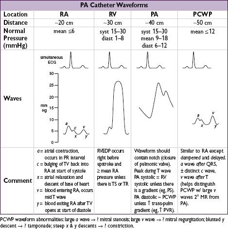
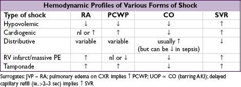
Tailored therapy in cardiogenic shock (Circ 2009;119:e391)
• Goals: optimize both MAP and CO while ↓ risk of pulmonary edema
MAP = CO × SVR; CO = HR × SV (which depends on preload, afterload and contractility)
pulmonary edema when PCWP >20–25 (↑ levels may be tolerated in chronic HF)
• Optimize preload = LVEDV LVEDP LAP PCWP (NEJM 1973;289:1263)
goal PCWP ~14–18 in acute MI, ≤14 in acute decompensated HF
optimize in individual Pt by measuring SV w/ different PCWP to create Starling curve
↑ by giving NS (albumin w/o clinical benefit over NS; PRBC if significant anemia)
↓ by diuresis (qv), ultrafiltration or dialysis if refractory to diuretics
• Optimize afterload wall stress during LV ejection = [(~SBP × radius) / (2 × wall thick.)] and ∴ ∝ MAP and ∝ SVR = (MAP – CVP / CO); goals: MAP >60, SVR 800–1200
MAP >60 & SVR ↑: vasodilators (eg, nitroprusside, NTG, ACEI, hydral.) or wean pressors
MAP <60 & SVR ↑ (& ∴ CO ↓): temporize w/ pressors until can ↑ CO (see below)
MAP <60 & SVR low/nl (& ∴ inappropriate vasoplegia): vasopressors (eg, norepineph-rine [a, b], dopamine [D, a, b], phenylephrine [a] or vasopressin [V1] if refractory)
• Optimize contractility ∝ CO for given preload & afterload; goal CI = (CO / BSA) >2.2
if too low despite optimal preload & vasodilators (as MAP permits):
inotropes: eg, dobutamine (mod inotrope & mild vasodilator) or milrinone (strong inotrope & vasodilator, incl pulm), both proarrhythmic, or epi (strong inotrope & pressor)
mechanical support devices: eg, IABP, percutaneous or surgical VAD (left-sided, right-sided or both) or ECMO (Circ 2011;123:533)