
Marc L. Turner
Scottish National Blood Transfusion Service, Edinburgh, Scotland, UK
A variety of transmissible spongiform encephalo-pathies or prion diseases are described in animals and humans (Table 15.1). Scrapie, an endemic disease of sheep and goats, was first described over 250 years ago. Chronic wasting disease is spreading in deer and elk in the USA. Bovine spongiform encephalopathy (BSE) was first described in cattle in the UK in 1986, though in retrospect the first cases probably appeared as early as 1982 [1,2]. It remains unclear whether BSE arose from scrapie in sheep or from a sporadic case of prion disease in cattle, but it is thought that it was transmitted through the food chain via rendered meat and bonemeal. In the UK over 180 000 clinical cases of BSE have been described, with around 300 cases in other European countries, and occasional cases elsewhere in the world, probably related to exported UK cattle or meat and bonemeal. The UK epidemic peaked in 1992 and has now subsided as a result of a ban on the use of ruminant protein in cattle feed. However, mathematical projections suggest that 1–2 million infected cattle could have entered the human food chain before showing evidence of disease. Unlike scrapie, BSE has proved itself capable of crossing species barriers by infecting a number of other animals including exotic and domestic cats (feline spongiform encephalopathy) and exotic ruminants in zoos (exotic ungulate encephalopathy).
Table 15.1 Prion diseases.
|
Animals |
Human |
|
Scrapie |
Sporadic Creutzfeldt–Jakob disease |
|
Chronic wasting disease |
Kuru |
|
Transmissible mink encephalopathy |
Iatrogenic Creutzfeldt–Jakob disease |
|
Bovine spongiform encephalopathy |
Variant Creutzfeldt–Jakob disease |
|
Feline spongiform encephalopathy |
Familial Creutzfeldt–Jakob disease |
|
Exotic ungulate encephalopathy |
Gerstmann–Sträussler–Scheinker disease |
|
Fatal familial insomnia |
In humans several forms of prion disease have been described. Sporadic or classical Creutzfeldt–Jakob disease (CJD) was first described in the early 1920s. It presents at a median age of 68 years as a rapidly progressive dementia with a duration of illness of around 6 months. The incidence of CJD is around 1 per million per annum throughout the world, with no clear link to the incidence of prion disease in other animals.
In the 1950s, a form of prion disease called kuru was described in the Foré people of the highlands of Papua New Guinea. This disease presented at a much younger age, with cerebellar ataxia as a prominent feature and a more prolonged clinical course. Kuru was transmitted from person to person, probably through the cannibalistic funeral rites practised by the tribe at that time. It is informative to note that children died from kuru and that although cannibalistic feasts discontinued around 1959–1960, there are still occasional patients presenting with the clinical disease. This points to a very wide range of incubation periods, with an upper limit of 40–50 years or perhaps even beyond the normal human lifespan.
In the 1980s a number of iatrogenic transmissions of CJD were described. These fell broadly into two groups. Direct central nervous systems (CNS) transmission due to contaminated neurosurgical instruments, EEG electrodes and dura mater grafts led to a rapidly progressive dementia reminiscent of sporadic CJD after a short incubation period of around 2 years and death within about 6 months of presentation. Peripheral transmission from cadaveric pituitary-derived growth- and follicle-stimulating hormone gave rise to a clinical picture reminiscent of kuru, with a prolonged incubation period of some 13–15 years.
Finally, a number of familial forms of CJD have been described including familial CJD, Gerstmann–Sträussler–Scheinker (GSS) disease and fatal familial insomnia (FFI), which arise due to polymorphisms in the gene for prion protein (PrP).
Prion diseases are therefore interesting from an aetiological perspective in that they can arise spontaneously, are transmissible and can also arise due to genetic polymorphism.
Variant CJD
The UK government instituted routine surveillance for CJD in 1989 in response to the BSE epidemic, with the aim of monitoring any change in the incidence or pattern of disease in the UK population. In 1995 the first cases of variant CJD were described. The clinical features differ from those of sporadic CJD. Patients are younger, with a median age at presentation of 28 years (range 12–74 years). They often present with behavioural change, such as depression and anxiety, or with dysaesthesia. Untreated, the disease progresses to cerebellar ataxia, involuntary movements, dementia and death over a period of 7–38 months. In the UK 176 cases of variant CJD have been described thus far, though the incidence of new cases appears to be falling. Elsewhere there have been 27 cases described in France, 5 in Spain, 4 in the Republic of Ireland, 3 in the USA and the Netherlands, 2 in Portugal, Canada and Italy, and 1 in Japan, Saudi Arabia and Taiwan. Two of the American and Irish patients, and one each of the French, Canadian and Taiwan patients had spent a considerable time in the UK, whereas the others did not, and probably contracted the disease in their own countries. Though original estimates of the number of people who may eventually develop the disease gave very high upper limits, the recent downturn in the number of new cases in the UK has led to a revised prediction of just over 70 cases. However, retrospective studies of appendix samples in the UK recorded suggest a prevalence of subclinical disease of around 1/2000 (95% confidence interval (CI), 1/1250–1/3500). These individuals should be assumed to be at risk of passing infection to others via contaminated surgical instruments or blood transfusion.
A considerable amount of epidemiological, clinical, neuropathological and experimental data now supports the view that variant CJD is the same strain of disease as BSE [4] and that these are different from the prion strains that give rise to other forms of CJD in humans or scrapie and chronic wasting disease in animals.
Aetiology and pathophysiology
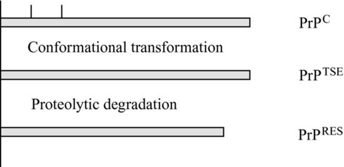
Prion diseases are associated with a change in the secondary structure of PrP. PrP is a widely expressed 30–35 kDa glycoprotein with two N-linked oligosaccharides. It is normally linked to the cell membrane by a glycosylphosphatidylinositol (GPI) anchor, though transmembrane anchorage has also been described. The normal secondary structure of PrP contains around 40% α-helices and 3% β-pleated sheets, with the membrane-distal part of the molecule largely unstructured. The development of prion disease is associated with a change in the secondary structure of the PrP glycoprotein, with an increase in the proportion of β-pleated sheets to some 40–50% of the molecule largely at the expense of the unstructured region (PrPTSE) (Figure 15.1). This changes the physicochemical characteristics of the molecule, giving it increased resistance to both physical and biological degradation. In vitrotreatment with proteinase-K removes the membrane-distal part of the molecule, but is unable to digest the 30–32 kDa core (PrPRes). PrPTSE accumulates in vivo, leading to the deposition of amyloid plaques. The pathophysiology of the disease remains debated. Some authorities propose the presence of a small DNA molecule associated with PrPTSE (termed a virion), but this has not yet been identified and the infectious agent does appear to be resistant to physical conditions that would normally degrade DNA. The prion hypothesis proposes that the abnormal isoform of the protein is itself the infectious agent, changing the structure of the normal form either through heterodimer formation or though a physicochemical process of nuclear polymerization.
Fig 15.1 The prion hypothesis. PrPC (top) is a 30–35 kDa glycoprotein with two N-linked glycosylation sites, anchored by glycosylphosphatidylinositol to the cell membrane, with 40% α-helix and 3% β-pleated sheet. Prion diseases are associated with conformational change in the secondary structure, with an increase in the amount of β-pleated sheet to some 40–50% of the molecule (PrPTSE) (middle). This changes the physicochemical and biological properties of the molecule, rendering it resistant to degradation by enzymes such as proteinase-K (PrPRES) (bottom).
Accumulation of amyloid plaques consisting of PrPTSE leads to the classical neuropathological features of neuronal death, astrogliosis and spongiform degeneration of the CNS (Plates 15.1 and 15.2 in the plate section). In sporadic, iatrogenic and familial forms of CJD, abnormal PrP accumulation appears to be confined to the CNS. In variant CJD, abnormal PrPTSE accumulation has been demonstrated in follicular dendritic cells (FDCs) in the tonsil, spleen, cervical, mediastinal, paraaortic and mesenteric lymph nodes and gut-associated lymphoid tissue of the appendix up to 2 years prior to the onset of clinical disease (Plate 15.3 in the plate section). This observation is consistent with what we know about the pathophysiology of transmission of prion diseases by peripheral routes in experimental animals.
Fig 15.2 Likely impact of a putative variant CJD assay with 99% sensitivity and specificity.

Experimental peripheral transmission of scrapie strains in murine models leads to the presence of infectivity and/or PrPTSE in the spleen and lymph node from a very early stage of infection, well before detection of infectivity of PrPTSEin the CNS. Interestingly, immunosuppression and splenectomy have long been known to decrease the efficiency of peripheral transmission, whereas irradiation and thymectomy do not. A series of experiments has demonstrated that mice with severe combined immunodeficiency are resistant to peripheral but not central prion challenge and that sensitivity is regained after allogeneic bone marrow transplantation. Similarly, PrP-negative mice with a PrP-positive CNS implant can be infected only by peripheral transmission following PrP-positive allogeneic bone marrow transplant, whereas PrP-positive mice develop resistance to peripherally transmitted disease following PrP-negative bone marrow transplant. Detailed knockout experiments have demonstrated that Rag 1, Rag 2 and μMT knockout mice are resistant to peripheral challenge whereas CD4, CD8, β-microglobulin and perforin knockout models display normal sensitivity. These data led to the suggestion that B-lymphocytes were essential to peripheral transmission whereas T-lymphocytes were not. However, B-lymphocytes are also essential for FDC survival and more recent studies have demonstrated that PrP-positive FDCs are essential to peripheral transmission whereas PrP-positive B-lymphocytes are not. Indeed, peripheral transmission can be inhibited even by temporary FDC inactivation by lymphotoxin β-receptor blockade and also by depletion of complement receptors. These data convincingly support the seminal role of FDCs in the early stages of peripheral transmission.
Assessing peripheral blood infectivity in animal models
It has been demonstrated that PrP is present in the peripheral blood of normal individuals at a concentration of 100–300 ng/mL [5], with the majority found in platelets and plasma. PrPTSE accumulation has recently been demonstrated in the peripheral blood of humans with clinical variant CJD [6], though it has not proved possible to detect infectivity in this context, probably because of limitations in the volumes of blood that can be inoculated into experimental animals and the species barrier between man and rodents. Most of the information on peripheral blood infectivity comes from animal experiments where it has proved possible to demonstrate infectivity in the peripheral blood of sheep and rodents with experimental scrapie and BSE, and in rodents with experimental GSS, during both the clinical and incubation phases of disease [7–10]. However, no infectivity has been demonstrated in natural scrapie in sheep and goats, natural transmissible mink encephalopathy or natural or experimental BSE in cattle. The reason for these differences is not clear. Levels of peripheral blood infectivity have been investigated in the Fukuoka-1 strain of GSS in experimental mice and have been found to be on the order of 100 infectious units/mL during the clinical phase of disease and 5–10 infectious units/mL during the incubation period. A fourfold to fivefold higher level of infectivity was demonstrated in the buffy coat (containing the leucocytes and platelets) compared with plasma. Plasma itself showed a 10-fold higher concentration of infectivity compared with any of the Cohn fractions in an experimental fractionation system. The distribution of infectivity in blood was similar during the incubation phase of disease. Similar findings have been described in the 263K scrapie hamster model, where a little infectivity is associated with (purified) red cells or platelets, around 40% is associated with leucocytes and the remainder is in the plasma. Indeed, more recent data from the same group suggests that washing the leucocytes removes most of the associated infectivity.
In a different model, sheep experimentally infected with BSE or scrapie by oral ingestion have been bled during the incubation and clinical phases of the disease and whole-blood donations administered intravenously to secondary recipients [11]. Up to 50% of the secondary recipients in both cohorts have subsequently developed the relevant prion disease, amounting to proof of the principle that certain forms of prion disease can be transmitted by blood transfusion.
Clinical transmission of CJD from the peripheral blood of patients
In humans, of 37 reported attempts to transmit sporadic CJD from peripheral blood of patients with clinical disease by intracerebral inoculation into rodents, there have been five positive reports. Interestingly, transmission of CJD from human peripheral blood to primates by intracerebral inoculation has not proved possible and this has thrown some doubt on the validity of the rodent data. Thus far, there have been no successful transmissions of variant CJD from human peripheral blood to rodents or primates.
There have been three anecdotal case reports of patients who have developed sporadic CJD some time after receiving blood components or plasma products. In none of these, however, has it been shown that the donors themselves developed CJD. In comparison a large number of epidemiological case-control, look-back and surveillance studies over the past 25 years have shown little evidence of increased risk of sporadic CJD in blood or plasma product recipients, even where a donor is known to have subsequently developed sporadic CJD. One recent study has suggested an increased risk of transfusion more than 10 years before the clinical onset in sporadic CJD patients, though the significance of this remains uncertain.
In contrast, there are now 18 patients identified with variant CJD who, in the past, were blood donors; 67 recipients of blood components and plasma products from these donors have been traced of whom 17 are alive and 50 deceased [12]. Thus far three of the recipients have developed clinical variant CJD and one has shown evidence of abnormal prion accumulation in the spleen and a cervical lymph node [13,14].
Strategies for risk containment
Blood services have felt it prudent to implement precautionary policies to contain the risk of transmission of variant CJD. However, such policies require careful evaluation, both in terms of likely efficacy in reducing the risk of secondary transmission by blood transfusion and in terms of the potential increase in other risks including that of blood shortages. Consideration also needs to be given to the cross-impact of different policies and the opportunity costs incurred.
Donor selection
The UK blood services use a number of criteria to exclude blood and tissue donors who may be at increased risk of sporadic, iatrogenic or familial CJD (Table 15.2). There are no epidemiological risk factors described thus far that would discriminate a high-risk group for development of variant CJD within the UK. For example, there is no evidence that veterinary surgeons, cattle farmers, abattoir workers or others with a high risk of exposure to infected bovine materials are at higher risk of developing variant CJD than the general population. In comparison, some individuals who have been vegetarians for prolonged periods have developed variant CJD. A number of countries have taken the precautionary step of excluding blood donors who have spent more than a defined period in the UK between the beginning of 1980 and the end of 1996. The defined period varies depending on the frequency and pattern with which indigenous donors visit the UK and the likely prevalence of subclinical variant CJD in the general population. These are factors that impact upon the efficacy of UK donor exclusion in terms of risk reduction and on the likely negative impact on the blood donor base.
Table 15.2 UK criteria for excluding blood and tissue donors who have, or may have had contact with, sporadic, iatrogenic, or familial CJD.
|
Obligatory |
|
Permanently exclude donors with CJD or other prion-associated disorder |
|
Permanently exclude anyone identified at high risk of developing a prion-associated disorder |
|
Recipients of dura mater, corneal or scleral grafts |
|
Recipients of human pituitary-derived extracts such as growth hormone and gonadotrophins |
|
Individuals at familial risk of prion-associated diseases |
|
This includes individuals who have had two or more blood relatives develop a prion-associated disease and individuals who have been informed that they are at risk following genetic counselling |
|
Exceptions |
|
Individuals who have had two or more blood relatives develop a prion-associated disease but who, following genetic counselling, have been informed that they are not at risk. This requires confirmation by the consultant with responsibility for donors |
Subsequent to the evidence of transmission of variant CJD by blood transfusion, the UK blood services moved in April 2004 to defer blood donors who have themselves received blood transfusions in order to reduce the risk that tertiary and higher order transmissions would lead to a self-sustaining outbreak. This led to the loss of approximately 5–10% of the donor base.
Importation of blood components
An alternative approach would be to source blood components from countries with a low incidence of BSE and variant CJD. It is impractical to source all red blood cell concentrates for the UK (some 2.5 million components per annum) from overseas volunteer nonremunerated donors. The short shelf life of platelet concentrates also mitigates against this approach. Consideration needs to be given to the risk of other infectious agents in the proposed alternative donor population and long-term security of supply. It is possible to source plasma from overseas since surplus clinical plasma is generated by red cell collection programmes and the product can be virus inactivated and cryopreserved for transportation with a 2-year shelf life.
In the UK it has been decided to import methylene blue-inactivated plasma for neonates and children born since 1st January 1996. Neonates in particular receive a proportionately high number of blood components due to prematurity and surgery for congenital disorders. They are likely to have a low primary exposure to BSE through the food chain and they have the longest prospective lifespan during which to develop clinical variant CJD should they become infected. More recently solvent-detergent fresh frozen plasma has been imported for patients exposed to large volumes of plasma (such as patients undergoing plasma exchange for thrombotic thrombocytopenic purpura).
Development of peripheral blood assays for donor screening
There is no conventional immune response to prion infection and no DNA has been detected in association with transmission of these diseases. Hence, conventional serological and molecular approaches to the development of peripheral blood assays, utilized to such good effect in screening for microbiological disease, are not applicable to prion diseases.
A number of nonspecific markers of CNS damage are known to be elevated in the peripheral blood of patients with CJD, including 14-3-3 and S100, but given that patients with CNS damage are excluded by donor selection criteria, it seems unlikely that these would have much to offer in the context of screening normal healthy blood donors.
Surrogate markers could allow exclusion of individuals at risk of development of variant CJD. Reduced transcription of erythyroid differentiation-associated factors (EDAF) has been described in the bone marrow and peripheral blood of scrapie-infected sheep and rodents and of BSE-infected cattle. However, these findings do not appear to have been borne out in humans.
The gold standard would be, of course, infectivity bioassays. However, not only is it impractical to use such an approach for primary screening but also, as noted above, infectivity is not detectable in the peripheral blood of patients with variant CJD despite the fact that it is clearly transmissible. This implies that total reliance will need to be placed on any in vitro assay since there may be no other way of establishing whether the test-positive individual is ‘truly’ infected or will develop clinical disease in the future.
Detection of PrPTSE in the peripheral blood is the only practical way forward, although there are a number of fundamental problems. First is the analytical sensitivity likely to be required. If one assumes infectivity in human blood to be on the order of 1–10 infectious units/mL during the incubation period of disease (an extrapolation from the rodent models) and that the ratio of infectivity to PrPTSE is similar to that seen in animal models, then the concentration of PrPTSE in infected peripheral blood will be on the order of 0.01–0.1 pg/mL (in the context of 100–300 ng/mL of PrPC). Moreover, there are uncertainties around the physicochemical form of PrPTSE in blood and indeed the exact relationship between PrPTSE and infectivity. Nevertheless, a number of assays are under development based on a combination of proteinase-K digestion, the use of chaotropic agents, high affinity ligands or monoclonal antibodies as capture or detection agents and/or in vitro amplification, which are beginning to approach the levels of sensitivity required. Some of these approaches use PrPTSE concentration steps to further increase the analytical sensitivity of the assay. A recent publication describes a prototype assay with an analytical sensitivity of 10−7 to 10−10 of variant CJD infected brain homogenate capable of detecting PrPTSE in 15 out of 21 blood samples from patients with clinical variant CJD [6].
A second problem is the validation of such assays given that normally this would involve samples from patients with the disease in question. Variant CJD assays will have to be validated using animal model systems and human blood spiked with homogenized prion-infected tissues, given the limited volumes of blood available from patients with variant CJD.
Moreover, the diagnostic sensitivity and specificity of an assay is dependent not only on its analytical features but also on the population under study. An assay that has a high level of specificity in the clinical context of a patient with suspected disease may have a very low level of specificity (i.e. have a high false-positive rate) in the context of healthy blood donors. This point is made in Figure 15.2, which illustrates the consequences of screening 1 million blood donors with an assay with 99% sensitivity and specificity. Assuming a prevalence of subclinical variant CJD of 1 in 10 000 (as an example), around 99 infected individuals would be detected (true positives), whilst 1 would be missed (false negative). The majority of donors would of course be true negatives, but a sizeable minority (just under 10 000) would be falsely positive. This brings into perspective the requirement for confirmatory assays, based on different analytical principles. Even with confirmatory assays it may still be very difficult to predict whether a test-positive individual will ever go on to develop clinical variant CJD (and/or whether they are infective to others).
Test-positive individuals would have to be informed and deferred. The psychological and social impact on the donor and the overall impact on donor recruitment and retention should not be underestimated.
Component processing
Universal leucocyte reduction was introduced in the UK in July 1998, predicated on the thesis that if variant CJD infectivity was present in the peripheral blood, it was likely to be mainly associated with the mononuclear leucocyte population. Modern leucocyte-reduced filters remove 3–4 log10 of leucocytes with no evidence of selective subset removal or cellular fragmentation. However, experimental studies in rodents suggest that only 40–70% of the infectivity in peripheral blood is removed by leucocyte reduction, with little impact on plasma-associated infectivity as expected. Under most scenarios, sufficient infectivity would therefore remain to allow transmission to a recipient. Several companies are now developing prion removal devices that may be able to remove additional 3 log infectivity from red cells concentrates and thereby impact on transmission risk (Table 15.3) [15,16].
Table 15.3 Likely impact of leucocyte reduction and prion reduction devices on variant CJD infectivity and transmissibility.

Plasma products
Plasma product recall is not indicated if a blood donor develops sporadic CJD, based on the accumulated clinical evidence of a low risk of transmission in look-back and surveillance studies. In December 1997 the UK Committee for the Safety of Medicines recommended product recall if a donor became infected with variant CJD in view of the uncertainties surrounding transmissibility of the disease. In Autumn 1999 the use of UK plasma for fractionation was discontinued altogether because of the recognition that a significant number of cases of variant CJD among donors would lead to multiple recalls and critical product shortages irrespective of the transmissibility of the disease. Other European plasma fractionation centres continue to use their own plasma.
Most studies suggest a significant reduction in infectivity by the Cohn fractionation process. Studies with 263K and 301V spikes using Western blot, DELFIA and infectivity bioassays as readouts suggest that cold ethanol precipitation, depth filtration, ion-exchange chromatography and nanofiltration all give several log10 reductions in infectivity titre, though whether these steps are additive is unclear. Criticisms of these studies surround the use of homogenized brain as the spike because infectivity may not be in the same physicochemical form as that seen in naturally infected blood. The studies of Brown et al. referred to earlier, using plasma from mice infected with the Fukuoka 1 strain of GSS, have shown an overall reduction of up to 3–4 log10, though the starting levels of infectivity are low and so estimates of the reduction in infectivity by serial plasma processing steps are likely to be conservative.
A number of patients with variant CJD have donated plasma for product manufacture. The implicated batches have been identified and, where possible, the recipients traced, notified and managed as ‘at risk for public health purposes’. One patient with haemophilia has shown evidence of abnormal prion accumulation in the spleen following exposure to UK blood components and plasma products over an extended period of time [17]. However, no plasma product recipients have thus far developed clinical variant CJD [18].
Optimal use of blood components
There remains a need to reduce blood use and outdating both to manage the risk of unnecessary exposure to variant CJD and to reduce pressure on the blood supply at a time when a significant reduction in the number of blood donors due to the introduction of new donor selection or screening criteria is a real possibility. Key issues to be addressed include better evidence of the efficacy of current clinical transfusion practice, reduction in blood outdate and discard rates, and adoption of approaches for blood conservation.
Cell, tissue and organ transplantation
Though the level of infectivity associated with other cell and tissue products is unknown, in almost all cases the mass of tissue transplanted is sufficiently large that the concentration of infection required to effect transmission would be well below the sensitivity of current assays. A precautionary assumption should therefore be made that these tissues will also transmit infection should the donor be infected.
Key points
1. To date, there have been four cases of variant CJD prion transmission by red cell components.
2. The prevalence of subclinical disease may be significantly higher than that suggested by the incidence of clinical cases.
3. A number of precautionary measures have already been taken, including donor deferrals, universal leucocyte reduction and sourcing of plasma for fractionation from outside the UK.
4. Further precautionary measures are under consideration, including prion reduction filters and prion assays.
5. Such measures require careful evaluation in terms of likely efficacy, associated risks and opportunity costs.
References
1. Collee JG & Bradley R. BSE: a decade on. Part 1. Lancet 1997; 349: 636–641.
2. Collee JG & Bradley R. BSE: a decade on. Part 2. Lancet 1997; 349: 715–721.
3. Hilton DA, Ghani AC, Conyers L et al. Prevalence of lymphoreticular prion protein accumulation in UK tissue samples. J Pathol 2004; 203: 733–739.
4. Brown P, Will RG, Bradley R et al. Bovine spongiform encephalopathy and variant Creutzfeldt–Jakob disease: background, evolution and current concerns. Emerg Infect Dis 2001; 7: 6–16.
5. Macgregor I. Prion protein and developments in its detection. Transfusion 2001; 11: 3–14.
6. Edgeworth JA, Farmer M, Sicilia A et al. Detection of prion infection in variant Creutzfeldt–Jakob disease: a blood-based assay. Lancet 2011; 377: 487–493.
7. Brown P, Rohwer RG, Dunstan BC et al. The distribution of infectivity in blood components and plasma derivatives in experimental models of transmissible spongiform encephalopathy. Transfusion 1998; 38: 810–816.
8. Brown P, Cervenakova L, McShane LM et al. Further studies of blood infectivity in an experimental model of transmissible spongiform encephalopathy with an explanation of why blood components do not transmit Creutzfeldt–Jakob disease in humans. Transfusion 1999; 39: 1169–1178.
9. Brown P. The pathogenesis of transmissible spongiform encephalopathy: routes to the brain and the erection of therapeutic barriers. Cell Molec Life Sci 2001; 58: 259–265.
10. Brown P, Cervenakova L & Diringer M. Blood infectivity and the prospects for a diagnostic screening test in Creutzfeldt–Jakob disease. J Lab Clin Med 2001; 137: 5–13.
11. McCutcheon S, Richard A, Blanco A, Houston EF, de Wolf C, Tan BC, Smith A, Groschup MH, Hunter N, Hornsey VS, MacGregor IR, Prowse CV, Turner M & Manson JC. All clinically relevant blood components transmit prion disease following a single blood transfusion: a sheep model of vCJD. PLOS ONE 6(8): e23169. DOI: 10.1371/journal.pone.0023169.
12. Hewitt PE, Llewelyn CA, McKenzie J et al. Creutfeldt–Jakob disease and blood transfusion: results of the UK Transfusion Medicine Epidemiology Review study. Vox Sanguinis 2006; 91: 221–230.
13. Llewelyn CA, Hewitt PE, Knight RS et al. Possible transmission of variant Creutzfeldt–Jakob disease by blood transfusion. Lancet 2004; 363: 417–421.
14. Peden AH, Head MW, Ritchie DL et al. Preclinical vCJD after blood transfusion in a PRNP codon 129 heterozygous patient. Lancet 2004; 364: 527–529.
15. Gregori L, McCombie N, Palmer D et al. Effectiveness of leucoreduction for removal of infectivity of transmissible spongiform encephalopathies from blood. Lancet 2004; 364: 529–531.
16. Sowemimo-Coker SO, Demczyk CA, Andrade F & Baker CA. Evaluation of removal of prion infectivity from red blood cells with prion reduction filters using a new rapid and highly sensitive cell culture-based infectivity assay. Transfusion 2010; 50: 980–988.
17. Peden A, McCardle L, Head MW et al. Variant CJD infection in the spleen of a neurologically asymptomatic UK adult patient with haemophilia. Haemophilia 2010; 16: 286–304.
18. Zaman SMA, Hill FGH, Palmer B et al. The risk of variant Creutfeldt–Jakob disease amongst UK patients with bleeding disorders, known to have received potentially contaminated plasma products. Haemophilia 2011; 1–7. DOI: 10.1111/j.1365-2516.2011.02508.x.
Further reading
Blajchman MA, Goldman M, Webert KE et al. Proceedings of a Consensus Conference: the screening of blood donors for variant CJD. Transf Med Rev 2004; 18: 73–92.
Collinge J. Variant Creutzfeldt–Jakob disease. Lancet 1999; 354: 317–323.
Dodd RY. Prions and precautions: be careful for what you ask. Transfusion 2010; 50: 956–958.
Foster PR. Prions and blood products. Ann Med 2000; 32: 501–513.
Ludlam CA & Turner ML. Managing the risk of transmission of variant Creutzfeldt–Jakob disease by blood products. Br J Haematol 2005; 132: 13–24.
Peden A, Head MW, Jones M et al. Advances in the development of a screening test for vCJD. Expert Opinion on Medical Diagnostics 2008: 2: 207–219.
Turner ML (ed.). Creutzfeldt–Jakob Disease: Managing the Risk of Transmission by Blood, Plasma and Tissues. Bethesda, MD: AABB Press; 2006.
Turner ML & Ludlam CA. An update on the assessment and management of the risk of transmission of variant Creutzfeldt–Jakob disease by blood and plasma products. Br J Haematol 2008: 144: 14–23.
Zhou S, Fang CT & Schonberger LB. Transfusion transmission of human prion diseases. Transf Med Rev 2008; 22: 58–69.