
Rebecca Cardigan1 & Stephen Thomas2
1NHS Blood and Transplant, Cambridge, UK
2NHS Blood and Transplant, Brentwood, UK
Whole blood and its processing to components
Guidelines from the UK, Council of Europe and AABB define a blood donation as 450 mL ± 10% of blood collected into citrate anticoagulant also containing phosphate and dextrose. There are no absolute indications for transfusion of whole blood and the vast majority of blood units collected are processed to components – red cell and platelet concentrates, and plasma. Such plasma is suitable for either fractionation to plasma derivatives or freezing as whole fresh frozen plasma (FFP).
Component production from whole blood consists of centrifugation to separate out plasma and cells of different density, followed by manual or automated transfer of components from the primary collection pack to transfer packs. Collection and transfer packs are manufactured as a single closed unit to maintain sterility.
Whole blood donations from which platelets are to be harvested must be held and processed at 20–24°C, but, for other donations, pre-processing storage and centrifugation can be at either 22 or 4°C. Some countries hold all blood overnight at 22°C prior to component production, which yields components of acceptable quality.
There remains a small but finite risk of transmission of viruses via single-unit or small pool blood components. Techniques for pathogen inactivation of FFP and platelets are now available in Europe and are under development for red cells. These are discussed in the appropriate sections below.
Collection of components by apheresis
Apheresis involves separation of the blood into components during collection on specially designed equipment, the harvesting of specific blood elements and return of the rest of the blood to the donor. Because there is less loss of iron, plasma and platelet apheresis donors can donate monthly, and plateletpheresis permits collection of 1–3 adult doses/procedure, depending on the donor. Apheresis has been regarded as a more risky procedure than whole blood donation and tended to be undertaken only in donor clinics with trained nursing and medical staff available. However, apheresis equipment has developed into small portable machines drawing only a low extracorporeal volume so that they can be used safely on mobile sessions. Such equipment can be programmed flexibly to collect red cells and platelets or plasma, with double red cell collection being another option. An advantage of red cell collection by apheresis is that the haematocrit and haemoglobin content is much more consistent and predictable than in those produced from whole blood donations. In addition, double red cell collections could reduce the number of donors to whom recipients are exposed, which may be particularly relevant for transfusion-dependent and paediatric patients. Thus, the distinction between whole blood donation and apheresis is becoming less, and it is likely that ‘near donor processing’ will expand in future years.
Regulations, specifications and quality monitoring
Specifications for the key parameters of each component type are generally set out in a national guideline, such as those published by the UK Transfusion Services or AABB. European Guidelines published by the Council of Europe are not legally binding, but intended to promote improvements in practice. However, in 2005, the EU directive 2002/98/EC, Setting standards of quality and safety for the collection, testing, processing, storage and distribution of human blood and blood components, became legally binding in the UK as the Blood Safety and Quality Regulations (BSQR) 2005. In the UK, compliance of Blood Establishments with UK Guidelines and the BSQR is regulated by the Medicines and Healthcare Regulatory Authority (MRHA) – for more details see Chapter 17.
Many countries sample a proportion of blood components for quality monitoring to assess compliance with set specifications. The proportion tested is usually determined by statistical process control, but would typically be about 1% of components produced. Statistical process control identifies systems that are capable or performing well and also highlights trends towards poor performance at an early stage so that corrective action can be put in place to address the problem.
Leucocyte reduction of blood components
Many developed countries (although notably not the USA) have implemented universal leucocyte reduction (LR) of blood components. In the UK and Ireland, the risk that variant Creutzfeldt–Jakob disease (vCJD) might be transmissible by blood, and in particular by leucocytes, was the major factor in this decision in 1998 (Chapter 15). In other countries, additional benefits, such as a reduction in immune-related complications and removal of cell-associated viruses, were considered equally important. Adverse immunological effects attributed to leucocytes include HLA alloimmunization, transfusion-associated graft-versus-host disease (TA-GvHD) and immunosuppression, which in turn may lead to increased postoperative sepsis and tumour recurrence. However, universal LR may remove some of the beneficial effects of transfusion-induced immunomodulation, such as improved survival of transplanted kidneys, and suppression of Crohn's disease. These are discussed in detail in Chapter 10.
Production of leucocyte-reduced blood components
LR is completed prior to component storage while the cells are still intact, usually within 48 hours of donation. For whole blood donations this is achieved by filtration, whereas an LR step by centrifugation/elutriation is integral to some apheresis technologies. Most whole blood LR filters remove >2 logs of platelets in addition to >4 logs of leucocytes; therefore, only FFP and red cells can be produced by centrifugation of leucocyte-reduced whole blood (Figure 21.1). To produce platelet concentrates, each component (red cells, plasma or platelets) must be filtered after their separation from whole blood (Figure 21.1). The same processing options apply for non-leucocyte-depleted components, except that the filters are omitted. LR results in a 10–15% loss of volume of whole blood or processed component, but has minimal adverse effects on the quality of blood components.
Fig 21.1 (a) Production of red cell concentrates (RCC) and plasma from blood donations. (b) Production of red cell concentrates, platelet concentrates and plasma from blood donations.

The specification for leucocyte-reduced blood components reflects the current capability of LR systems, the fact that only a fraction of components are tested for residual leucocytes and that the limit of sensitivity of current counting methods by flow cytometry is around 0.3 × 10⁶/U. Specifications set by the UK, Council of Europe and AABB appear to be different, but are in fact broadly similar (Table 21.1). Despite advances in technology, LR systems occasionally fail. The risk that an LR system will result in blood components being issued that fail to meet the required specification for residual leucocytes is dependent on a number of factors: the capability of the LR system, potential manufacturing defects in the LR filter or pack system, the proportion of components that are tested for residual leucocytes and donor-related causes. Although most donor-related causes of filter failure are poorly understood, it is known that donors with a sickle cell trait are more likely to either block LR filters or fail to remove leucocytes effectively, and 100% of these donations are therefore usually assessed for residual leucocytes.
Table 21.1 Specifications for leucocyte-reduced blood components.

Removal of cell-associated viruses and prions by leucocyte reduction
Viruses associated with different leucocyte subtypes include cytomegalovirus (CMV), mainly in monocytes, and other DNA herpes viruses such as Epstein–Barr virus and human herpes virus 8 (in B cells) and T-cell viruses, such as human T-cell lymphotropic virus (HTLV) I and II. Most studies of pre-storage LR have demonstrated its efficacy in preventing transfusion-transmitted CMV, but a recent study has suggested that if enough patients are studied, a small increase in risk might emerge. Bedside filtration appears to be unreliable in this regard. The Council of Europe, the AABB and the British Committee for Standards in Haematology all consider that components leucocyte-reduced before storage are equivalent in safety to those tested as CMV seronegative. A recent review by the Advisory Committee on the Safety of Blood, Tissues and Organs (SaBTO) in the UK has concluded that LR should be considered to offer sufficient CMV protection for most CMV negative patients, with a number of notable exceptions where the risks of transmission remain the same but the consequences of a transmission could be more severe [1]. Information on removal of other viruses by LR is limited, although one study of HTLV-I removal showed incomplete clearance of virus from some asymptomatic carriers [2]. Studies in a rodent red cell transfusion model of vCJD suggest that LR only reduces infectivity by <50% [3] and LR does not prevent transmission of infectivity in a sheep model [4].
Table 21.2 Specification and typical values for volume and haemoglobin content for leucocyte-reduced (LR) red cell components.
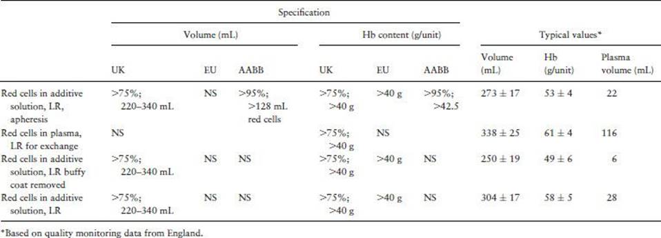
Red cell components (for specifications, see Table 21.2)
For the vast majority of red cell components, an additive solution is introduced following separation, to achieve a haematocrit of 50–70% and maintain red cell quality during storage. Red cells used for intrauterine transfusions (IUT) and exchange transfusion of neonates are normally stored or reconstituted in plasma. The most important changes that occur during storage are loss of intracellular potassium and a reduction in red cell recovery following transfusion. Red cell concentrates in additive solution have a 35–42-day shelf life (depending on the how the red cells are produced and the storage solution), at a controlled temperature of 2–6°C. Red cells which are stored only in plasma have a 21–35-day shelf life. To minimize the possibility of bacterial proliferation and maintain viability, red cells should be removed from refrigeration as little as possible. For patients with severe febrile or anaphylactic reactions to red cells or those with immunoglobulin A (IgA) deficiency, red cells are washed and resuspended in saline or an approved additive solution. The objective of washing is to remove as much plasma as possible, as such reactions can be due to antibodies to plasma proteins. At least one closed system for cell washing is now available, which allows red cells to be stored after washing, albeit with a shortened shelf life. Red cells from donors with rare phenotypes or from occasional patients with multiple red cell alloantibodies, for whom provision of compatible donor blood is extremely difficult, can be stored frozen for 30 years. Prior to transfusion, frozen red cells are thawed and washed to remove the cryoprotectant used to store them.
Pathogen inactivation in red cells
Red cells present a challenge for photochemical pathogen inactivation methods due to the high degree of light absorption by haemoglobin. Two systems are currently in development and show some promise. The first, the Intercept system, uses a compound that does not depend on light activation, instead using a long incubation period (18 hours) to allow binding of a linker to the DNA, preventing pathogen replication. The in vitro quality and in vivo recovery of the red cells appears to be satisfactory at the end of their shelf life [5]. However, this was also true for a previous version of the same system, for which phase III chronic transfusion studies were suspended. This was due to antibodies being detected in a small number of recipients – these reacted with red cells after, but not before, pathogen inactivation, raising concern that the treatment step resulted in the formation of neoantigens on red cells. The manufacturer has since modified the system to reduce this risk and phase III trials are currently underway. An alternative system, Mirasol, is being developed to treat whole blood, using riboflavin and a high dose of UV light. It is initially being developed for military use and studies on components produced from inactivated whole blood are ongoing, with the recovery of red cells in vivo shown to be acceptable [6]. It is likely that it will be 2–5 years before either system for red cells is licensed for routine use.
Prion reduction in red cells
Since leucocyte reduction alone is unlikely to render units free of PrPsc, there is considerable interest in alternative methods to reduce the risk of transmission of vCJD by transfusion. Filters that remove prion protein from red cell concentrates are well advanced in their development, with the P-Capt filter developed by PRDT in collaboration with Macopharma now licensed in Europe. As yet, there are no prion removal filters for whole blood, platelets or single donor plasma. The P-Capt filter requires prior LR, so is associated with a further 10–15% loss of haemoglobin. Prion removal and LR may be combined into one filter in the future, with more than one company working on such an approach. On the basis of current working assumptions on levels of infectivity and prevalence of infection in the UK population, it is predicted that at least 3 log removal of infectivity (in addition to LR) would be needed to provide clinical benefit in terms of preventing transmission of vCJD. The P-Capt prion removal filter has been reported to remove 3–4 logs of infectivity from red cells spiked with scrapie-infected hamster brain and >1.2 log (to below the limit of detection) of infectivity from the blood of hamsters infected with scrapie [7]. The UK transfusion services have also commissioned an independent assessment of the efficacy of prion reduction, since evidence to date has been generated solely by the manufacturers. The P-Capt filter has been shown to have negligible effect on the in vitro quality of red cells, the expression of common red cell antigens or recovery of red cells following transfusion to healthy volunteers. In the UK, SaBTO has recommended that prion filtration be implemented for patients born after 1 January 1996, subject to satisfactory results from a clinical safety study in surgical patients. Post-marketing surveillance would be instigated to monitor unexpected reactions and alloimmunization, with baseline data being collected for 6 months prior to any implementation of prion filtration. The safety study (PRISM) is now complete, and no adverse effects on the safety of transfusion were attributed to the PCapt filter. However, SaBTO has deferred review of the recommendation to implement filtration until the independent evaluation of efficacy is complete. The outcome of the review is expected in 2013.
Platelet concentrates
Platelet production and storage
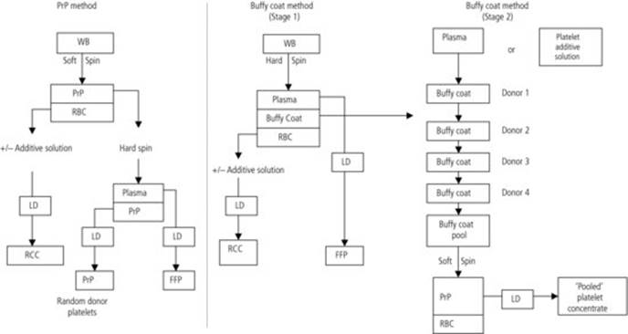
Platelets may be produced either from whole blood donations or by apheresis, in which platelets with or without plasma are collected and the red cells returned to the donor. Specifications for platelet yield and residual leucocyte count are similar for the two methods (see Table 21.3). Apart from exposing the patient to fewer donors and the possibility of HLA/HPA matching with the patient, apheresis platelets are not intrinsically of higher quality. Platelet production from whole blood may be carried out either from pooled buffy coats generated by bottom and top processing or from platelet-rich plasma (PRP) as an intermediate step (Figure 21.2). Buffy coat-derived platelets have long been favoured in Europe, are standard in the UK and have recently been adopted in Canada, while the PRP method is standard in the USA. LR by filtration may be routinely incorporated into either process. An adult therapeutic dose of platelets (2.5–3.0 × 10¹¹) can be manufactured from buffy coats, or by the PRP method, from four to six whole blood donations. In contrast, with the appropriate selection of donors, 1–3 adult doses can be harvested from a single donor during one apheresis collection procedure.
Table 21.3 Specification and typical values for volume and platelet content for LR platelet components.

Platelets are stored with agitation in incubators set at 20–24°C for 5 days, which may be extended to 7 days if a bacterial screening or pathogen inactivation method is used. Platelet concentrates should never be placed in the refrigerator as this impairs the recovery and survival of platelets following transfusion. With pre-storage LR and modern storage packs, platelets stored for 7 days in plasma maintain their in vitro function well. Apheresis platelets stored in plasma for 7 days have shown acceptable recovery and survival and several countries are performing additional clinical studies to assess the functionality of platelets stored beyond 5 days. During storage, platelets undergo a fall in pH due to accumulation of lactate, show increased surface expression of activation markers such as P-selectin (CD62P) and change from discoid to round. Many different laboratory assays have been advocated to monitor development of this so-called ‘platelet storage lesion’ but few have been demonstrated to correlate with in vivo survival. pH remains the only quantitative change that must be monitored routinely and must be above 6.2–6.4 at outdate. Visual inspection to look for the ‘swirling’ effect of discoid platelets has been recommended, but this is highly subjective and changes only when the platelets have been grossly damaged.
Fig 21.2 Production of platelet components from whole blood.
For patients with severe anaphylactic-type reactions, which are usually due to plasma proteins, it is possible to prepare platelets to be virtually plasma free by centrifuging the platelet concentrate, removing the supernatant plasma and replacing it with storage medium. The component is sometimes referred to as ‘washed platelets’, although there is no actual wash step, as this is unnecessary and may lead to platelet activation. Platelet storage media differ from red cell additive solutions in containing some or all of potassium, magnesium, acetate, citrate, phosphate, gluconate and chloride. In Europe, platelets in 100% storage medium have only a 24-hour shelf life, although it is possible that this could be extended using newer solutions containing glucose that are in development. A different component, containing approximately 70% storage medium and 30% plasma is now in production in a number of countries. This strategy makes more plasma available for fractionation, appears to reduce minor allergic reactions and allows a normal shelf life. Clinical data to day 7 and beyond are limited. These solutions have great potential, but require careful validation, which may need to include volunteer and patient studies.
Pathogen reduction in platelets
In order to mitigate the risk of bacterial growth in platelet concentrates stored at 20–24°C and other viral risks, a number of technologies have been developed using UV light, with or without specific additives, to inactivate pathogens. The mode of action of these technologies is to modify nucleic acid, thus preventing replication. This technology has no effect on prions, which lack nucleic acid.
Amotosalen, which belongs to a group of naturally occurring compounds called psoralens, is employed in one system for pathogen inactivation of platelet concentrates (Intercept, S-59). Amotosalen/UV-A treatment results in a high degree of killing of the major transfusion-transmitted viruses HIV, HCV and HBV, and intracellular pathogens such as CMV and HTLV. Following the treatment step, it is necessary to remove the amotosalen from the component prior to storage. A number of randomized clinical trials have been performed and have shown that amotosalen-treated platelets, whether prepared by apheresis or from pooled buffy coats, are effective in preventing haemorrhage in thrombocytopenic patients with haematological malignancies. However, platelet increments and intertransfusion intervals were less favourable than in control patients, raising the possibility that increased numbers of platelet units might be required to support such patients [8,9]. This question can be answered only by further large-scale clinical studies. A recent study reported an increased risk of bleeding in the treatment group, although the study was open label and not powered with bleeding as the primary endpoint [10].
An alternative system for pathogen reduction in platelets, called Mirasol, has been developed using riboflavin (vitamin B2) and UV light. A removal step following treatment is not needed. Platelets treated using this system show good in vitro quality, and acceptable recovery and survival in healthy subjects. However, as with Intercept-treated platelets, a clinical trial of the component showed lower count increments in the treatment group and more frequent transfusions being necessary, although noninferiority was not demonstrated [11]. A third system in development – the Theraflex system – uses no additive and exposure to UV-C light. In vitro studies and recovery and survival studies in healthy volunteers appear acceptable, and phase II/III clinical safety and efficacy studies are now in progress [12].
Both the Intercept and Mirasol systems are licensed in Europe, but not by the FDA in the USA. They are in routine use in some European countries, but not yet the UK. In addition to their inactivation of pathogens, inactivation of leucocytes offers an alternative to irradiation of components to prevent TA-GvHD. Pathogen inactivation of platelets would obviate the need for irradiation and CMV testing of components and for some systems permit a 7 day shelf life; this would simplify platelet stock management and reduce wastage. Their broad range of activity against viruses would also be expected to confer protection against new emerging infections that may be transmitted by transfusion. However, until a red cell pathogen inactivation system is also available, some of these benefits cannot be realized in full.
Fresh frozen plasma
Definition and specification
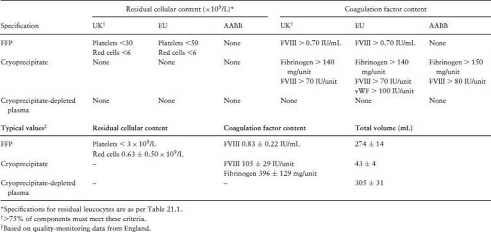
FFP is the plasma from a single donation, usually 250–300 mL, which has been frozen soon after collection without pooling. FFP can also be derived from apheresis collections, in 300 or 600 mL volumes. It is used primarily as a source of multiple coagulation factors in situations such as massive transfusion, disseminated intravascular coagulation and liver disease (Chapters 26 and 27). The permitted shelf life (12 months to 7 years) depends on the storage temperature. In Europe, FFP must be monitored for levels of factor VIII (Table 21.4). Although most FFP is prescribed for patients with normal or elevated factor VIII levels, it is selected for quality-monitoring purposes, as it is labile and hence sensitive to exposure to adverse conditions. FFP is thawed (in a protective overwrap to prevent bacterial contamination) in a waterbath, a purpose-designed microwave oven or dry heat source. Once thawed, FFP should be used as soon as possible since the levels of labile coagulation factors decline during further storage. Some countries permit thawed plasma to be used for up to 5 days if it is labelled as a different component.
Table 21.4 Specifications and typical values for residual cellular and coagulation factor content of frozen plasma components.
Pathogen inactivation
Four systems for producing pathogen-inactivated FFP are now available and licensed in Europe. Three are suitable for single donor plasma: methylene blue (MB), amotosalen and riboflavin. The other, solvent–detergent (SD) treatment, is applied to pools of plasma. All methods offer good virus protection, but all are associated with loss of clotting factors [13]. The key features of pathogen inactivated FFP are shown in Table 21.5.
Table 21.5 Comparison of pathogen inactivation methods for plasma.
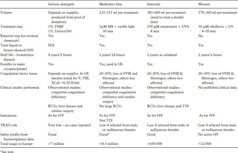
MB is a phenothiazine dye which, when exposed to white light, generates reactive oxygen species that damage nucleic acids, preventing viral replication. Treatment is applied to single units of plasma and requires prior removal of leucocytes by filtration or freeze–thawing. The MB is contained in or added to the integral pack system, mixed with the plasma and then placed on a light box for activation. The MB is removed using an adsorption filter prior to final storage of the component, leaving residual MB concentrations of <0.3 μM. At these concentrations, no toxicity has been demonstrated or is predicted. Glucose 6 phosphate dehydrogenase deficiency is not a contraindication to use of this product. The amotosalen and riboflavin treatment systems are similar to those described for platelets.
SD treatment can be applied only to pools of several hundred ABO-identical units; as the treatment destroys the lipid envelope of red cells, no RhD matching is required. Exposure to SD destroys the lipid envelope of HIV, HBV and HCV, and no such transmissions have been reported. Non-lipid-coated viruses such as parvovirus B19 and hepatitis A are not specifically inactivated, but their titre may be reduced in downstream processing. In addition, plasma pools with high genomic titres of these viruses are rejected and pools contain specified levels of viral antibodies, which may be at least partially protective. No increase in clinical cases of hepatitis A virus or B19 in SD FFP recipients is evident. Concerns have been expressed in the United States that SD FFP might be associated with increased thrombotic risk in certain clinical situations, attributed to loss of proteins C and S during SD treatment. However, these complications have not been prominent in recipients of SD FFP manufactured by the European method, which results in greater preservation of these proteins. A variant of SD FFP in which ABO groups are mixed is in development. By neutralization of A and B substances by anti-A and anti-B, it is intended to produce a ‘universal FFP’ that could be given to patients of any ABO group. A lyophylized SD FFP product is also in development.
The French regulatory authority have recently announced a phased withdrawal of MB FFP in response to haemovigilance data suggesting that severe allergic reactions are more common than to untreated or SD FFP. Their investigations suggest that in some cases these appear to be reactions to MB itself rather than plasma proteins. Other European countries that use MBFFP have not observed higher rates of allergic reactions to MB FFP compared with untreated FFP.
vCJD and FFP
In animal studies, plasma was found to contain infective prion. Therefore, a precautionary measure for the UK was announced in 2002 whereby FFP for children born on or after 1 January 1996 would be imported from a country with a low risk of vCJD. This was implemented in 2004, with plasma coming from volunteer donors extensively tested for transfusion-transmitted viruses. Plasma is treated with the MB pathogen inactivation process following arrival in the UK. In 2005, the use of imported MB FFP was extended to patients under the age of 16, and in 2012 the recommendation was made to continue its supply to those born on or after 1 January 2012.
Cryoprecipitate and cryosupernatant
Cryoprecipitate is manufactured by slowly thawing single units of FFP overnight at 4°C. This precipitates out the so-called cryoproteins, namely factor VIII, fibrinogen, fibronectin and factor XIII. By removing most of the supernatant plasma (‘cryosupernatant’), a component providing a high concentration of these clotting factors is obtained (Table 21.4). Although originally developed for factor VIII deficiency (haemophilia A), most cryoprecipitate is now prescribed to treat congenital or acquired hypofibrinogenaemia, usually in the context of liver disease, disseminated intravascular coagulation or massive transfusion. An adult dose of 10–12 packs is generally indicated once the fibrinogen level falls below 0.5–1.0 g/L. Some countries pool 5 units of cryoprecipitate to facilitate its administration. An alternative product would be a virus-inactivated fibrinogen concentrate, but in many countries this is only licensed for congenital deficiency and clinical studies demonstrating efficacy of either cryoprecipitate or fibrinogen concentrate for acquired deficiency are lacking. Cryosupernatant has been used successfully as a replacement fluid in plasma exchange procedures for thrombotic thrombocytopenic purpura (TTP). It was thought to have theoretical advantages over FFP, possibly because it lacks the highest molecular weight multimers of the von Willebrand factor, although recent studies suggest that FFP is equally effective.
Virus inactivation of cryoprecipitate and cryosupernatant
Production of cryoprecipitate from SD FFP has been performed experimentally. Such cryoprecipitate contains insufficient von Willebrand factor to treat patients with von Willebrand's disease, but acceptable levels of fibrinogen. Cryoprecipitate produced from MB plasma is routinely used in the UK for patients under the age of 16. Intercept and Mirasol-treated plasma can also be used to produce cryoprecipitate.
Granulocytes for transfusion
The use of transfused granulocytes is uncommon. They are sometimes used for severely neutropenic patients (granulocyte count <0.5 × 10⁹/L) with focal bacterial or fungal infection refractory to antimicrobial therapy, but there are difficulties in obtaining sufficient functional cells from donors and administering them frequently enough to the patient. Granulocytes can either be collected by apheresis or produced from whole blood. Animal studies suggest that >1 × 10¹⁰ granulocytes once or twice daily are required to treat an adult, but apheresis usually produces no more than 0.5 × 10¹⁰/dose unless donors are stimulated with granulocyte colony-stimulating factor (G-CSF). Therefore, unstimulated apheresis granulocytes are not suitable for adults. There has been renewed interest in the use of granulocytes by studies of granulocyte colony-stimulating factor (G-CSF) mobilized granulocytes collected by apheresis. Administration to the donor of a single subcutaneous injection of 10 μg/kg G-CSF plus oral dexamethasone 8 mg 12–24 hours prior to apheresis raises the peripheral leucocyte count to >25 × 109/L. This, coupled with starch sedimentation, allows collection of a therapeutic dose of granulocytes of 5–20 × 108 granulocytes/kg body weight of the recipient. This can result in a measurable rise in the peripheral granulocyte count in the patient and recovery of migrated cells from saliva. Clinical trials of such granulocytes are ongoing. At present, use of G-CSF for granulocyte collection is permitted in volunteer donors unrelated to the patient in the USA but not in other countries.
Some European countries transfuse buffy coats as a source of granulocytes. A dose of 1 × 10¹⁰ can be achieved from 10 buffy coats. A pooled granulocyte component made from 10 buffy coats has been developed in the UK and its safety assessed in clinical studies. The main advantage of the pooled component over standard buffy coats is a reduction in red cell contamination and volume, and that it is issued as a single unit.
All granulocyte preparations should be released for issue as soon as possible after collection, which may mean that certain time-consuming screening assays such as HCV genome testing cannot be done prior to release. They must be gamma irradiated to prevent TA-GvHD and should be administered to the patient without delay. If a short period of storage is unavoidable, this should be at 22°C without agitation. Because of red cell contamination, a red cell cross-match should be performed.
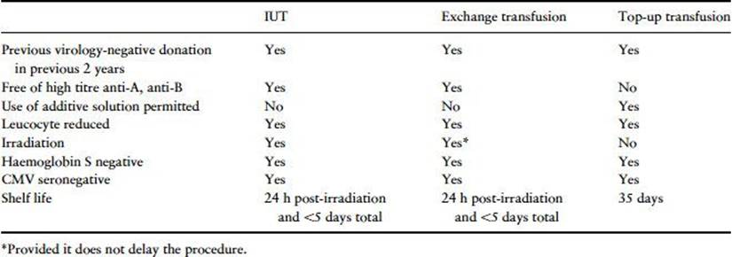
Table 21.6 UK specifications for red cells for intrauterine transfusion (IUT), exchange/large volume transfusions and ‘top-up’ transfusions for neonates.
Components for intrauterine transfusion and for neonates and infants
General requirements (see Table 21.6)
Cellular components for these recipients should be selected or processed to reduce the risk of CMV transmission. Components other than those in additive solution must be free of clinically significant red cell antibodies, including high titre anti-A and anti-B. Irradiation is required for intrauterine and exchange transfusions providing it does not unnecessarily delay the procedure. Red cells for ‘top-up’ transfusions need be irradiated only if there has been a previous intrauterine transfusion (IUT), in severe T lymphocyte deficiency syndromes or if the component is prepared from a family member. Family donations are not encouraged except in rare cases of fetomaternal alloimmunization where the infant's requirements cannot be met from donor blood. Although components are leucocyte depleted at source, they should still be administered through a 170–200-μm filter to remove any microaggregates formed during storage.
Intrauterine/exchange/large-volume transfusion of neonates
Red cells are given in utero to treat severe fetal anaemia due to haemolytic disease of the fetus and newborn (HDFN) or parvovirus B19 infection. Red cells for IUT are prepared from plasma-reduced blood to a haematocrit of 0.7–0.85. In cases of fetomaternal alloimmunization to platelets, transfusions of selected platelets (usually HPA-1a and HPA-5b negative) may be given in utero (Chapter 5). Apheresis of genotyped donors can be used to produce a hyperconcentrated platelet for this indication. Alternatively, platelets can be concentrated from a whole blood donation.
Exchange transfusion is undertaken to treat hyperbilirubinaemia due to either haemolytic disease or prematurity (Chapter 32). Either whole blood or partially packed red cells with a haematocrit of 0.5–0.6 may be used. Red cells in additive solution are not recommended by some paediatricians for exchange transfusion, because of concerns regarding the adverse effects of mannitol. Some countries use red cells in additive solution for large-volume transfusion of neonates and infants without adverse effect (see Chapter 32). To reduce the risk of hyperkalaemia, red cells for exchange or IUT should be administered within 24 hours of irradiation and by the end of day 5 from donation.
Top-up transfusions for neonates
Premature neonates are among the most heavily transfused patients in any hospital. Most red cell transfusions are given to replace repeated samples taken for laboratory testing. As each infant may require multiple small transfusions, adult packs are split into four to eight ‘paedipacks’ of 30–60 mL, which can be allocated to one infant for the duration of transfusion dependence. Such a strategy reduces donor exposure considerably. For these small-volume transfusions, red cells in additive solution may be used, up to their normal shelf life. Studies of RhEpo in premature infants have not convincingly shown a reduction in transfusion requirements.
Platelet concentrates and FFP
Platelets are most simply prepared from apheresis donations. Multiple aliquots can be allocated to the same infant if required. An alternative strategy for platelets is to prepare a platelet concentrate from a single buffy coat or from a unit of whole blood using the PRP method. These components are generally used for sick babies with multiple coagulation defects. Platelets from a panel of HPA-1a and 5b-negative donors can be used in suspected cases of neonatal alloimmune thrombocytopenia (Chapter 5).
Key points
1. There are no recognized indications for the transfusion of whole blood and therefore blood is separated into its components for transfusion (red cells, plasma and platelets).
2. Blood components can be produced from whole blood donations or collected directly from the donor by apheresis technology.
3. In the UK and some other countries, all blood components are leucocyte-reduced but others such as the USA have a variable proportion of leucocyte-reduced blood components.
4. Systems are now available in Europe to inactivate pathogens in plasma or platelet components prior to storage.
5. Filters have been developed that are designed to remove prion protein from red cells to reduce the risk of transmission of vCJD.
References
1. Advisory Committee on the Safety of Blood, Tissues and Organs. Position statement on CMV transmission by blood transfusion. http://www.dh.gov.uk/health/about-us/public-bodies-2/advisory-bodies/sabto/.
2. Pennington J, Taylor GP, Sutherland J, Davis RE, Seghatchian J, Allain JP & Williamson LM. Persistence of HTLV-I in blood components after leukocyte depletion. Blood 2002; 100: 677–681.
3. Gregori L, McCombie N, Palmer D et al. Effectiveness of leucoreduction for removal of infectivity of transmissible spongiform encephalopathies from blood. Lancet 2004; 364: 529–531.
4. McCutcheon S, Alejo Blanco AR, Houston EF, de Wolf C, Tan BC, Smith A, Groschup MH, Hunter N, Hornsey VS, MacGregor IR, Prowse CV, Turner M & Manson JC. All clinically-relevant blood components transmit prion disease following a single blood transfusion: a sheep model of vCJD. PLoS One 2011; 6(8): e23169. Epub 17 August 2011.
5. Cancelas JA, Dumont LJ, Rugg N, Szczepiorkowski ZM, Herschel L, Siegel A, Pratt PG, Worsham DN, Erickson A, Propst M, North A, Sherman CD, Mufti NA, Reed WF & Corash L. Stored red blood cell viability is maintained after treatment with a second-generation S-303 pathogen inactivation process. Transfusion 2011; 51: 2367–2376.
6. Cancelas JA, Rugg N, Fletcher D, Pratt PG, Worsham DN, Dunn SK, Marschner S, Reddy HL & Goodrich RP. In vivo viability of stored red blood cells derived from riboflavin plus ultraviolet light-treated whole blood. Transfusion 2011; 51: 1460–1468.
7. Gregori L, Gurgel PV, Lathrop JT, Edwardson P, Lambert BC, Carbonell RG, Burton SJ, Hammond DJ & Rohwer RG. Reduction in infectivity of endogenous transmissible spongiform encephalopathies present in blood by adsorption to selective affinity resins. Lancet 2006; 368: 2226–2230.
8. Snyder E, McCullough J, Slichter SJ, Strauss RG, Lopez-Plaza I, Lin JS, Corash L & Conlan MG; SPRINT Study Group. Clinical safety of platelets photochemically treated with amotosalen HCl and ultraviolet A light for pathogen inactivation: the SPRINT trial. Transfusion 2005; 45: 1864–1875.
9. van Rhenen D, Gulliksson H, Cazenave JP, Pamphilon D, Ljungman P, Klüter H, Vermeij H, Kappers-Klunne M, de Greef G, Laforet M, Lioure B, Davis K, Marblie S, Mayaudon V, Flament J, Conlan M, Lin L, Metzel P, Buchholz D & Corash L; euroSPRITE trial. Transfusion of pooled buffy coat platelet components prepared with photochemical pathogen inactivation treatment: the euroSPRITE trial. Blood 2003; 101: 2426–2433.
10. Kerkhoffs JL, van Putten WL, Novotny VM, Te Boekhorst PA, Schipperus MR, Zwaginga JJ, van Pampus LC, de Greef GE, Luten M, Huijgens PC, Brand A & van Rhenen DJ; Dutch–Belgian HOVON Cooperative Group. Clinical effectiveness of leucoreduced, pooled donor platelet concentrates, stored in plasma or additive solution with and without pathogen reduction. Br J Haematol 2010; 150: 209–217.
11. Mirasol Clinical Evaluation Study Group. A randomized controlled clinical trial evaluating the performance and safety of platelets treated with MIRASOL pathogen reduction technology. Transfusion 2010; 50: 2362–2375.
12. Mohr H, Steil L, Gravemann U, Thiele T, Hammer E, Greinacher A, Müller TH & Völker U. A novel approach to pathogen reduction in platelet concentrates using short-wave ultraviolet light. Transfusion 2009; 49: 2612–2624.
13. Rock G. A comparison of methods of pathogen inactivation of FFP. Vox Sanguinis 2011; 100: 169–178.
Further reading
American Association of Blood Banks. Standards for Blood Banks and Transfusion Services, 27th edn. Bethesda, MD: AABB Press; 2011.
British Committee for Standards in Haematology. Guidelines for the use of platelet transfusions. Br J Haematol 2003; 122: 10–23.
British Committee for Standards in Haematology. Transfusion guidelines for neonates and older children. Br J Haematol 2004; 124: 433–453.
British Committee for Standards in Haematology. Guidelines for the use of fresh-frozen plasma, cryoprecipitate and cryosupernatant. Br J Haematol 2005; 126: 11–28.
Council of Europe. Guide to the Preparation, Use and Quality Assurance of Blood Components, 16th edn. Strasbourg: Council of Europe Publishing; 2010.
McClelland DBL (ed.). Handbook of Transfusion Medicine, 4th edn. London: The Stationery Office; 2007.
United Kingdom Blood Transfusion Services/National Institute for Biological Standards and Control. Guidelines for the Blood Transfusion Services in the United Kingdom, 7th edn. London: The Stationery Office; 2005. Available at: www.transfusionguidelines.org, UK.
Webert KE, Cserti CM, Hannon J, Lin Y, Pavenski K, Pendergrast JM & Blajchman MA. Proceedings of a Consensus Conference: pathogen inactivation-making decisions about new technologies. Transfus Med Rev 2008; 22: 1–34.