
Irene Roberts1, Naomi Luban2 & Helen V. New3
1Departments of Haematology and Paediatrics, Imperial College London, London, UK
2George Washington University School of Medicine and Health Sciences, Washington, DC, USA
3Department of Paediatrics, Imperial College Healthcare NHS Trust/NHS Blood and Transplant, London, UK
Introduction
Transfusion of children is a specialized area, particularly in the perinatal period, and requires close cooperation between experts in obstetrics and fetal medicine as well as neonatologists, paediatricians, haematologists, transfusion medicine specialists, nursing and laboratory staff. Recipients of fetal and neonatal transfusions are particularly vulnerable to potential side effects of transfusion, and a disproportionate number of errors occur during paediatric transfusion [1]. It is therefore important that staff involved understand the principles of paediatric special components, criteria for transfusion, how to prescribe and administer paediatric components, and the need for long-term follow-up when considering the consequences of transfusion.
This chapter briefly describes the transfusion management of the most important perinatal and paediatric disorders where specialized transfusion support is essential. The three sections (transfusion of the fetus, neonate and older child) discuss treatment of disorders including haemolytic disease of the fetus and newborn (HDFN), alloimmune and autoimmune thrombocytopenia, neonatal coagulopathy, transfusion support for children on paediatric intensive care and for those with haemoglobinopathies. While historical practice has been based mostly on expert opinion, there is an increasingly evidence-based approach in several areas. There is variation in neonatal component provision and transfusion practice between countries [2] and in general this chapter describes UK recommendations.
General issues
Adverse outcomes of paediatric transfusion
Reports to the UK national haemovigilance scheme, the Serious Hazards of Transfusion (SHOT) scheme, have suggested a disproportionate number of adverse outcomes of transfusion in paediatric patients compared to adults, particularly in infants and neonates [1]. A significant proportion of reports relate to transfusion errors such as transfusion of an incorrect blood component, including components lacking special requirements. For the neonatal and infant age groups, there have been relatively fewer reports of adverse reactions to transfusion than in older children, and it is likely that these are underreported, perhaps because they may have a more subtle presentation in these patients or are masked by intercurrent illness.
In the UK, a number of measures have been introduced to reduce the theoretical risk of transfusion transmission of variant CJD (vCJD) to all recipients, including universal leucocyte reduction of blood components, and there are increased measures for recipients born after 1 January 1996, such as providing imported FFP.
Recognized hazards of transfusion in neonates
Transfusion of neonates has become safer, especially with the more widespread use of satellite packs. The most important recognized hazards of transfusion in neonates are:
· infection: bacterial or viral;
· hypocalcaemia (more common in neonates than in infants or children);
· volume overload;
· citrate toxicity;
· rebound hypoglycaemia (high glucose levels from blood additives);
· hyperkalemia (from large-volume transfusion);
· thrombocytopenia (after exchange transfusion);
· transfusion-associated graft-versus-host disease (TA-GvHD) (if nonirradiated blood components are given to those at risk; see below and Chapter 11).
There are theoretical concerns over potential toxicity of adenine and mannitol if given in large volumes to neonates [3], but a randomized trial showed apparent safety of these additives in neonatal cardiac surgery [4], and they are widely used for neonatal cardiac surgery in the UK with no evidence of adverse effects. Several retrospective studies have also reported an association between red blood transfusions and subsequent necrotizing enterocolitis (NEC) in neonates [5,6], but proof of a causal association requires prospective studies.
Strategies to minimize transfusion risk in neonates
a. Provision of special components for the fetal/neonatal age group (principles and UK practice):
· Reduction of infection risk:
· cytomegalovirus (CMV)-safe: for UK, seronegative ‘accredited’ donors who have donated at least twice in the previous 2 years; for the United States, leucocyte-reduced blood may be considered as CMV-safe without in addition being tested as CMV seronegative; the precise efficacy of combining both leucocyte reduction and CMV-seronegativity to prevent transfusion transmission of CMV to neonates is not known, and there is a current observational study to try to define this in neonates <1500 g birth weight [7];
· for UK, pathogen-inactivated FFP imported from a country with low risk of vCJD (also for all recipients born after 1 January 1996).
· Reduction of risk of morbidity from antibodies in donor plasma:
· screen donors for high titre anti-A, anti-B and atypical antibodies;
· for neonates who are not group O, do not use group O FFP and avoid group O platelets where possible.
· Reduction of risk of hyperkalaemia in large-volume transfusion (e.g. neonatal exchange):
· use fresh blood (<5 days old).
· Reduction of risk of toxicity from additives in large-volume transfusion:
· use citrate-phosphate-dextrose (CPD) for intrauterine transfusion (IUT) and neonatal exchange transfusions; saline-adenine-glucose-mannitol (SAG-M) for other large volume and top-up transfusions (in order to reduce the plasma content of the units and hence the theoretical risk of transfusion transmission of vCJD). Some countries resuspend red cells for neonatal exchange in plasma.
· Provision of haematocrit appropriate to clinical situation:
· high (0.70–0.85) for IUT, to reduce volume overload; intermediate (0.5–0.6) for neonatal exchange transfusion; broader range (0.5–0.7) for neonatal top-up transfusion.
· Irradiation of cellular components to prevent TA-GvHD for at-risk patients (see below).
b. The following strategies have been shown to reduce the need for red cell transfusion and/or donor exposure in neonates:
· development and implementation of transfusion guidelines;
· minimizing iatrogenic blood sampling;
· prevention and treatment of haematinic deficiencies (iron and folic acid);
· use of dedicated satellite packs (‘paedipacks’);
· judicious use of erythropoietin (see below);
· autologous cord blood transfusion through delayed clamping of the cord at delivery.
Prescription and administration of blood for neonates and children
There are frequent reports of overtransfusion of neonates and small children due to the prescription of blood components in ‘units’ rather than ‘mL’. It is therefore recommended that transfusion prescriptions for these patients should be calculated in mL/kg and written up as a precise volume over a given time. Small-volume administration sets are available for transfusions to neonates in order to reduce the proportion of dead space in the giving set.
The fetus and neonate: crossmatching and general considerations
Management of the fetus and neonate in specific transfusion situations is discussed in detail below. The general principles for transfusion are summarized here:
· Prior to the first transfusion, samples should be obtained from the mother for ABO, RhD grouping and antibody screening and from the fetus/neonate for ABO, RhD and direct antiglobulin test (DAT) (plus an antibody screen if no maternal sample is available).
· In the fetus/neonate, the ABO group is determined on the cells only (as most infants do not produce anti-A and anti-B until 3–6 months of age).
· Red cells that are ABO compatible with maternal and neonatal plasma, RhD identical with neonate or negative should be used (note if exchange or ‘top-up’ transfusion is required for HDFN due to ABO incompatibility, group O red cells with low titre anti-A and -B or group O red cells suspended in AB plasma should be used).
· Group O blood is acceptable; units with high titre anti-A/anti-B must be excluded.
· If the mother's blood group is unknown, blood for the fetus/neonate should be crossmatched against the baby's plasma.
· If no atypical antibodies are present in the maternal (or infant) sample, and if the DAT of the infant is negative, crossmatching is not necessary for the first 4 months of postnatal life. It is preferable to use the maternal sample for the antibody screen: any significant antibodies may be at higher levels and therefore more easily detectable than in the infant plasma and it will also be easier to obtain sufficient sample.
· If the antibody screen or DAT is positive, full serological investigation and compatibility testing are necessary.
· Electronic crossmatch should only be used where it is controlled by an appropriate algorithm that takes into account maternal group and antibody screen and the baby's group and DAT. Red cells (and platelets if given) should be CMV safe and leucocyte reduced.
· Note that alloantibody formation is rare in the fetus and neonate and is usually associated with massive transfusion or with the use of fresh or whole blood.
· Gamma-irradiation of cellular blood components to reduce the risk of TA-GvHD is recommended for:
· IUTs;
· transfusions to neonates previously transfused in utero;
· exchange transfusions as long as gamma-irradiation would not result in a delay in transfusion;
· transfusions from a family member;
· neonates with known or suspected inherited T-lymphocyte immune deficiencies (e.g. severe combined immunodeficiency).
These precautions are due to the immaturity of the fetal and neonatal immune system, which may lead to a reduced ability to reject transfused allogeneic lymphocytes, immune tolerance and the persistence of donor lymphocytes for up to 6–8 weeks after exchange transfusion.
Specific transfusion situations
Fetal transfusion
Fetal transfusions are a particularly specialized area of transfusion medicine, undertaken by fetal medicine specialists with the support of haematologists and transfusion medicine and laboratory staff. Intrauterine transfusions of red cells are given for the prevention and treatment of fetal anaemia, most commonly for HDFN and parvovirus infection (see Table 32.1 for causes of fetal anaemia). Intrauterine platelet transfusions may be given as part of the management of neonatal alloimmune thrombocytopenia (NAIT) (Chapter 5). Issues surrounding the investigation and management of suspected HDFN or NAIT are commonly encountered on neonatal units.
Table 32.1 Principal causes of fetal and neonatal anaemia.
|
Impaired red cell production · Diamond–Blackfan anaemia · Congenital infection, e.g. parvovirus, CMV · Congenital Dyserythropoietic Anaemia · Pearson's syndrome |
|
Haemolytic anaemias · Alloimmune: haemolytic disease of the fetus and newborn (Rh, ABO, Kell, other) · Autoimmune, e.g. maternal autoimmune haemolysis · Red cell membrane disorders, e.g. hereditary spherocytosis · Red cell enzyme deficiencies, e.g. pyruvate kinase deficiency · Some haemoglobinopathies, e.g. α thalassaemia major, HbH disease · Infection, e.g. bacterial, syphilis, malaria, CMV, toxoplasma, Herpes simplex |
|
Anaemia due to haemorrhage · Occult haemorrhage before or around birth, e.g. twin-to-twin, fetomaternal · Internal haemorrhage, e.g. intracranial, cephalhaematoma · Iatrogenic: due to frequent blood sampling |
|
Anaemia of prematurity · Due to impaired red cell production, impaired erythropoietin production and reduced red cell lifespan · Hb nadir usually 6.5–9 g/dL |
|
Causes in italics commonly present in the fetus; other causes may present during fetal life but neonatal presentation is more common. |
Haemolytic disease of the fetus and newborn
Red cell antibody testing in pregnancy
The three factors essential in the pathogenesis of HDFN are:
· maternal red cell alloantibodies that cross the placenta;
· fetal red blood cells that express antigens against which the antibodies are directed; and
· antibodies that are able to mediate red cell destruction.
Clinically relevant alloantibodies are almost always immunoglobulin G (IgG) and are reactive at 37°C. Women develop these antibodies as a result of previous transfusions, previous pregnancies or both. Identification of such antibodies is the main goal of antenatal screening.
The objectives of red cell antibody testing in pregnancy are to:
· identify red cell alloantibodies that are present at booking or develop during pregnancy;
· identify the pregnancy at risk of fetal or neonatal HDFN as a result of antibodies;
· identify the fetus requiring treatment in utero or in the neonatal period;
· identify RhD negative women who require anti-D prophylaxis (around 16% of women are RhD negative);
· ensure swift provision of compatible blood for obstetric emergencies (also see Chapter 26).
Red cell serology at the booking visit
At the booking visit, which should take place before the 16th week of pregnancy, all women should have their ABO and RhD group determined and should be screened for red cell alloantibodies. If red cell antibodies are detected at the booking visit and/or if there is a history of HDFN, the antibodies should be identified, quantified and monitored as outlined below. It is particularly important to monitor women with anti-D, anti-c and anti-K since these antibodies may be associated with severe HDFN affecting the fetus. Even if no red cell alloantibodies are detected at booking, all pregnant women should be retested at 28 weeks' gestation. Further testing of women without detectable antibodies is unnecessary since immunization later in pregnancy is unlikely to result in antibody levels sufficient to cause HDFN requiring treatment.
Partial D and weak D
Du (weak D) individuals, rather than having 10 000–100 000 RhD proteins on the surface of each red cell, as is usually the case, have 50–5000 per red cell. This low antigen density may be difficult to detect but as they have minimal or no structural RhD abnormality they are regarded as D positive, do not form immune anti-D and therefore do not require prophylaxis with anti-D. Individuals who have significant structural abnormalities of the RhD antigen with part of the protein missing are described as having partial D status, e.g. DVI. Partial D individuals can make anti-D against the epitopes of RhD that they lack if they are exposed to normal RhD antigens. Therefore partial D individuals shouldreceive anti-D prophylaxis and D negative red cell transfusions as if they were D negative. It is important that reagents for D grouping do not detect DVI (so that these individuals group as D negative).
ABO antibodies
There is no need to test for ABO immune antibodies in antenatal samples as their presence is not predictive of HDFN and such antibodies very rarely cause significant haemolysis in utero.
Samples at delivery
In the case of babies born to women with clinically significant red cell alloantibodies (see below), a DAT should be carried out on cord blood. If it is positive, a red cell eluate may help identify the red cell antibody. Infants born to mothers with clinically significant antibodies should be monitored for 48–72 hours for the presence of haemolysis (see below for management of HDFN in the neonate).
A DAT should not be routinely performed on all D-positive infants of D-negative mothers as maternal prophylactic anti-D may result in a positive DAT in the baby. This antibody binding should not cause destruction of the baby's red cells, but the report of a positive DAT may result in unnecessary additional investigations and concern.
D-negative mothers with no previously detected anti-D should have prophylactic anti-D administered if the infant is D positive. A Kleihauer test should also be carried out on all such women to assess the requirement for additional anti-D (see below).
Clinically relevant red cell alloantibodies
The main antibodies implicated in HDFN are:
· Rh group – D, c, C, e, E, Ce and Cw;
· Kell group – K1, K2 and Kpa;
· Duffy group – Fya;
· Kidd group – Jka.
Red cell alloantibodies not implicated in HDFN include anti-Lea and anti-Leb, anti-Lua, anti-P, anti-Xga and anti-Gerbish.
The antibodies most commonly implicated in severe to moderate HDFN are anti-D, anti-c and anti-K, and these are the three most likely to cause problems in the fetus as well as the neonate. Anti-K inhibits erythropoiesis as well as causing haemolysis.
Anti-D is the commonest cause of HDFN. This is because anti-D is highly immunogenic and a significant proportion of women are D negative (16%). Most anti-D antibodies are IgG1 or IgG1 plus IgG3. The presence of IgG3 alone, which has 100 times the destructive ability of IgG1, is uncommon and rarely associated with HDFN in utero, but can cause severe postnatal manifestations of HDN.
Anti-c is found most commonly in women with the R1R1 genotype (CDe/CDe), which occurs in 20% of pregnant women. Such women also have the propensity to make anti-E. HDFN due to anti-E is both less common and less severe. However, anti-E and anti-c in combination cause more severe HDFN than either antibody alone. Note that in such cases only the anti-E is detectable in eluates from cord blood red cells.
Anti-K1 is the most common red cell alloantibody outside the ABO and Rh system. K1 is the principal antigen of the Kell blood group system and is highly immunogenic; 5% of K1-negative individuals will produce anti-K1 if transfused with K1-positive blood. K1 has around twice the potency of c and E and 20 times the potency of Fya. Anti-K1 often causes severe HDFN; the haemolytic anaemia is compounded by suppression of erythropoiesis by anti-K1 inhibiting the growth of erythroid progenitor cells. Anti-K titres can be an unreliable predictor of the severity of HDFN. Therefore it is important to identify the fetuses at risk of HDFN by determining the fetal Kell genotype in all mothers with anti-K1 whose partners are heterozygous for K1 (since only 50% of such fetuses will be K1-positive). Moderate to severe HDFN may also be caused by anti-K2 (anticellano) and anti-Kpa.
A number of other red cell alloantibodies have also been reported to cause HDFN of variable severity, e.g. anti-U. These initially present with a positive indirect antiglobulin test (IAT) in maternal serum; therefore all women with a positive IAT should have further investigation to identify any clinically relevant red cell alloantibodies.
Serological monitoring of pregnant women with anti-D, anti-c or anti-K
Women with these antibodies should be tested at monthly intervals to 28 weeks gestation and subsequently at fortnightly intervals. All samples should be checked in parallel with the previous sample. The women should be referred to a specialist fetal medicine unit if the antibody reaches a critical level and/or if it rises significantly. In addition, those women who have previously had a baby affected by HDFN should be referred before 20 weeks gestation for assessment irrespective of the antibody level.
For anti-D and anti-c, the antibodies can be quantified, and increases of 50% or more compared with the previous sample are significant irrespective of gestation. Anti-D levels of 4–15 IU/mL are associated with a moderate risk of HDFN, and should trigger referral to a specialist unit, and a level of >15 IU/mL indicates a high risk of hydrops fetalis. For anti-c, levels of 7.5–20 IU/mL are associated with a moderate risk of HDFN and a level of >20 IU/mL indicates a risk of severe HDFN. Paternal phenotyping provides further information on the risk to the fetus. Fetal RhD and c typing on maternal blood are reliable and should be performed where the father is heterozygous and there is a risk of HDFN either from previous history or the antibody levels.
For anti-K, the antibody titres may not accurately reflect the degree of fetal anaemia although samples should be titred for comparison during pregnancy. Amniocentesis is also not a good indicator of the severity of fetal anaemia since anaemia due to anti-K results from a combination of haemolysis and red cell hypoplasia. All women with anti-K should be referred to a specialist fetal medicine unit early in pregnancy unless the father is confirmed K negative, and fetal K typing should also be used if it is available.
Management of HDFN in the fetus
Fetal monitoring of ‘at risk’ pregnancies
The aims are to prevent hydrops developing in utero and to time delivery so that the baby has the best chance of survival. Fetal monitoring includes the following:
· Weekly Doppler ultrasonography of the fetal middle cerebral artery should be carried out to identify fetal anaemia; this is reliable up to 36 weeks gestation.
· Regular ultrasound scans should be carried out for fetal growth, hepatosplenomegaly and/or hydrops.
· Where expertise for fetal Doppler ultrasonography is unavailable, amniocentesis may be used to measure amniotic fluid bilirubin as an indirect measure of fetal haemolysis; the bilirubin is plotted on a graph of Liley zones modified by Whitfield in order to predict the severity of HDFN and plan management.
· Fetal blood sampling should be carried out if severe HDFN before 24 weeks gestation is suspected, if there is a rapid rise in maternal antibody or if there has been a previous intrauterine death due to HDFN (fetal blood sampling carries a 1–3% fetal loss rate and may cause fetomaternal haemorrhage with further sensitization).
· IUT should be carried out if anaemia is severe and delivery is not possible due to extreme prematurity.
Intrauterine transfusion (IUT)
The aims of IUT are to:
· prevent or treat fetal hydrops before the fetus can be delivered;
· enable the pregnancy to advance to a gestational age that will ensure survival of the neonate (in practice, up to 36–37 weeks) with as few invasive procedures as possible.
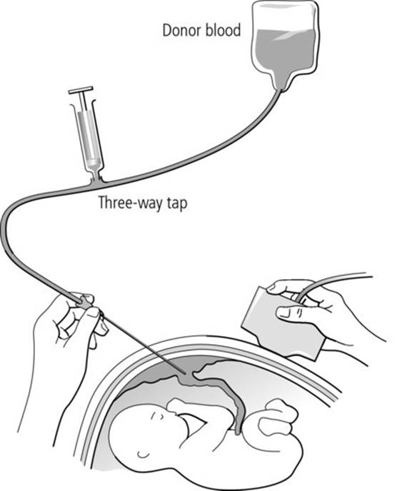
These are achieved by starting the transfusion programme as late as safely possible but before hydrops develops and maximizing the intervals between transfusions by transfusing as large a volume of red cells as is considered safe. Transfusions may be intravascular, intraperitoneal or intracardiac. All transfusions are carried out with ultrasound guidance (Figure 32.1). During transfusion the point of the needle and fetal heart should be watched closely for signs of needle displacement, cardiac tamponade and bradycardia. The fetal loss rate associated with IUT is around 1–3% but is higher when the fetus is hydropic. IUT is generally indicated when the haematocrit falls to below 0.25 between 18 and 26 weeks gestation or to less than 0.3 after 26 weeks gestation. The aim of the transfusion is to raise the haematocrit to around 0.45 and repeat transfusion is often necessary after 2–3 weeks.
Fig 32.1 Intrauterine transfusion. Reproduced from Practical Transfusion Medicine 3rd Edition.
Intrauterine transfusion: component specification
· Plasma-reduced red cells with a haematocrit of 0.7–0.85.
· The red cells should be 5 days old or less, in CPD anticoagulant and sickle screen negative.
· The red cells should be group O (low titre haemolysin) or ABO identical with the fetus (if known), D negative and red cell antigen negative for the identified maternal antibody (in practice, if the mother has anti-c, R1R1 red cells are used). K-negative blood is recommended to reduce maternal alloimmunization risks.
· An IAT-crossmatch compatible with maternal serum and negative for the relevant antigen(s) determined by maternal antibody status should be carried out.
· Red cells for IUT should always be irradiated because of the risk of TA-GVHD.
· Red cells for IUT should be warmed to 37°C immediately prior to transfusion and transfused at a rate of 5–10 mL/minute.
Management of HDFN in the neonate
The severity of HDFN varies considerably from a hydropic infant with gross hepatosplenomegaly who needs immediate exchange transfusion to mild jaundice with or without anaemia.
The following tests should be carried out at delivery from all suspected cases:
· ABO and RhD group;
· DAT;
· serum unconjugated bilirubin;
· full blood count, reticulocyte count and blood film.
Affected babies should be monitored by checking their bilirubin and haemoglobin every 6 hours. A rising bilirubin level may require treatment with exchange transfusion (see below) and/or phototherapy depending upon gestational age, postnatal age and birth weight (action charts are available for guidance; see Further reading, NICE, 2010). Phototherapy should be given from birth to all Rh-alloimmunized infants with haemolysis as the bilirubin can rise steeply after birth and this expectant approach will prevent the need for exchange transfusion in some infants. Phototherapy devices vary in efficacy and should have a minimum irradiance over the appropriate wavelengths [8]. Some studies have shown that administration of IVIG to neonates with HDFN reduces the need for exchange transfusion and it may be used for babies with HDFN where the bilirubin continues to rise significantly despite phototherapy [9].
‘Late’ anaemia presents at a few weeks of age in some babies with milder haemolytic disease who do not require exchange transfusion and in babies who have had earlier intrauterine transfusions; ‘top-up’ transfusion may be required. The blood film may show evidence of ongoing haemolysis and the anaemia is aggravated by the normal postnatal suppression of erythropoiesis.
Special features of HDFN due to ABO antibodies
· ABO haemolytic disease occurs only in offspring of women of blood group O and is confined to the 1% of such women that have high titre IgG antibodies.
· Haemolysis due to anti-A is more common (1 in 150 births) than anti-B.
· Hyperbilirubinaemia may be severe but anaemia is usually mild or absent.
· The blood film shows very large numbers of spherocytes with little or no increase in nucleated red cells.
· The DAT is usually, but not always, positive.
· It is not usually necessary to test for IgG antibodies in the plasma or on the red cells as the diagnosis can usually be easily made from the blood film appearances and positive DAT.
· Severe HDFN requiring exchange transfusion occurs in only 1 in 3000 births.
· If an exchange transfusion is required, this should be with group O red cells, with low titre anti-A and -B or with group O red cells suspended in AB plasma.
Principles of prevention of HDFN
Sensitization of D-negative pregnant women can be largely prevented by the combination of routine antenatal and postnatal anti-D prophylaxis. A dose of anti-D immunoglobulin of 125 IU (25 mg) given intramuscularly (IM) suppresses immunization by 1 mL of D-positive red cells (note that in the UK the dose of anti-D is given in IU whereas in other countries it is expressed in milligrams). While anti-D is extremely effective as prophylaxis, it cannot reverse immunization once it has occurred and has no effect on the development of non-D antibodies.
Antenatal anti-D prophylaxis
UK recommendations are for anti-D to be offered routinely to all nonsensitized D-negative pregnant women between 28 and 34 weeks of pregnancy as prophylaxis against sensitization by unrecognized small-volume fetomaternal haemorrhages. Variable prophylaxis regimens are used in the UK, either a dose of at least 500 IU (100 mg) at both 28 and 34 weeks or single dose of 1500 IU at 28–30 weeks.
Additional anti-D prophylaxis is given for potentially sensitizing events:
· amniocentesis, cordocentesis, chorionic villus sampling;
· other in utero therapeutic intervention/surgery;
· external cephalic version;
· fall/abdominal trauma;
· antepartum haemorrhage;
· ectopic pregnancy;
· intrauterine death (associated with chronic fetomaternal haemorrhage);
· miscarriage;
· therapeutic termination of pregnancy.
For potentially sensitizing events up to 12 weeks of pregnancy, anti-D (250 IU) is given for therapeutic termination of pregnancy but not for uncomplicated miscarriage or mild painless vaginal bleeds. Between 12 and 20 weeks, 250 IU anti-D is given following any potentially sensitizing events for nonsensitized mothers. For events after 20 weeks, in addition the volume of the fetomaternal haemorrhage (FMH) should be assessed (see below): for bleeds up to 4 mL at least 500 IU anti-D should be given, with higher doses for bleeds >4 mL. Anti-D should be given within 72 hours of the sensitizing event. However, it may still be beneficial up to 10 days after the event.
Postnatal anti-D prophylaxis
Delivery of the baby is the most common time for fetal red cells to enter the maternal circulation and potentially cause sensitization. Post-delivery, the baby's blood group and D type should be checked, and if the baby is D positive an estimation of the volume of fetomaternal bleed should be performed on a maternal sample. If the baby is D positive, anti-D should be given within 72 hours of delivery. For an FMH of ≤4 mL, the anti-D dose is at least 500 IU in the UK (1000–1500 IU, 200–300 mg in the USA and some European countries) and following administration no further testing or follow-up is required. For larger FMHs, e.g. following traumatic births, Caesarean sections and manual removal of the placenta, higher doses are given depending on the volume of the bleed (see below).
Assessment of FMH
The acid elution (Kleihauer) test may be used for screening and initial quantification of an FMH. The principle of the Kleihauer test is that HbF-containing fetal red cells resist acid elution and therefore on staining appear dark pink in comparison to the unstained HbA-containing ‘ghost’ cells. By counting the numbers of pink-staining HbF-containing cells in several low power fields, having ascertained that the cells on the film are at a sufficient density, an initial estimate of the size of FMH can be made. Current UK guidelines [10] recommend that if <10 fetal cells are seen in 25 low power fields in the semi-quantitative Kleihauer test it can be assumed that the volume of FMH is <2 mL, but if there are ≥10 fetal cells accurate quantification is required. This may be performed by Kleihauer using higher power cell counting, but should also be confirmed by quantification of D-positive cells by flow cytometry. (Note that maternal hereditary persistence of fetal haemoglobin (HPFH) may cause a false positive Kleihauer to maternal HbF-containing red cells.)
Large fetomaternal bleeds
FMH ≥4 mL are considered to be ‘significant’ bleeds; 0.8% of women have an FMH of greater than 4 mL and 0.3% greater than 15 mL at delivery. If the bleed is ≥4 mL but less than the volume covered by the standard anti-D dose in use:
· a repeat test for fetal cells should be carried out on the mother 72 hours after the initial anti-D injection (if given IM, at 48 hours if intravenous) to confirm that the dose was sufficient;
· if fetal cells are still present and the baby is confirmed as D positive, repeat flow cytometry quantitation should be undertaken and further anti-D given to cover the remaining fetal cells; repeat testing for fetal cells should be performed at 72 hours with further anti-D and testing if necessary until no fetal cells are detected.
If the bleed is ≥4 mL and greater than the FMH volume that would be covered by the standard anti-D dose given, additional anti-D is required and there should be follow-up at 72 hours to check for clearance of fetal cells as above.
· The additional dose of anti-D required is 125 IU/mL for each additional mL of fetal red cells not already covered.
· For large bleeds, intravenous anti-D may be considered following specialist advice, and there is a different dose calculation.
Anti-D is not indicated in the following circumstances:
· patients who are already sensitized;
· those classified as weak D (e.g. Du);
· if the infant is D negative;
· for women not capable of child-bearing (following transfusion of D-positive blood);
· for complete abortions <12 weeks gestation if there has been no surgical treatment.
It is possible that determination of fetal D type by molecular typing of free fetal DNA will be in widespread use for all pregnant D-negative women in the future to avoid giving unnecessary anti-D to women with a D-negative fetus.
Fetal platelet transfusion – neonatal alloimmune thrombocytopenia (NAIT)
NAIT is analogous to HDFN, with maternal alloantibodies to antigens on fetal platelets causing immune destruction both in utero and postnatally (for details see Chapter 5). Alloantibodies to HPA-1a, HPA-5b and HPA-3a account for almost all cases of NAIT, the commonest being anti-HPA-1a (80–90% of cases). The main clinical problem in NAIT is intracranial haemorrhage, occurring in 10% of cases with long-term neurodevelopmental sequelae in 20% of survivors. The diagnosis of NAIT is made by demonstrating platelet alloantibodies in maternal plasma and incompatibility between parental platelet antigen genotypes (see Chapter 5).
Management of pregnancies at risk for NAIT (see also Chapter 5)
Prenatal management of NAIT remains controversial and all pregnancies should be monitored in a specialist fetal medicine centre with experience of NAIT.
· The principal options that have been used are an invasive approach using fetal transfusion with HPA-compatible platelets or a noninvasive approach relying on treatment of the mother with intravenous IgG and/or steroids. The latter approach is now recommended in most cases because of the risks associated with fetal blood sampling and transfusion.
· Platelets for intrauterine transfusion need to be HPA compatible with maternal antibody and hyperconcentrated to a platelet count of at least 2000 × 109/L. They must also be irradiated.
Management of neonate with suspected NAIT
· The platelet count must be monitored for at least 72 hours after birth as it may continue to fall during this time.
· Severely thrombocytopenic babies (platelets <30 × 109/L) with suspected or confirmed NAIT should be transfused with HPA-compatible platelets (HPA-1a/5b negative are available ‘off the shelf’ from transfusion centres in the UK).
· Babies with an intracranial haemorrhage in association with NAIT should have their platelet count maintained above 50 × 109/L with HPA-compatible platelets.
· If there is ongoing severe thrombocytopenia, intravenous IgG (total dose 2 g/kg over 2–5 days) may reduce the need for platelet transfusions until spontaneous recovery occurs 1–6 weeks after birth. However, it is only effective in about 75% of cases and the response is delayed for 24–48 hours.
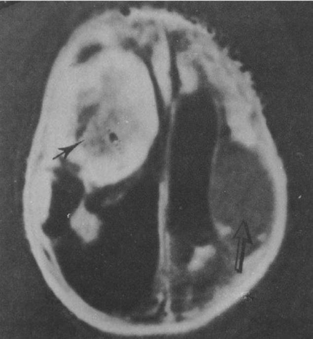
· All babies with severe thrombocytopenia due to NAIT should have a cranial ultrasound to look for evidence of intracranial haemorrhage (Figure 32.2).
Fig 32.2 MRI studies: inversion recovery sequence showing subacute haematoma (black arrow) and chronic haematoma (open arrow). Adapted from de Vries LS, Connell J, Bydder GM et al. Recurrent intracranial haemorrhages in utero in an infant with alloimmune thrombocytopenia. Case report. Br J Obstet Gynaecol 1988; 95(3): 299–302.
Neonatal transfusions
Red cells
Red cell transfusions to neonates may be:
· large volume, occurring once/twice:
· exchange transfusion (80–200 mL/kg);
· surgery, e.g. cardiac, extracorporeal membrane oxygenation (ECMO);
· small volume top-ups (most common, 10–20 mL/kg) occurring often through neonatal hospitalization.
Neonatal exchange transfusions
Exchange transfusion is used to treat severe anaemia at birth, particularly in the presence of heart failure and severe hyperbilirubinaemia. In the case of anaemia alone, a single volume (80–100 mL/kg) exchange is sufficient. For hyperbilirubinaemia, such as in HDFN, the aim is to remove both antibody-coated red cells and excess bilirubin. Double-volume exchange (160–200 mL/kg) gives the best reduction in bilirubin (50%) and removes 90% of the infant's circulating RhD-positive cells. The pH of reconstituted whole blood or plasma-reduced red cells up to 5 days post-collection and storage used in exchange transfusion is around 7.0, which does not cause acidosis in the infant.
Exchange transfusion is a specialist procedure and should be undertaken only by experienced staff. The incidence of HDFN is declining partly due to the introduction of routine antenatal anti-D prophylaxis, so expertise in exchange transfusions is also reduced. Neonatal units should have access to written protocols. As neonatal red cells for exchange have a limited shelf life and the clinical situation can change rapidly, it is important that there is good liaison between laboratory staff, clinical haematologists and neonatologists to ensure timely provision of this specialized resource.
Indications for exchange transfusion
Hyperbilirubinaemia
Transfusions are indicated:
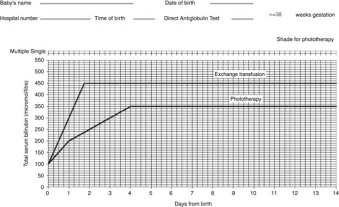
· when the serum bilirubin level indicates it is a necessity, at a threshold depending on the gestational and postnatal age of the baby (see the example of UK recommended treatment threshold graphs in Figure 32.3) and/or
· when there are clinical features and signs of acute bilirubin encephalopathy.
Fig 32.3 Treatment threshold graph for term babies with neonatal jaundice. From: National Institute for Health and Clinical Excellence (2010) CG 98 Neonatal jaundice. London: NICE. Available from www.nice.org.uk/guidance/CG98. Reproduced with permission.
Anaemia
Evidence of anaemia is shown:
· when the cord haemoglobin is less than 8 g/dL.
Exchange transfusion in the neonate: component specification
· Plasma-reduced red cells with a haematocrit of 0.5–0.6 are recommended as packed cells may have a haematocrit up to 0.75 and cause a very high post-exchange haematocrit.
· The red cells should be less than 5 days old, collected into CPD anticoagulant, sickle screen negative, Kell negative. Some countries use reconstituted whole blood with red cells resuspended in plasma.
· The most recent British Committee for Standards in Haematology (BCSH) guidelines [11] state that red cells for neonatal exchange transfusion should be gamma-irradiated (and transfused within 24 hours of irradiation); gamma-irradiation is essential in the case of neonates who have previously received IUT and in all other cases is advisable unless to do so would lead to clinically relevant delay.
· Red cells for exchange should be warmed to 37°C immediately prior to transfusion.
Other large-volume neonatal transfusions
Large-volume transfusions, where the volume transfused approximates to the neonatal blood volume, are used for cardiac surgery and occasionally other surgery including for NEC. In the UK, the component provided for this situation has the same specification as for neonatal top-up transfusions and should be used within 5 days of donation (24 hours post-irradiation if required) in order to reduce the risk of hyperkalaemia in the recipient.
Neonatal resuscitation for severe anaemia post-delivery rarely requires a ‘large-volume transfusion’ and many centres keep blood suitable for neonatal top-up transfusions on the labour ward for this indication.
Neonatal ‘top-up’ transfusions
Small-volume ‘top-up’ transfusions are commonly given to low birth weight preterm babies, with up to 80% of those weighing <1500 g at birth receiving at least one transfusion. These infants become anaemic in early life due to repeated iatrogenic losses, reduced red cell life span, low endogenous erythropoietin and a hyporegenerative bone marrow (see Table 32.2 for a summary of indications for transfusion). Strategies to limit transfusion including point-of-care testing have had limited success to date. Other attempts at delayed cord clamping and autologous placental transfusion have had a minor impact on allogeneic transfusion with potential risks [12].
Table 32.2 Indications for neonatal ‘top-up’ transfusions. Adapted from British Committee for Standards in Haematology guidelines [18].
|
Clinical situation |
Transfuse at |
|
· Anaemia in the first 24 hours |
Hb <12 g/dL |
|
· Neonate receiving mechanical ventilation |
Hb <12 g/dL |
|
· Acute blood loss |
10% blood volume lost |
|
· Oxygen dependency (not ventilated) |
Hb <8–10 g/dL |
|
· Late anaemia, stable patient (off oxygen) |
Hb 7 g/dL |
Two recent randomized trials to investigate optimal transfusion triggers have had contradictory outcomes. These trials compared restrictive versus liberal transfusion triggers for preterm infants (Iowa trial [13]; PINT trial [14]). Both had similar restrictive transfusion thresholds, but the Iowa liberal thresholds were higher than those for PINT. At short-term follow-up, the Iowa trial showed more frequent adverse neurological outcomes and incidence of apnoeas in the restrictive transfusion group, but that was not the case for the PINT trial. At 18–21 month follow-up in the PINT trial, there was a statistically significant cognitive delay for those in the restrictive group [15], supporting the suggestion that liberal transfusions may be neuroprotective. However, at an average 12-year follow-up of the Iowa group by MRI, the brain volumes of the liberally transfused but not the restrictive group were significantly smaller than controls, giving the opposite conclusion to PINT [16]. Overall, the use of restrictive thresholds only resulted in modest reductions in exposure to transfusion. A recent Cochrane review has concluded that it is prudent not to use triggers outside the restrictive and liberal thresholds in the studies described above until further trials are completed [17].
Guidelines for triggers for ‘top-up’ transfusion have been devised by committees in a number of countries, including the UK, Canada and the USA. Since there has been little objective evidence to guide practice, these guidelines are based on clinical experience and represent consensus views. Table 32.2 summarizes the indications for ‘top-up’ transfusions agreed by the BCSH neonatal and paediatric guidelines [18] and used in many UK neonatal intensive care units. These UK guidelines are in general closer to the restrictive than the liberal thresholds in the recent trials.
Neonatal ‘top-up’ transfusions: component specification
· Small-volume ‘top-up’ transfusions can be given without further testing provided that there are no atypical maternal antibodies in maternal/infant serum and the infant's DAT is negative.
· Hct 0.5–0.7.
· CMV seronegative in the UK.
· The red cells should be ≤35 days old (in SAG-M).
· ‘Paedipacks’ (aliquotted donations from a single unit) should be used wherever possible for repeated transfusions to minimize donor exposure.
· The volume of a neonatal ‘top-up’ transfusion is usually 10–20 mL/kg.
Role of erythropoietin in reducing neonatal red cell transfusion
There have been numerous clinical trials of erythropoietin for the prevention or amelioration of neonatal anaemia, particularly anaemia of prematurity, since endogenous erythropoietin production is low in preterm babies for the first 6–8 weeks of life. These trials show that erythropoietin (250 units/kg/day 3 times per week for the first 6 weeks of life) can reduce red cell transfusions in well preterm babies but has a negligible effect on transfusion requirements of sick preterm babies, particularly those of less than 26 weeks gestation at birth. In practice, this means that erythropoietin has a limited role in neonates as it works best in those that need it least.
Therefore most neonatal units no longer use erythropoietin routinely. The situations in which erythropoietin can be useful are:
· in neonates whose parents refuse permission to use blood components;
· to prevent ‘late anaemia’ in babies with HDFN.
T antigen activation
Severe haemolytic transfusion reactions are occasionally seen in neonates or young children transfused with adult blood or fresh frozen plasma containing anti-T antibodies. This may be due to exposure or ‘activation’ of the T antigen on neonatal red cells, usually as a result of infection with clostridia, streptococci or pneumococci and/or in association with NEC. Up to 25% of infants with NEC have T antigen activation but so do many healthy neonates and haemolysis is extremely rare [19]. Therefore, although there remains some controversy, the majority of centres worldwide consider that no special provision for neonates with NEC is necessary and neither screen neonates for T activation nor donors for high titre anti-T.
Neonatal platelet transfusions
Neonatal thrombocytopenia
Severe thrombocytopenia (platelets <50 × 109/L) occurs in ∼2–5% of neonates on neonatal units. It is much more common in sick preterm infants, 30–40% of whom will develop thrombocytopenia in the first 4 weeks of life. Causes of neonatal thrombocytopenia are shown in Table 32.3. The most common cause presenting in the first few days of life is that associated with intrauterine growth restriction or maternal hypertension; however, the most important cause of severe thrombocytopenia (platelets < 50 × 109/L) at birth is neonatal alloimmune thrombocytopenia (NAIT).
Table 32.3 Causes of neonatal thrombocytopenia.
|
Early onset (< 72 hours after birth) |
|
Placental insufficiency (PET, IUGR, diabetes) |
|
NAIT |
|
Birth asphyxia |
|
Perinatal infection (group B strep, E. coli, listeria) |
|
Congenital infection (CMV, toxoplasmosis, rubella) |
|
Maternal autoimmune (ITP, SLE) |
|
Severe Rhesus HDFN |
|
Thrombosis (renal vein, aortic) |
|
Aneuploidy (trisomy – 21, 18, 13) |
|
Congenital/inherited (TAR, Wiskott–Aldrich) |
|
Late onset (>72 hours after birth) |
|
Bacterial and fungal sepsis |
|
Necrotizing enterocolitis |
|
Congenital infection (CMV, toxoplasmosis, rubella) |
|
Maternal autoimmune (ITP, SLE) |
|
Congenital/inherited (TAR, Wiskott–Aldrich) |
|
The most common causes are in bold type. |
Investigation of neonatal thrombocytopenia
In most cases the following tests will identify the diagnosis.
· Full blood count and film. The combination of erythroblastosis, high Hb, mild neutropenia without neutrophil left shift suggests that the cause is intrauterine growth restriction and/or maternal hypertension. However, neutrophil left shift and toxic granulation with more severe thrombocytopenia suggests that the cause is bacterial infection with or without DIC.
· Congenital infection screen. The most common congenital infection associated with neonatal thrombocytopenia is CMV.
Screening for NAIT should be carried out in any case of severe thrombocytopenia (platelets <50 × 109/L) presenting in the first week of life unless there is very clear evidence of acute infection.
Neonatal thrombocytopenia due to maternal ITP
· Around 10% of infants of mothers with ITP or SLE develop neonatal thrombocytopenia secondary to transplacental passage of maternal platelet autoantibodies.
· Fetal platelet counts cannot be reliably predicted from maternal platelet counts nor from platelet serology.
· Thrombocytopenia is usually mild and intracranial haemorrhage occurs in less than 1% of at-risk babies.
· Platelet counts of babies born to mothers with ITP or SLE should be checked at birth and monitored daily for 2–3 days if below 200 × 109/L at birth.
· If the baby is well, treatment is unnecessary unless the platelet count falls below 20 × 109/L.
· Severe thrombocytopenia (platelets <20 × 109/L): treatment with intravenous IgG (single dose 1 g/kg, repeated if necessary) is usually effective.
· Cranial ultrasound to look for intracranial haemorrhage should be performed in all neonates with severe thrombocytopenia.
· Platelet transfusion is reserved for life-threatening haemorrhage and should be given in conjunction with intravenous IgG.
Indications for platelet transfusion in neonates
Published guidelines for neonatal platelet transfusion acknowledge the lack of evidence on which to base recommendations and aim for a safe approach. Suggested guidelines based on clinical experience are shown in Table 32.4. Some evidence suggests that prophylactic platelet transfusions are not required for healthy neonates until the platelet count falls to 20–30 × 109/L. However, a higher trigger level (50 × 109/L) should be used for babies with the greatest risk of haemorrhage, especially extremely low birth weight neonates (<1000 g) in the first week of life.
Table 32.4 Guidelines for platelet transfusion in neonatal thrombocytopenia.
|
· Platelet count <30 × 109/L in otherwise well infants, including NAIT if no evidence of bleeding and no family history of intracranial haemorrhage · Platelet count <50 ×109/L in infants with: clinical instability concurrent coagulopathy birth weight <1000 g and age <1 week previous major bleeding (e.g. GMH-IVH) current minor bleeding (e.g. petechiae) planned surgery or exchange transfusion platelet count falling and likely to fall below 30 NAIT if previous affected sibling with ICH · Platelet count <100 × 109/L in infants with: major bleeding |
A recent prospective observational study of outcomes of neonates with platelets below 60 × 109/L showed that although a third of these patients developed thrombocytopenia of <20 × 109/L, only 9% developed major haemorrhage [20]. Those patients with major haemorrhage were mostly <28 weeks gestational age within the first 14 days of life. Prospective randomized trial data are required to inform future guidance for the most appropriate prophylactic platelet transfusion thresholds.
Neonatal platelet transfusion: component specification
· ABO and RhD identical or compatible.
· HPA compatible in infants with NAIT.
· CMV seronegative in UK.
· Apheresis, produced by standard techniques without further concentration.
· Irradiated if appropriate.
· Volume transfused usually 10–20 mL/kg.
Neonatal FFP and cryoprecipitate transfusion
There is much uncertainty around the appropriate use of FFP in neonates. A recent UK-wide survey of FFP transfusion practice showed that 42% of infant FFP transfusions were given as prophylaxis for abnormal coagulation in the absence of bleeding [21].
Definition of neonatal coagulopathy
Neonatal coagulopathy can be difficult to define. Plasma concentrations of different coagulation proteins mature at different rates and neonates have a different balance of procoagulant and anticoagulant proteins compared to older children. This results in different postnatal and gestational age-related coagulation ranges in the first months of life, particularly for the activated partial thromboplastin time (APTT), although overall neonatal haemostasis may be functionally as effective as in adults. There is a particular danger of misinterpreting neonatal results reported as the activated partial thromboplastin time ratio (APTR), calculated by dividing the neonatal APTT result by the midpoint of the local adult range, as these may appear inappropriately abnormal. Moreover, most laboratories rely on previously published neonatal ranges due to the difficulties in obtaining locally derived ranges in this age group. As the published ranges are likely to be using different analysers and reagents from the local laboratory, this needs to be taken into account when interpreting individual results.
Causes of haemorrhage in the newborn
In well infants the most common causes of bleeding are:
· vitamin K deficiency (haemorrhagic disease of the newborn);
· inherited disorders, particularly haemophilias;
· NAIT.
In sick infants the most common causes are:
· DIC – secondary to perinatal asphyxia, necrotizing enterocolitis or, less commonly, sepsis;
· liver disease.
Vitamin K deficiency bleeding (haemorrhagic disease of the newborn) (VKDB)
Vitamin K is necessary for posttranslational carboxylation of coagulation factors II, VII, IX and X and of the natural anticoagulants protein C and protein S. Levels of vitamin K and of all of these factors are physiologically low at birth. This physiological deficiency can be exacerbated by breast feeding, prematurity and liver disease, resulting in haemorrhagic disease of the newborn, often referred to as vitamin K deficiency bleeding (VKDB). Treatment of VKDB depends on the severity of bleeding. Mild cases should be given vitamin K (1 mg) intravenously or subcutaneously as this increases levels of active vitamin K deficiency coagulation factors within a few hours; where there is significant bleeding FFP may be given in addition to vitamin K.
There are three patterns of VKDB:
· Early VKDB presents in the first 24 hours of life, usually with severe haemorrhage. It is caused by severe vitamin K deficiency in utero, usually as a result of maternal medication that interferes with vitamin K, e.g. anticonvulsants.
· Classical VKDB presents at 2–7 days old in babies who have not received prophylactic vitamin K at birth. The risk is increased in breast-fed babies and in those with poor oral intake. The incidence in babies not receiving vitamin K supplementation is 0.25–1.7%.
· Late VKDB occurs 2 to 8 weeks after birth. It usually presents with sudden intracranial haemorrhage in an otherwise well, breast-fed term baby or in babies with liver disease.
Vitamin K prophylaxis
For prevention of neonatal vitamin K deficiency, both the American Academy of Pediatrics and the Department of Health in the UK recommend vitamin K supplementation at birth. Some studies have suggested a link between intramuscular vitamin K at birth and later childhood malignancies. Although other studies have not confirmed the link with malignancy, the controversy is unlikely to be resolved unequivocally in the short term. Current recommendations are that babies are given 1 mg vitamin K intramuscularly and if parents decline this they should be offered oral vitamin K, requiring multiple doses.
Indications for FFP and cryoprecipitate
Guidelines for the use of FFP, cryoprecipitate and albumin in neonates have been published by national committees in a number of countries. The guidelines aim to minimize their risks in the newborn both by the use of pathogen-inactivated products and by limiting their use for a small number of clinical indications. It is important to monitor the clinical and laboratory outcome of plasma transfusions.
The only indications for FFP in neonates recommended in the BCSH guidelines [18] and supported by evidence are: DIC, VKDB and inherited deficiencies of coagulation factors. Prophylactic FFP administered to preterm neonates at birth does not prevent intraventricular haemorrhage or improve outcome at 2 years of life. Similarly, FFP is not superior to other colloid or crystalloid solutions as a volume replacement solution in standard neonatal practice and there is no evidence to support its use to ‘correct’ the results of abnormal coagulation screens.
Neonatal FFP and cryoprecipitate: component specifications
· Current BCSH guidelines state that FFP for transfusion to neonates should preferably be of the same ABO group or group AB. Group O FFP should only be given to patients of group O.
· Plasma components may be standard or pathogen inactivated (although pathogen inactivated is not available in all countries). In the UK, single-donor FFP and cryoprecipiate for patients born after 1 January 1996 is imported from countries with low risk of vCJD. Plasma is inactivated by methylene blue with approximately 30% loss of activity of factors VIII, XI and fibrinogen as a result. Some hospitals in the UK use pooled solvent-detergent-treated imported FFP as an alternative (see Chapter 21 for further details).
· The FFP volume transfused is usually 10–20 mL/kg.
Neonatal neutropenia
Normal neutrophil levels vary with postnatal age, falling in healthy babies from around 5–10 × 109/L at birth to 2–6 × 10⁹/L by the end of the first week of life. Neutropenia is therefore variably defined depending on postnatal age: less than 2.0 × 10⁹/L at birth and less than 1.0 × 10⁹/L from one week of age. The most common causes are neutropenia secondary to intrauterine growth restriction or maternal hypertension and neutropenia secondary to severe sepsis. The presence of neutrophil left shift and toxic granulation in a neutropenic neonate suggests acute bacterial infection.
Alloimmune neonatal neutropenia (see Chapter 5)
· This is analogous to HDFN: there is maternal sensitization to fetal neutrophil antigens during pregnancy.
· The most common implicated antibodies are anti-NA1 and anti-NA2.
· The estimated incidence of neonatal alloimmune neutropenia is 3% of live births but most cases are mild and asymptomatic and the diagnosis may be missed.
· Infants with severe neonatal alloimmune neutropenia develop severe cutaneous, respiratory or urinary tract infection.
· Treatment is with antibiotics and, if necessary, granulocyte colony-stimulating factor (rarely needed).
Granulocyte transfusions in neonates
There is no good evidence of the benefit of granulocyte transfusions for the treatment of neonatal infection. Both granulocyte colony-stimulating factor and granulocyte-macrophage colony-stimulating factor can be used to increase the neutrophil count in neutropenic neonates, but there is no clear evidence that this improves outcome.
Transfusion in older infants and children
General points
Although most children never require blood transfusion, there are several groups who are frequently transfused, including those on a paediatric intensive care unit (PICU) or undergoing cardiac surgery or ECMO, those with inherited transfusion-dependent disorders, such as thalassaemia major, and those undergoing intensive chemotherapy for haematological malignancies. For some of these patients, including those with thalassaemia major and sickle cell disease, bone marrow transplantation (BMT) or cord blood transplantation is a possible future treatment. Therefore all such children for whom BMT is a possible option should receive CMV-safe blood components. All children on regular transfusions should be vaccinated against hepatitis B as early as possible. Those on chronic transfusion therapy, particularly those with haemoglobinopathies, but also those with Congenital Dyserythropoietic Anaemia, aplastic anaemia and other bone marrow failure syndromes, should have an extended red cell phenotype (see below) performed prior to, or as soon as possible after, commencing regular transfusions. For chronically transfused paediatric patients, monitoring growth and development is an important outcome measure of efficacy.
Formula for calculating red cell transfusion volume in children
Several different formulas for calculating transfusion volume in children are in widespread use. Most formulas commonly used in the UK are based on the increase in Hb or haematocrit required and a ‘transfusion factor’. The transfusion factor used varies from 3 to 5. There is a lack of evidence from prospective randomized trials to show which transfusion factor best predicts the rise in Hb/haematocrit and whether the same factor should be applied to all groups of children. Some recent retrospective studies suggest a transfusion factor of 5 better predicts the Hb/haematocrit in critically ill children but transfusion factors of 3 or 4 appear satisfactory for most children on long-term red cell transfusion. An example using a transfusion factor of 3 for packed red cell transfusion is shown here:

A calculation for transfusion volume used in the USA is

The normal rate of red cell transfusion is around 5 mL/kg/h.
Transfusion on paediatric intensive care unit (PICU)
There has been a recent randomized controlled trial of red cell transfusion in stable critically ill children on PICU (TRIPICU [22]), comparing a restrictive Hb trigger (7 g/dL) versus a liberal one (9.5 g/dL). There was no difference in the primary outcome (including mortality and new or progressive multiple organ dysfunction syndrome), suggesting that a restrictive transfusion strategy is reasonable for this group of patients.
Cardiac surgery and ECMO
Transfusion is frequently used in paediatric cardiac surgery including for priming of bypass circuits and treatment of postoperative bleeding and coagulopathy post-bypass. Transfusion practice in paediatric cardiac surgery is very variable, with major differences in the use of components across centres. There is a lack of recent large studies providing evidence for the age of blood to be used, anticoagulant and component support or for appropriate transfusion triggers in this situation, although the TRIPICU study [22] suggested that noncyanotic postoperative cardiac patients can be transfused at Hb of 7 g/dL without adverse outcome. ECMO is used post-bypass and for other situations of cardiopulmonary dysfunction/collapse. As with cardiac bypass, there is limited data to support specific blood component administration/transfusion triggers.
Paediatric massive transfusion
Trauma centres are increasingly setting up paediatric massive transfusion protocols in parallel with adult protocols, with fixed ratios of red cell to plasma transfusions, although there is little specific evidence for paediatric patients in this area.
Leukaemia, chemotherapy and BMT
Many aspects of the transfusion of children with leukaemia/cancer or undergoing haemopoietic stem cell transplantation (HSCT) are managed in a similar manner to adults. However, there are some that deserve particular consideration in the paediatric situation.
Platelet transfusion in children undergoing chemotherapy or HSCT
· Indications for platelet transfusion in children are consensus based; those developed by the BCSH are shown in Table 32.5. In general, in noninfected, well children a platelet count of 10 × 109/L can be used as a transfusion trigger but higher thresholds are used for children who are sick and/or bleeding.
· The optimal platelet count for routine lumbar punctures for children on treatment for leukaemia is uncertain, but current recommendations are 20–40 × 109/L.
· Platelets should be ABO-compatible where possible because of the risk of haemolysis.
· Platelets should be RhD compatible and D negative girls should receive D negative platelets whenever possible, and if not should be administered anti-D.
· A transfusion of 10–20 mL/kg is given to children under 15 kg and an apheresis unit (maximum 300 mL) for children over 15 kg.
Table 32.5 Indications for platelet transfusion in children with thrombocytopenia.
|
Platelet count <10 × 109/L |
|
Platelet count <20 × 109/L and one or more of the following: |
|
severe mucositis |
|
DIC |
|
anticoagulant therapy |
|
platelets likely to fall < 10 × 109/L before next evaluation |
|
risk of bleeding due to a local tumour infiltration |
|
Platelet count 20–40 × 109/L and one or more of the following: |
|
DIC in association with induction therapy for leukaemia |
|
extreme hyperleucocytosis |
|
prior to lumbar puncture or central venous line insertion |
|
DIC, disseminated intravascular coagulation. |
Granulocyte transfusion in children undergoing chemotherapy or HSCT
· There is no evidence to support the use of prophylactic granulocyte transfusions.
· Empirical data from some studies support their use where there is severe bacterial or fungal infection in neutropenic children, including SCT, but they increase the risk of platelet refractoriness.
· Granulocytes for transfusion should be ABO, RhD and crossmatch compatible.
· Granulocytes for all recipients should always be irradiated and be CMV serologically negative.
HSCT donors
· Children who act as HSCT donors for their sibling(s) may require blood transfusion to cover blood lost during the procedure; however, this is avoided wherever possible. Allogeneic blood transfused to the donor during the bone marrow harvest should always be irradiated.
· Peripheral stem cell collections by apheresis are routinely performed in children in specialized centres.
Autologous donation of red cells is now no longer undertaken except in exceptional circumstances.
Transfusion support for children with haemoglobinopathies (also see Chapter 29)
Thalassaemia major
By definition all patients with thalassemia major are transfusion-dependent. Transfusion therapy is determined by the degree of anaemia and evidence of failure to thrive. Most children start transfusion when their haemoglobin drops below 6 g/dL.
Current BCSH [18] and Thalassaemia International Federation [23] guidelines recommend transfusion:
· to maintain an average Hb of 12 g/dl;
· to maintain a pretransfusion Hb of 9–10.5 g/dL;
· to prevent marrow hyperplasia, skeletal changes and organomegaly by inhibiting erythropoiesis;
· extended red cell phenotyping should be carried out before starting transfusions;
· transfusion requirements should be adjusted to accommodate growth;
· splenectomy may be considered if hypersplenism develops and causes a sustained increase in red cell requirements;
· iron chelation therapy should be considered after 10 transfusions and started once the ferritin is >1000 ng/mL (if possible starting after the age of 2 years because of desferrioxamine toxicity);
· since BMT and cord blood transplantation are the only cure, families should be offered HLA typing of siblings as possible bone marrow donors and/or cryopreservation of HLA-matched sibling cord blood.
Sickle cell disease
Red cell transfusion in children with sickle cell disease should not be routine but reserved for specific indications (Table 32.6). Extended red cell phenotyping before the first transfusion is very important because up to 35% of patients otherwise develop red cell alloimmunization and may be very difficult to crossmatch. The majority of antibodies are in the Rh or Kell systems and may be transient and very difficult to detect, leading to a risk of delayed transfusion reactions.
Table 32.6 Indications for transfusion in sickle cell disease.
|
· ‘Top-up’ |
splenic sequestration* |
|
hepatic sequestration* |
|
|
aplastic crises* |
|
|
· Exchange transfusion |
chest syndrome* stroke* |
|
mesenteric syndrome (abdominal crisis) |
|
|
multisystem organ failure |
|
|
· Hypertransfusion |
stroke (to prevent recurrence)* |
|
primary stroke prevention (raised TCD velocity)* |
|
|
renal failure (to prevent/delay deterioration) |
|
|
chronic sickle lung disease |
|
|
· Surgery |
selected patients pre-operatively (e.g. joint replacement) |
|
*proven value TCD -- Transcranial Doppler |
|
Indications for ‘top-up’ transfusion in sickle cell disease
Indications include splenic or hepatic sequestration and aplastic crisis. The aim is to raise the haemoglobin to the child's normal steady state (the haemoglobin should never be raised acutely to >10 g/dL since this is likely to cause an increase in blood viscosity).
Indications for exchange transfusion in sickle cell disease
· Acute chest syndrome
· Mesenteric (abdominal) syndrome
· Stroke
· Selected patients preoperatively
· Multiorgan failure
The aim is to reduce sickling and increase oxygen carriage without an increase in viscosity
Indications for hypertransfusion in sickle cell disease
· Prevent recurrence of stroke (i.e. secondary prevention of stroke)
· Prevent the development of stroke in children with sickle cell disease with Doppler evidence of cerebrovascular infarction/haemorrhage in the absence of clinical evidence of stroke (i.e. primary prevention of stroke)
· Delay or prevent deterioration in end organ failure (e.g. chronic sickle lung)
The aims are to maintain the percentage of HbS below 25% and the Hb between 10 and 14.5 g/dL.
Indications for preoperative transfusion in sickle cell disease
The BCSH guidelines are based on observational studies and one large randomized controlled study as there are few other available data. On the basis of these guidelines and recent information from the TAPS trial [24]:
· Top-up transfusion (Hb 8–10 g/dL) is as effective as exchange transfusion and may be safer.
· Although minor, low risk procedures (e.g. grommet insertion) and moderate risk surgery (e.g. tonsillectomy, laparascopic cholecystectomy) may be undertaken without transfusion in some patients, the recent TAPS study [24] showed that patients with HbSS undergoing low and moderate risk surgery had an increased risk of adverse events without transfusion; the authors recommended on the basis of their results that preoperative transfusion to a haemoglobin of about 10 g/dL should be part of the standard management of patients with HbSS for low and moderate risk surgery.
· Exchange transfusion should be performed preoperatively for major procedures such as hip/knee replacement, organ transplantation, eye surgery and considered for major abdominal surgery.
Practical aspects of transfusion in sickle cell disease
· Extended red cell phenotyping (for Rh K, Fy, Jk, MNS and U) should be carried out; this should be done before the first transfusion and may be usefully arranged at an outpatient clinic follow-up during the first year of life.
· The R0 blood group (cDe/cDe) is common in patients of African or Caribbean origin: all R0 patients should receive C-negative, E-negative blood (i.e. rr or R0).
· The use of sickle trait positive blood should be avoided by testing donor blood for HbS.
· During exchange transfusion in the acute situation, a total exchange of 1.5–2 times blood volume is required to achieve an HbS level of 20% or less; this may take 2–3 procedures if carried out manually. Automated exchange using a cell separator allows the exchange to be completed as a single procedure. The volume of packed cells (in mL) for each exchange is weight (kg) × 30.
· Normal saline (not FFP or albumin) should be used as volume replacement at the beginning of the exchange prior to starting venesection to avoid dropping the circulating blood volume.
Key points
1. Special components are available for fetal and paediatric transfusion to improve transfusion safety and optimize efficacy.
2. Transfusion management of HDFN in the fetus and neonate is complex and requires careful multidisciplinary input.
3. Neonatal red cell top-up transfusions have been the subject of recent trials but the outcomes are not clear-cut; current guidelines are in general closer to the restrictive than liberal thresholds in the trials.
4. Neonatal thrombocytopenia is common in neonatal units; there is a lack of evidence about appropriate triggers for prophylactic platelet transfusions and these are the subject of a current randomized trial.
5. Neonatal coagulopathy is difficult to define in practice and a significant proportion of FFP transfusions are given prophylactically to infants with presumed abnormal coagulation tests in the absence of bleeding.
6. There are specific recommendations for red cell transfusions for older children on PICU and for platelet transfusions for those undergoing chemotherapy.
7. Children with thalassaemia need careful transfusion management to allow normal growth and development and to minimize iron overload.
8. Transfusions for children with sickle cell disease may be given for acute complications or as part of a chronic hypertransfusion programme for primary or secondary prevention of stroke.
References
1. Stainsby D, Jones H, Wells AW et al. Adverse outcomes of blood transfusion in children: analysis of UK reports of the serious hazards of transfusion scheme 1996–2005. Br J Haematol 2008; 141: 73–79.
2. New HV, Stanworth SJ, Engelfriet CP et al. Neonatal transfusions. Vox Sanguinis 2009; 96: 62–85.
3. Luban NLC, Strauss RG & Hume HA. Commentary on the safety of red cells preserved in extended-storage media for neonatal transfusions. Transfusion 1991; 31: 229–235.
4. Mou SS, Giroir BP, Molitor-Kirsch EA et al. Fresh whole blood versus reconstituted blood for pump priming in heart surgery in infants. N Engl J Med. 2004; 351: 1635–1644.
5. Blau J, Calo JM, Dozor D et al. Transfusion-related acute gut injury: necrotizing enterocolitis in very low birth weight neonates after packed red blood cell transfusion. J Pediatr 2011; 158: 403–409.
6. Christensen RD. Association between red blood transfusions and necrotizing enterocolitis. J Pediatr 2011; 158: 349–350.
7. Josephson CD, Castillejo MI, Caliendo AM et al. Prevention of transfusion-transmitted cytomegalovirus in low-birth weight infants (≤1500 g) using cytomegalovirus-seronegative and leukoreduced transfusions. Transfus Med Rev 2011; 25: 125–132.
8. Bhutani VK; Committee on Fetus and Newborn, American Academy of Pediatrics. Phototherapy to prevent severe neonatal hyperbilirubinemia in the newborn infant 35 or more weeks of gestation. Pediatrics 2011; 128: e1046–1052.
9. American Academy of Pediatrics Subcommittee on Hyperbilirubinemia. Management of hyperbilirubinemia in the newborn infant 35 or more weeks of gestation. Pediatrics 2004; 114: 297–316.
10. British Committee for Standards in Haematology. Guidelines for the estimation of fetomaternal haemorrhage; 2009. Available at: http://www.bcshguidelines.com/documents/BCSH_FMH_bcsh_sept2009.pdf.
11. British Committee for Standards in Haematology. Guidelines on the use of irradiated blood components. Br J Haematol 2010; 152: 35–51.
12. Strauss RG & Widness JA. Autologous or allogeneic: cord blood red blood cells still are investigational. Transfus Med Rev 2012; 26: 91–92.
13. Bell EF, Strauss RG, Widness JA et al. Randomized trial of liberal versus restrictive guidelines for red blood cell transfusion in preterm infants. Pediatrics 2005; 115: 1685–1691.
14. Kirpalani H, Whyte RK, Andersen C et al. The Premature Infants in Need of Transfusion (PINT) study: a randomized, controlled trial of a restrictive (low) versus liberal (high) transfusion threshold for extremely low birth weight infants. J Pediatr 2006; 149: 301–307.
15. Whyte RK, Kirpalani H, Asztalos EV et al. Neurodevelopmental outcome of extremely low birth weight infants randomly assigned to restrictive or liberal hemoglobin thresholds for blood transfusion. Pediatrics 2009; 123: 207–213.
16. Nopoulos PC, Conrad AL, Bell EF et al. Long-term outcome of brain structure in premature infants: effects of liberal vs restricted red blood cell transfusions. Arch Pediatr Adolesc Med 2011; 165: 443–450.
17. Whyte R & Kirpalani H. Low versus high haemoglobin concentration threshold for blood transfusion for preventing morbidity and mortality in very low birth weight infants. Cochrane Database Syst Rev 2011; 11: CD000512.
18. British Committee for Standards in Haematology. Transfusion guideline for neonates and older children. Br J Haematol 2004; 124: 433–453.
· 2005 amendment to the guidelines on transfusion for neonates and older children. Br J Haematol 2006; 136: 514–516.
· 2007 amendment to the transfusion guidelines for neonates and older children (specification of imported FFP). Available at: http://www.bcshguidelines.com/documents/FFP_neonate_Amendment_1_17_Oct_2007.pdf.
19. Boralessa H, Modi N, Cockburn H, Malde R, Edwards M, Roberts I & Letsky E. RBC T activation and hemolysis in a neonatal intensive care population: implications for transfusion practice. Transfusion 2002; 42: 1428–1434.
20. Stanworth SJ, Clarke P, Watts T et al. Prospective, observational study of outcomes in neonates with severe thrombocytopenia. Pediatrics. 2009; 124: e826–834.
21. Stanworth SJ, Grant-Casey J, Lowe D et al. The use of fresh-frozen plasma in England: high levels of inappropriate use in adults and children. Transfusion 2011; 51: 62–70.
22. Lacroix J, Hebert PC, Hutchison JS et al. Transfusion strategies for patients in pediatric intensive care units. N Engl J Med 2007; 356: 1609–1619.
23. Cappellini M-D, Cohen A, Eleftheriou A et al. Guidelines for the clinical management of thalassaemia, 2nd edn revised; 2008. Available at: http://www.thalassaemia.org.cy/pdf/Guidelines_2nd_revised_edition_EN.pdf.
24. Howard, J, Malfroy, M, Llewelyn, C et al. Pre-operative transfusion reduces serious adverse events in patients with sickle cell disease (SCD): results from the Transfusion Alternatives Preoperatively in Sickle Cell Disease (TAPS) randomised controlled multicentre clinical trial. Blood (ASH annual meeting abstracts) 2011; 118: 9.
Further reading
Aher S & Ohlsson A. Late erythropoietin for preventing red blood cell transfusion in preterm and/or low birth weight infants. Cochrane Database Syst Rev 2006; 3: CD004868.
British Committee for Standards in Haematology. Guideline for blood grouping and antibody testing in pregnancy; 2006. Available at: http://www.bcshguidelines.com/documents/antibody_testing_pregnancy_bcsh_07062006.pdf.
British Committee for Standards in Haematology. Guideline on the administration of blood components; 2009. Available at: http://www.bcshguidelines.com/documents/Admin_blood_components_bcsh_05012010.pdf.
Egbor M, Knott P & Bhide, A. Red-cell and platelet allommunisation in pregnancy. Best Pract Res Clin Obstet Gynaecol 2012: 26; 119–132.
National Institute for Health and Clinical Excellence. CG 98 Neonatal Jaundice. London: NICE; 2010. Available at: www.nice.org.uk/guidance/CG98.
Patra K, Storfer-Isser A, Siner B, Moore J & Hack M. Adverse events associated with neonatal exchange transfusion in the 1990s. J Pediatr 2004; 144: 626–631.
Puckett RM & Offringa M. Prophylactic vitamin K for vitamin K deficiency bleeding in neonates. Cochrane Database Syst Rev 2000; 4: CD002776.