
Cristina V. Navarrete
NHS Blood and Transplant, Colindale Centre and University College London, London, UK
Introduction
The genes coding for human leucocyte antigens (HLAs) are located on the short arm of chromosome 6, spanning a distance of approximately 4 Mb. This genomic region is divided into three subregions [1, 2].
· Class I subregion contains genes coding for the heavy (α) chain of the classical (HLA-A, -B and -C) and nonclassical (HLA-E, -F and -G) class I molecules. The nonclassical major histocompatibility complex class I chain-related gene A and gene B (MICA and MICB) have also been mapped to this subregion, centromeric to the HLA-B gene (Figure 4.1).
· Class II subregion contains the classical HLA-DR, -DQ and -DP genes and the nonclassical HLA-DMA, -DMB, -DOA and -DOB genes. The low-molecular-mass polypeptide genes LMP2 and LMP7, TAP1 and TAP2transporters and the Tapasin (Tpn) genes involved in the processing, transport and loading of HLA class I antigenic peptides are also located in this subregion (see Figure 4.1).
· Class III subregion lies between the other two subregions and contains genes coding for a diverse group of proteins, including complement components (C4Bf), tumour necrosis factor (TNF) and heat-shock proteins (HSPs).
Fig 4.1 Map of the human leucocyte antigen complex. HSP, heat-shock protein; TNF, tumour necrosis factor. Based on Trowsdale and Campbell, 1997, in Dominique Charron (ed.), HLA Genetic Diversity of HLA Functional and Medical Implication, Vol. 1, pp. 499–504.
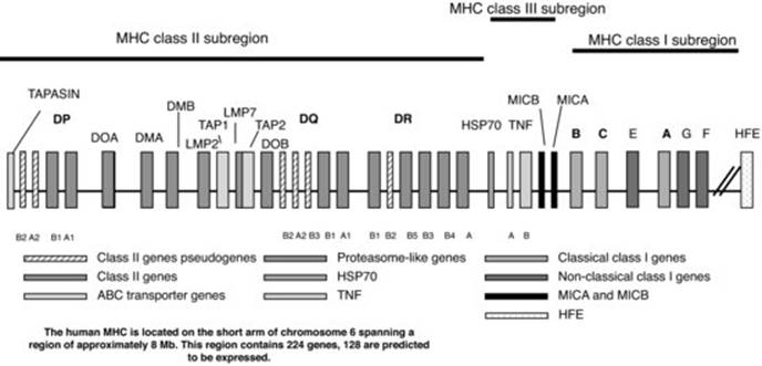
The nonclassical class I-like gene HFE has been mapped to a locus located 4 Mb telomeric to HLA-F. Single point mutations in this gene are associated with the development of hereditary haemochromatosis (HH).
HLA class I genes
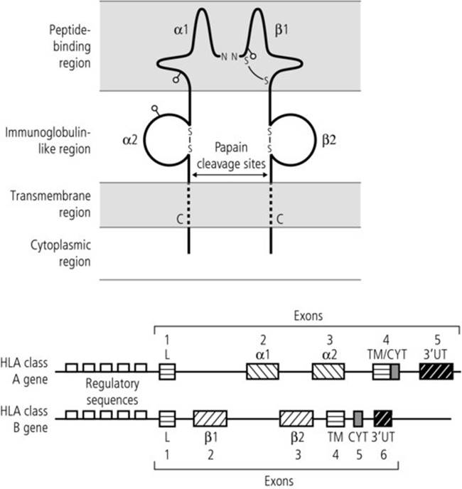
The HLA class I genes are classified according to their structure, expression and function as classical (HLA-A, -B and -C) and nonclassical (HLA-E, -F and -G). Both classical and nonclassical HLA class I genes code for a heavy (α) chain, of approximately 43 kDa, noncovalently linked to a nonpolymorphic light chain, the β2-microglobulin of 12 kDa, which is coded for by a gene on chromosome 15. The extracellular portion of the heavy chain has three domains (α1, α2 and α3) of approximately 90 amino acids long. These domains are encoded by exons 2, 3 and 4 of the class I gene, respectively. The α1 and α2 domains are the most polymorphic domains of the molecule and they form a peptide-binding groove that can accommodate antigenic peptides approximately eight to nine amino acids long.
The exon/intron organization of the nonclassical HLA class I genes (E, F and G) is very similar to the classical class I genes, but they have a more restricted polymorphism. The MICA and MICB gene products, however, do not bind β2-microglobulin and do not present antigenic peptides.
A schematic representation of the classical HLA class I gene and molecule is shown in Figure 4.2.
Fig 4.2 HLA class I molecule. β2-m, β2-microglobulin.

HLA class II genes
The classical HLA class II DR, DQ and DP A and B genes code for heterodimers formed by noncovalently associated α and β chains of approximately 34 and 28 kDa, respectively. The expressed α and β chains consist of two extracellular domains and a transmembrane and cytoplasmic domains. The α1/β1 and α2/β2 domains are encoded by exon 2 and exon 3 of the class II gene, respectively. The majority of the polymorphism is located in the β1 domain of the DR molecules and in the α1 and β1 domains of the DQ and DP molecules. Similarly to the class I molecules, these domains also form a peptide-binding groove. However, in the case of the class II molecules (DR), the groove is open at both sides and it can accommodate antigenic peptides of varying size, although most of them are approximately 13–25 amino acids long. A schematic representation of the HLA class II gene and molecule is shown in Figure 4.3.
Fig 4.3 HLA class II molecule.
The nonclassical HLA class I DMA, DMB, DOA and DOB genes have a similar structure to the classical class II genes, but show limited polymorphism.
Genetic organization and expression of HLA class II genes
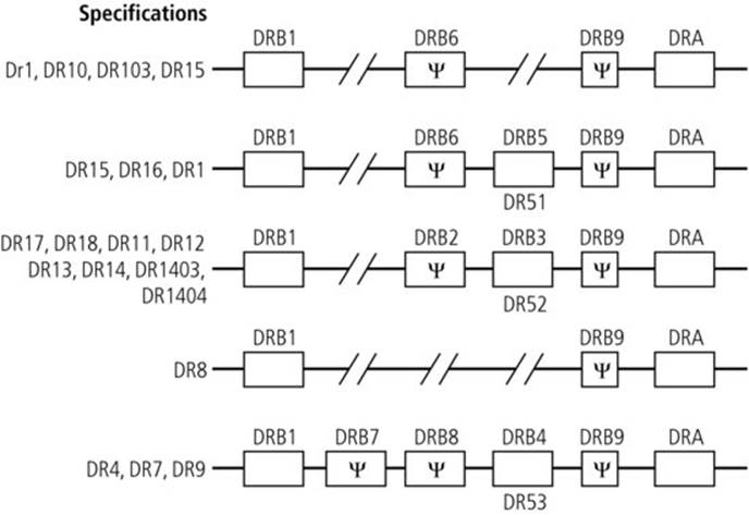
There is one DRA gene of limited polymorphism and nine DRB genes, of which B1, B3, B4 and B5 are highly polymorphic and B2, B6 and B9 are pseudogenes. The main serologically defined DR specificities (DR1–DR18) are determined by the polymorphic DRB1* gene. The number of DRB genes expressed in each individual varies according to the DRB1 allele expressed (Figure 4.4). There are a few exceptions to this pattern of gene expression, e.g. a DRB5 gene has been found to be expressed with some DR1 alleles. Some nonexpressed or null DRB5 and DRB4 genes have also been identified. In contrast to the DRB genes, there are two DQA and three DQB genes, but only the DQA1 and DQB1 are expressed and both are polymorphic. Similarly, there are two DPA and two DPB genes, but only the DPA1 and DPB1 are expressed and both are polymorphic.
Fig 4.4 Expression of HLA-DRB genes.
Expression of HLA molecules
The classical HLA class I molecules (A, B, C) are expressed on the majority of tissues and blood cells, including T and B lymphocytes, granulocytes and platelets. Low levels of expression have been detected in endocrine tissue, skeletal muscle and cells of the central nervous system. HLA-E and -F are also expressed on most tissues tested, but HLA-G shows a more restricted tissue distribution and to date HLA-G products have only been found on extravillous cytotrophoblasts of the placenta and mononuclear phagocytes. MICA and MICB molecules are expressed on fibroblasts, endothelial, intestinal and tumour epithelial cells.
The HLA class II molecules are constitutively expressed on B lymphocytes, monocytes and dendritic cells, but can also be detected on activated T lymphocytes and activated granulocytes. It is not clear whether they are also present on activated platelets. HLA class II expression can be induced on a number of cells such as fibroblasts and endothelial cells as the result of activation and/or the effect of certain inflammatory cytokines, such as interferon (IFN)-γ, TNF and interleukin (IL)-10.
Both classical and nonclassical HLA molecules can also be found in soluble forms and it has been suggested that they may play a role in the induction of peripheral tolerance.
Genetics
HLA genes are codominantly expressed and are inherited in a Mendelian fashion. One of the main features of the HLA genes is their high degree of polymorphism and the strong linkage disequilibrium (LD) in which they segregate. LD is a phenomenon where the observed frequency of alleles of different loci segregating together is greater than the frequency expected by random association. Whereas some of the polymorphism and the patterns of LD are expressed with similar frequencies in all populations, others are unique to some population groups. For example, HLA-A2 is expressed at a relatively high frequency in most population groups studied so far, whereas B53 is found predominantly in Black people.
The genetic region containing all HLA genes on each chromosome is termed the haplotype. Some HLA haplotypes are also found across different ethnic groups, e.g. HLA-B44-DR7, whereas others are unique to a particular population, e.g. HLA-B42-DR18 in Black Africans. This characteristic is particularly relevant for the selection of HLA-compatible family donors for patients requiring solid organ or haemopoietic stem cell (HSC) transplantation.
Function of HLA molecules
The main function of HLA molecules is to present antigenic peptides to T cells and this requires a fine interaction between the HLA molecules, the antigenic peptide and the T-cell receptor. A number of costimulatory molecules (e.g. CD80 and CD86) and adhesion molecules such as ICAM-1 (CD54) and LFA-3 (CD58) also contribute to these interactions.
The HLA class I molecules are primarily, but not exclusively, involved in the presentation of endogenous antigenic peptides to CD8 cytotoxic T cells. Both the classical and nonclassical HLA class I molecules also interact with a new family of receptors present on natural killer (NK) cells [3]. Some of these receptors, which are polymorphic and differentially expressed, have an inhibitory role whereas others are activating. The killer-activating and killer-inhibitory receptors belong to two distinct families: the immunoglobulin superfamily called killer immunglobulin receptors (KIRs) and the C-type lectin superfamily CD94-NKG2. The interaction between the inhibitory receptors and the relevant HLA ligand results in the prevention of NK lysis of the target cell. Thus, NK cells from any given individual will be alloreactive towards cells lacking their corresponding inhibitory KIR ligands, e.g. tumour or allogeneic cells. In contrast, NK cells will be tolerant to cells from individuals who express the corresponding KIR ligands. The MICA and MICB molecules, which are induced by stress, are polymorphic but do not have a peptide binding groove and nor do they bind β2m. These molecules also interact with the NK activatory receptor NKG2D and with γδT cells.
The LMP2 and LMP7 genes are thought to improve the capacity of the proteosomes to generate peptides of the appropriate size and specificity to associate with the class I molecules whereas TAP1 and TAP2 are primarily involved in the transport of the proteosome-generated peptides to the endoplasmic reticulum, where they associate with the class I molecules.
Classical HLA class II molecules are mostly involved in the presentation of exogenous antigenic peptides to CD4 helper T cells. Once activated, these CD4 cells can initiate and regulate a variety of processes leading to the maturation and differentiation of cellular (CD8 cytotoxic T cells) and humoral effectors (such as antibody production by plasma cells). Activated effectors also secrete proinflammatory cytokines (IL-2, IFN-γ, TNF-α) and regulatory cytokines (IL-4, IL-10 and transforming growth factor-β).
The main function of HLA-DM molecules is to facilitate the release of the class II-associated invariant chain (Ii) peptide from the peptide-binding groove of the HLA-DR molecules so that the groove can be loaded with the relevant antigenic peptide and this function is modulated by the DO molecules [4].
Identification of HLA gene polymorphism
The HLA polymorphisms were initially defined using serological and cellular techniques. With the development of gene cloning and DNA sequencing, it is now possible to perform a detailed analysis of these genes at the single nucleotide level. This analysis has shown the existence of certain locus-specific nucleotide sequences in both coding (exons) and noncoding (introns) regions of the genes and also the existence of regions of nucleotide sequences that are common to several alleles of the same and/or different loci. The DNA sequencing of a number of HLA alleles of various loci has also demonstrated that the majority of the variation is located in the α1 and α2 domain of the class I molecules and in the α1 and β1 domain of the class II molecules. These are called hypervariable regions.
Based on this information, a number of techniques have been developed to characterize these polymorphisms. Most of the described techniques make use of the polymerase chain reaction (PCR) to amplify the specific genes or region to be analysed. These techniques include PCR-SSP (PCR sequence-specific priming), PCR-SSOP (PCR sequence-specific oligonucleotide probing) and DNA sequencing-based typing (SBT).
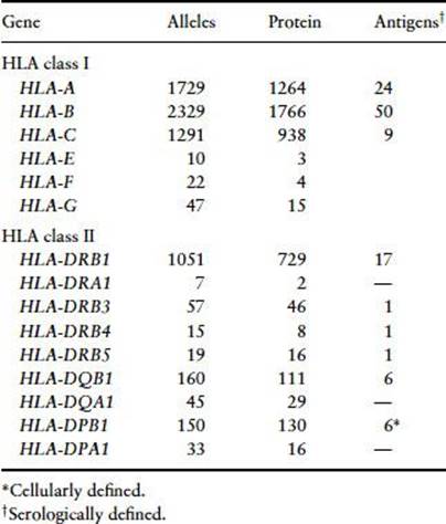
The number of recognized serologically defined antigens and DNA-identified HLA alleles is shown in Table 4.1 and can be accessed from http://hla.alleles.org/nomenclature/stats.html.
Table 4.1 Number of recognized HLA antigens/alleles. Adapted from Marsh et al. [5]. Taken from http://hla.alleles.org/nomenclature/stats.html. Accession date 07/12/11.
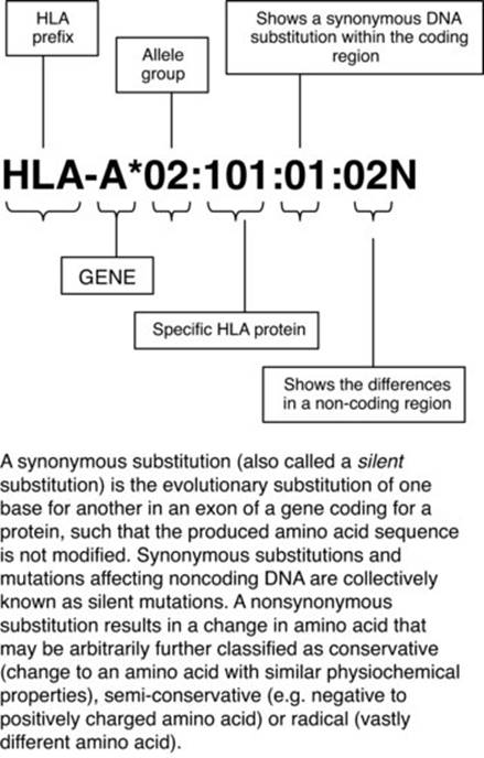
Due to the complexity of the HLA polymorphism and to the vast number of new alleles defined each year, a revised nomenclature has now been implemented [5,6] (see Figure 4.5). In this revised version optional suffixes may be added to an allele to indicate its expression status. Alleles that have been shown not to be expressed, ‘Null’ alleles, have been given the suffix ‘N’. An allele with ‘Low’ cell surface expression when compared with normal levels is indicated by the suffix ‘L’. Suffix ‘S’ is used to describe an allele specifying a protein that is expressed as a soluble ‘secreted’ molecule but is not present on the cell surface. A ‘C’ suffix describes an allele product that is present in the ‘Cytoplasm’ but not on the cell surface. An ‘A’ suffix indicates an ‘Aberrant’ expression and a ‘Q’ suffix is used when the expression of an allele is ‘Questionable’.
Fig 4.5 An example for a current HLA nomenclature. Taken from Nunes E, Heslop H, Fernandez-Vina M et al.Definitions of histocompatibility typing term. Blood 2011; 118: e180–e183 [6].
PCR sequence-specific priming
This technique involves the use of primers designed to anneal with DNA sequences unique to each allele and locus. The detection of the PCR-amplified product is then carried out by running the amplified product on an agarose gel. This technique allows the rapid identification of the HLA alleles in individual samples since the readout of this method is the presence or absence of the product for which specific primers were used. However, although this is a very rapid procedure, many PCR reactions have to be set up per sample in order to detect most of the defined alleles, e.g. at least 24 reactions for low resolution DR typing. Furthermore, for PCR-SSP typing the DNA sequence of the alleles must be known since novel unknown sequences may not always be detected.
PCR sequence-specific oligonucleotide probing
In this technique, the gene of interest is amplified using primers designed to anneal with DNA sequences common to all alleles of the loci of interest. The amplified PCR product is then immobilized on to support (e.g. nylon) membranes and the specificity of the products analysed by reacting the membranes with labelled allele-specific oligonucleotides. By scoring the probes that bind to specific regions, it is possible to assign the HLA type.
A modification of this technique, called reverse blot, involves the binding of the PCR-amplified product to labelled probes immobilized on membranes (strips) or plates. More recently, Luminex analysers are used to read the binding of the DNA to beads coated with an oligonucleotide probe. Reverse SSOP is useful when large numbers of samples need to be HLA-typed, e.g. bone marrow or cord blood donors.
DNA sequencing-based typing
DNA sequencing involves the denaturation of the DNA to be analysed to provide a single-strand template. Sequencing primers, exon- or locus-specific, are then added and the DNA extension is performed by the addition of Taq polymerase in the presence of excess nucleotides. The sequencing mixture is divided into four tubes, each of which contains specific dideoxyribonucleoside triphosphate (ddATP). When these are incorporated into the DNA synthesis, elongation is interrupted with chain-terminating inhibitors. In each reaction, there is random incorporation of the chain terminators and therefore products of all sizes are generated. The sequencing products are detected by labelling the nucleotide chain inhibitors with radioisotopes and, more recently, with fluorescent dyes. The products of the four reactions are then analysed by electrophoresis in parallel lanes of a polyacrylamide–urea gel and the sequence is read by combining the results of each lane using an automated DNA sequencer. In HLA SBT, some ambiguous results can be obtained with heterozygous samples and these may need to be retested by using PCR-SSP or reverse PCR-SSOP. SBT permits high resolution HLA typing, which is known to be important in the selection of HLA-matched HSC unrelated donors.
A major advantage of all DNA-based techniques is that no viable cells are required to perform HLA class I and II typing. Furthermore, since all the probes and primers are synthesized to order, there is a consistency of reagents used, allowing the comparison of HLA types from different laboratories. However, although serological typing is being rapidly replaced by DNA-based typing techniques, serological reagents may still be required for antigen expression studies.
The advantages and disadvantages of the various techniques described above are given in Table 4.2.
Table 4.2 Advantages and disadvantages of DNA-based techniques.
|
Technique |
Advantages |
Disadvantages |
|
Sequence-specific oligonucleotide probing (SSOP) |
Needs only one pair of genetic primers; fewer reactions to set up |
Different temperatures required for each probe |
|
Sequence-specific priming (SSP) |
Provides rapid typing with higher resolution than SSOP |
Too many sets of primers are needed to fulfil HLA type |
|
Sequencing-based typing (SBT) |
Provides the highest level of resolution Able to identify new alleles |
Not easy to perform |
|
Difficult to interpret |
Requires DNA sequence data to compare results Slower than rSSOP and SSP Limited throughput analyses High cost |
More recently a new approach to perform high resolution and high throughput HLA typing involving massive parallel clonal sequencing strategies and next-generation sequencing (NGS) platforms has been described. These NGSs are able to produce a large volume of HR HLA data.
Formation of HLA antibodies
HLA-specific antibodies are induced by pregnancy, transplantation, blood transfusions and planned immunizations. The affinity, avidity and class of the antibodies produced depend on various factors, including the route of immunization, the persistence and type of immunological challenge and the immune status of the host. Cytotoxic HLA antibodies can be identified in approximately 20% of human pregnancies. The antibodies produced are normally multispecific, high titre, high affinity and of the IgG class. Although these HLA IgG antibodies can cross the placenta, they are not harmful to the fetus. Antibodies produced following transplantation are mostly IgG, although rarely HLA IgM antibodies have been identified. In contrast, the majority of HLA antibodies found in multitransfused patients are multispecific IgM and IgG and are mostly directed at public epitopes. The introduction of leucocyte-reduced blood components (see Chapter 21) may lead to a reduction in alloimmunization in naive recipients, but it may not be very effective in preventing alloimmunization in already sensitized recipients, i.e. women who have become immunized as a result of pregnancy.
The deliberate immunization of healthy individuals to produce HLA-specific reagents is nowadays difficult to justify ethically. However, planned HLA immunization is still carried out in some countries to treat women with a history of recurrent miscarriages. These women are immunized with white cells from their partners or a third party to attempt to induce an immunomodulatory response that results in the maintenance of the pregnancy.
Detection of HLA antibodies
HLA antibodies are responsible for some of the serious immunological reactions to the transfusion of blood and blood components and play a pivotal role in the rejection of solid organ transplants.
A number of techniques to detect HLA antibodies have been developed. These include the complement-dependent lymphocytotoxicity (LCT) test, enzyme-linked immunosorbent assay (ELISA) and flow cytometry and, more recently, a Luminex-based technique [7,8].
Complement-dependent cytotoxicity test
The complement-dependent cytotoxicity (CDC) test, developed by Terasaki and McClelland (1964) [9], involves mixing equal volumes of serum and cells to allow the binding of the specific antibody to the target cell followed by the addition of rabbit complement. Complement-fixing antibodies reacting with the HLA antigen present on the cell surface lead to the activation of complement via the classical pathway and result in the disruption of the cell membrane. The lysed cells are then detected by adding ethidium bromide (EB) and the live cells are identified by adding acridine orange (AO) at the end of the incubation period. Live cells stained with AO when exposed to ultraviolet (UV) light appear green, whereas lysed cells allow the entry of EB, which binds to DNA, and they appear red under UV light. The reactions are scored by estimation of the percentage of dead cells in each well after establishing baseline values in the negative and positive as follows: 0–10% (one background cell death, negative), 11–20% (two doubtful negative), 21–50% (four weak positive), 51–80% (six positive) and 81–100% (eight strong positive).
The CDC assay, however, does not discriminate between HLA and non-HLA cytotoxic lymphocyte-reactive antibodies including autoantibodies. However, the majority of lymphocytotoxic autoantibodies are IgM and can be identified by screening the serum with and without dithiothreitol (DTT). The addition of DTT to the serum results in the breakdown of the intersubunit disulfide bonds in the IgM molecule, leading to the loss of cytotoxicity due to IgM. Prolonged exposure or excess DTT can lead to the breakdown of intramolecular disulfide bonds in the IgG molecules and also inactivate complement, but this can be inhibited by the addition of cystine.
The presence of lymphocytotoxic autoreactive antibodies in itself is not thought to be of clinical significance in solid organ transplant recipients or in patients immunologically refractory to random donor platelet transfusions.
Since the CDC test only detects cytotoxic HLA-specific antibodies, other techniques such as the ELISA or flow cytometry are needed to detect noncytotoxic HLA-specific antibodies.
Enzyme-linked immunosorbent assay
ELISA-based methods have often been the technique of choice for antibody detection, particularly where there has been a requirement for testing large numbers of samples. In this technique, a pool of purified HLA antigens is immobilized on a microwell plate, directly or via an antibody directed against a nonpolymorphic region of the HLA antigen or against the β2-microglobulin. Antibodies directed against the nonpolymorphic region of the HLA class I molecule, i.e. the α3 domain, are used to immobilize the specific HLA antigen to the microwell, ensuring that the more polymorphic α1 and α2 domains are available for antibody binding. HLA-specific antibodies bound to the immobilized antigens are then detected with an enzyme-linked secondary antibody which, upon addition of specific substrate, catalyses a colour change reaction that is detected in an ELISA reader.
In order to detect HLA specificity, each specific HLA antigen is isolated from an individual cell or cell line. Large panels of cells are cell lines used to purify these antigens in order to cover all the major HLA specificities at least once. A number of commercial kits are now available to screen for HLA antibodies and to define their specificities.
One of the main advantages of this ELISA technique is that they detect HLA-specific antibodies since it relies on the binding of the antibodies to wells coated with pools of solubilized or purified HLA antigens.
Flow cytometry
In this technique, cells and serum are incubated to allow the binding of the antibody to the target antigen. The bound antibody is then detected by using an antibody against human immunoglobulin labelled with a fluorescent marker such as fluorescein isothiocyanate or R-phycoerythrin. At the end of the incubation period, the cells are passed through the laser beam of the flow cytometer to identify the different cell populations based on their morphology/granularity and on the fluorescence. Normally, test sera with median fluorescence values greater than the mean + 3SD of the negative controls are considered positive, but each laboratory needs to establish its own positive and negative cut-off point values. By using a second antibody against cell-specific markers such as CD3 or CD19, it is possible to identify T- or B-cell reactivity.
The main advantages of flow cytometric techniques are the increased sensitivity when compared with LCT- and ELISA-based techniques and the detection of non-complement-fixing antibodies, allowing early detection of sensitization. However, one of the disadvantages is that it also detects non-HLA lymphocyte-reactive antibodies that are of unclear clinical relevance.
The use of flow cytometric techniques was initially investigated as an alternative cross-match technique and was shown to be more sensitive than previously described techniques. The increased sensitivity may be due to the fact that it detects both cytotoxic and noncytotoxic antibodies, some of which may be HLA specific.
Luminex
This technique uses fluorochrome-dyed polystyrene beads coated with specific HLA antigens. The precise ratio of these fluorochromes creates 100 distinctly coloured beads, each of which is coated with a different antigen. The beads are then incubated with the patient's serum and the reaction is developed using a PE-conjugated antihuman IgG (Fc-specific) antibody. The positive or negative reactions are then read using a Luminex analyser, which can distinguish between up to 100 different beads sets in a single tube. Luminex is the most sensitive technique currently available for the detection of HLA antibodies. Most recently, this technology has been further improved by the introduction of beads coated with single antigens, which improve the identification of antibody specificities that were not previously detected. In this technique, the beads can be coated with either HLA antigens for antibody screening or HLA oligonucleotide probes for HLA typing.
The CDC test and flow cytometry are the two main techniques used to perform antibody cross-matching between the patient's serum and the potential donor's cells in the solid organ transplant setting.
Clinical relevance of HLA antigens and antibodies
Although the main role of the HLA molecules is to present antigenic peptides to T cells, HLA molecules can themselves be recognized as foreign by the host T cells by a mechanism known as allorecognition. Two pathways of allorecognition have been identified, direct and indirect.
In the direct pathway, the host's T cells recognize HLA molecules (primarily class II) expressed on donor tissues, e.g. tissue dendritic cells and endothelial cells. Indirect allorecognition involves the recognition by the host T cells of donor-derived HLA class I and II antigenic peptides presented by the host's own antigen-presenting cells. Because of this mechanism, HLA antigen incompatibility is one of the main barriers to success of solid organ or HSC transplantation and also results in the strong alloimmunization seen in patients following transplantation or blood transfusion [10].
Solid organ transplantation
Matching for HLA-A, -B and -DR antigens is an important factor influencing the outcome of solid organ transplantation and particularly renal transplants. The application of the PCR-based techniques has allowed the identification of molecular differences between otherwise serologically identical HLA types of donor and recipient pairs, particularly in the HLA-DRβ1 chain. Correlation of these results with graft survival has shown a higher kidney graft survival rate when recipients and donors are HLA-DR identical by serological and molecular techniques than when they were HLA-DR identical by serological but not molecular methods (87% versus 69%) [11].
The presence of circulating HLA-specific antibodies directed against donor antigens in renal and cardiac recipients has been associated with hyperacute rejection of the graft. It is therefore important that these antibodies are detected and identified as soon as the patient is registered on the transplant waiting list to ensure that incompatible donors are not considered for transplantation [12]. Antibodies against the MICA and HLA DP antigens also seem to influence graft outcome, suggesting the possible need to screen for these antibodies in patients awaiting transplantation.
Furthermore, the appearance of donor-specific antibodies after transplantation has been associated with graft rejection, indicating the importance of posttransplant antibody monitoring for some groups of patients.
HLA and haemopoietic stem cell transplantation
HLA-DR incompatibility is one of the main factors associated with the development of acute graft-versus-host disease (aGVHD) but mismatches at the HLA-A and -B alleles, and to lesser extent HLA-C alleles, are also independent risk factors, particularly when using matched unrelated donors. Although HSC transplantation between HLA-identical siblings ensures matching for all HLA-A, -B, -C, -DR and -DQ genes, acute GVHD still develops in about 20–30% of these patients. This is probably due to the effect of untested HLA antigens, such as DP, or minor histocompatibility antigens in the activation of donor T cells. However, patients receiving grafts from HLA-matched unrelated donors have a higher risk of developing GVHD than those transplanted using an HLA-identical sibling [13].
The use of DNA-based methods has provided a unique opportunity to improve the HLA matching of patients and unrelated donors and to reduce the development of GVHD. However, it has been shown that the increased GVHD seen as a result of HLA mismatch is also associated with lower relapse rates, probably due to a graft-versus-leukaemia (GVL) response associated with the graft-versus-host response. On the other hand, the use of T-cell-depleted marrow, which has successfully decreased the incidence of GVHD, has resulted in an increased incidence of leukaemia relapse. Thus, it appears that mature T cells in the marrow, which may be responsible for GVHD, may also be involved in the elimination of residual leukaemic cells. Conversely, the rate of graft rejection is significantly higher in recipients of an HLA-mismatched transplant than in those receiving a transplant from an HLA-identical sibling (12.3 versus 2.0%) [13,14].
HSC transplantation using cord blood from HLA-matched and HLA-mismatched donors has now been associated with a reduced risk and severity of GVHD and with no increase in relapse rates. It is possible that the immaturity of the immunological effectors present in cord blood may contribute to the reduced GVHD without impairment of the GVL effect.
Graft failure, which is thought to be mediated by residual recipient T and/or NK cells reacting with major or minor histocompatibility antigens present in the donor marrow cells, has been shown to be associated also with antibodies reacting with donor's HLA antigens. Thus, rejection is particularly high in HLA-alloimmunized patients. However, in spite of these reports, HLA antibodies are more relevant in the posttransplant period, where highly immunized patients can develop immunological refractoriness to random platelet transfusions due to the presence of HLA antibodies. These patients require transfusions of HLA-matched platelets (see Chapter 27).
Blood transfusion
White cells and platelets present in transfused products express antigens that, if not identical to those present in the recipient, are able to activate T cells and lead to the development of antibodies and/or effector cells responsible for some of the serious complications of blood transfusion. Also, antibodies (and T cells) present in the transfused product may react directly with the relevant antigens in the recipient and lead to the development of a transfusion reaction. Amongst the transfusion reactions due to the presence of antibodies in the recipient are a febrile nonhaemolytic transfusion reaction (FNHTR) and immunological refractoriness to random platelet transfusions [15,16].
Although the occurrence of FNHTR has been commonly associated with the presence of HLA (and to a lesser extent HPA (human platelet antigen) or HNA (human neutrophil antigen)) antibodies in the recipient reacting with white blood cells or platelets present in the transfused product, it has recently been described that FNHTR may also be triggered by the direct action of cytokines such as IL-1β, TNF-α, IL-6 and/or by chemokines such as IL-8, which are found in transfused products.
Immunological refractoriness to random platelet transfusions is primarily due to the presence of HLA and, to a lesser extent, HPA and high titre ABO alloantibodies in the patient and reaction with the transfused incompatible platelets leading to the lack of platelet increments after the transfusion. Following the introduction of universal leucodepletion, the proportion of multitransfused patients with HLA antibodies seems to have decreased to approximately 10–20% and these patients are, in general, previously sensitized transplanted or transfused recipients and multiparous women.
The development of transfusion-related acute lung injury (TRALI) has been associated with the transfusion of blood components containing HLA and HNA antibodies able to recognize the relevant antigen(s) on recipient white cells and triggering an immunological reaction leading to the accumulation of neutrophils in the lungs and oedema. TRALI has sometimes been associated with the presence of HLA or HNA antibodies in recipients reacting with transfused leucocytes and/or to interdonor antigen–antibody reactions in pooled platelets.
Transfusion-associated (TA) GVHD, which is a rare but often severe and fatal transfusion reaction, is the result of immunocompetent HLA-matched T lymphocytes present in blood or blood components reacting with HLA and/or minor histocompatibility antigens present on the recipient cells. TA-GVHD occurs primarily in immunosuppressed individuals, although it can also occur in immunocompetent recipients. The diagnosis of TA-GVHD depends on finding evidence of donor-derived cells, chromosomes or DNA in the blood and/or affected tissues of the recipient.
HLA and disease
HLA genes are known to be associated with a number of autoimmune and infectious diseases [17,18] and different mechanisms to explain these associations have been postulated, including linkage disequilibrium with the relevant disease susceptibility gene, the preferential presentation of the pathogenic peptide by certain HLA molecules and molecular mimicry between certain pathogenic peptides and host-derived peptides. A number of diseases associated with both HLA class I and II have been described in Table 4.3. More recently genome-wide association studies (GWAS) [19] using over a thousand single nucleotide polymorphisms (SNPs) located in the MHC have identified a number of these SNP in strong linkage disequilibrium with some of the HLA associated diseases such as RA and SLE.
Table 4.3 HLA-associated diseases.
|
HLA class I genes |
|
Birdshot chorioretinopathy: HLA-A29 |
|
Behçet's disease: HLA-B51 |
|
Ankylosing spondylitis: HLA-B27 |
|
Psoriasis: HLA-Cw6 |
|
Malaria: HLA-B53 |
|
HLA class II genes |
|
Rheumatoid arthritis |
|
HLA-DRB1*0401 |
|
HLA-DRB1*0404 |
|
HLA-DRB1*0405 |
|
HLA-DRB1*0408 |
|
HLA-DRB1*0101/0102 |
|
HLA-DRB1*1402 |
|
HLA-DRB1*1001 |
|
Narcolepsy: HLA-DQB1*0602/DQA1*0102 |
|
Coeliac disease: HLA-DQB1*0201/DQA1*0501 |
|
Neonatal allo-immune thrombocytopenia: HLA-DRB3*0101 |
|
Malaria: HLA-DRB1*1302/DQB1*0501 |
|
Insulin-dependent diabetes mellitus: HLA-DQB1*0302/DQA1*0301 |
|
HLA-linked diseases |
|
Haemochromatosis: (HLA-A3) HFE gene C282Y, H63D and S65C |
|
21-OH deficiency: (HLA-B47) 21-OH gene |
|
Abacavir hypersensititvy: B*5701 |
Hereditary haemochromatosis
Hereditary haemochromatosis (HH) is a clinical condition of iron overload caused by an inherited disorder in the genes involved in the metabolism of iron. HH is a common genetic disorder in Northern Europe, where between 1 in 200 and 1 in 400 individuals suffer from the disease, with an estimated carrier frequency of between 1 in 8 and 1 in 10. Clinical manifestations of HH include cirrhosis of the liver, diabetes and cardiomyopathy. Detection of asymptomatic iron overload is important since removal of excess iron by phlebotomy can prevent organ damage [20,21].
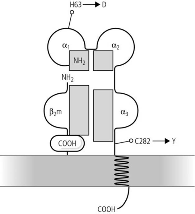
HH was originally described associated to HLA-A3, although this was not very specific since the majority of HLA-A3-positive individuals do not have HH. It was later found that mutations in the HFE gene located 3 Mb telomeric from the HLA-F gene was partly responsible for this condition. A number of mutations have now been identified and clinical data indicate that at least three of these mutations (C282Y, H63D and S65C) may predispose to and affect the clinical outcome of this condition. Over 90% of HH patients in the UK are homozygous for the mutation that replaces a cysteine (C) with a tyrosine (Y) at codon 282 in the HFE gene. The second and third mutations (H63D and S65C) are thought to be less important, although it may have an additive effect if inherited with the first mutation (Figure 4.6). Recent studies on blood donors have shown that approximately 1 in 280 donors is homozygous for the mutations.
Fig 4.6 HFE molecule. β2-m, β2-microglobulin.
A DNA-based technique to detect these three mutations simultaneously has now been developed and provides a simple, rapid and unambiguous definition of these mutations.
Neonatal alloimmune thrombocytopenia
Neonatal alloimmune thrombocytopenia (NAIT) is a serious condition in the newborn and is due to fetomaternal incompatibility for HPAs (see also Chapters 5 and 26). More than 80% of cases occur in women who are homozygous for the HPA-1b allele. Although the majority of cases are associated with the presence of HPA-1a antibodies, about 15% of cases are due to anti-HPA-5b. The production of HPA-1a antibodies is strongly associated with the HLA-DRB3*0101 allele. However, only approximately 35% of HPA-1a-negative, DRB3*0101-positive women develop antibodies upon exposure to the antigen, suggesting that other genes or factors may be involved in the development of alloimmunization against HPA-1a.
Other diseases
Diseases in which the molecular mimicry mechanism has been postulated include ankylosing spondylitis and Klebsiella infection. However, the precise pathogenic mechanisms involved remain unknown. More recently, it has been shown that HLA genes are also involved in the response to certain drugs, such as the association of HLAB-57 and abacavir, a drug used in the treatment of HIV [22].
Key points
1. HLA molecules are crucial in the induction and regulation of immune responses and in the outcome of transplantation using allogeneic-related and -unrelated donors and are also responsible for some of the serious immunological complications of blood transfusion.
2. The main feature of HLA genes is their high degree of polymorphism and linkage disequilibrium and, depending on their molecular structure, expression and function, they are classified as classical or nonclassical.
3. The detection of HLA polymorphisms is currently performed using DNA-based techniques at various degrees of resolution depending on the clinical needs and relevance.
4. The techniques currently used to screen and define the specificity of HLA antibodies allow the discrimination of HLA and non-HLA cytotoxic and noncytotoxic antibodies.
5. HLA antibodies produced following transfusion, transplantation or pregnancy are responsible for some of the most serious complications of blood transfusion.
6. HLA matching and cold ischaemia time are the two most important factors influencing the outcome of renal transplantation.
7. In the HSC transplantation setting, HLA matching for HLA class I and II genes is essential to minimize the development of a GVHD.
8. HLA genes are involved in the pathogenesis of a variety of diseases either directly through the presentation of pathogenic peptides or indirectly through their linkage disequilibrium with the relevant disease susceptibility gene(s).
References
1. Campbell RD. The human major histocompatibility complex: a 4000-kb segment of the human genome replete with genes. In: KE Davies & SM Tilghman (eds), Genome Analysis, Vol. 5: Regional Physical Mapping. New York: Cold Spring Harbor Laboratory Press; 1993, pp. 1–33.
2. Horton R, Wilming L, Rand V et al. Gene map of the extended human MHC. Nat Rev 2004; 5: 889–899.
3. Parham P & McQueen KL. Alloreactive killer cells: hindrance and help for haematopoietic transplants. Nat Rev Immunol 2003; 3: 108–121.
4. Traherne JA. Human MHC architecture and evolution: implications for disease association studies. J Immunogenet 2008; 35: 179–192.
5. Marsh SG, Albert ED, Bodmer WF et al. Nomenclature for factors of the HLA system. Tissue Antigens 2010; 75: 291–455.
6. Nunes E, Heslop H, Fernandez-Vina M et al. Definitions of histocompatibility typing term. Blood 2011; 118: e180–e183.
7. Brown C & Navarrete C. HLA antibody screening by LCT, LIFT and ELISA. In: J Bidwell & C. Navarrete (eds), Histocompatibility Testing. London: Imperial College Press; 2000, pp. 65–98.
8. Howell WM, Carter V & Clark B. The HLA system: immunobiology, HLA typing, antibody screening and crossmatching techniques. J Clin Pathol 2010, 63: 387–390.
9. Terasaki PL & McClelland JD. Microdroplet assay of human serum cytokines. Nature 2000; 204: 998–1000.
10. Choo SY. Yonsei Med J. 2007; 48(1): 11–23.
11. Opelz G & Döhler B. Effects of human leucocyte antigen compatibility of kidney graft survival: comparative analysis of two decades. Transplantation 2007; 84: 137–143.
12. Dyer PA & Claas FHJ. A future for HLA matching in clinical transplantation. Eur J Immunogenet 1997; 24: 17–28.
13. Madrigal JA, Arguello R, Scott I & Avakian H. Molecular histocompatibility typing in unrelated donor bone marrow transplantation. Blood Rev 1997; 11: 105–117.
14. Petersdorf EW, Malkki M, Gooley TA et al. MHC haplotype matching for unrelated hematopoietic cell transplantation. PloS Med 2007; 4: 59–68.
15. Harrison J & Navarrete C. Selection of platelet donors and provision of HLA matched platelets. In: J Bidwell & C. Navarrete (eds), Histocompatibility Testing. London: Imperial College Press; 2000, pp. 379–390.
16. Brown CJ and Navarette CV. Clinical relevance of the HLA system in blood transfusion. Vox Sanguinis 2011; 101: 93–105.
17. Caillat-Zucman S. Molecular mechanisms of HLA association with autoimmune diseases. Tissue Antigens 2009; 73(1): 1–8.
18. Gambaro G, Anglani F & D'Angelo A. Association studies of genetic polymorphisms and complex disease. The Lancet 2000; 355: 308–311.
19. Metzker ML. Sequencing technologies – the next generation. Nature Rev Genet 2010; 11: 31–46.
20. Mura C, Raguenes O & Ferec C. HFE mutations analysis in 711 haemochromatosis probands: evidence for S65C implication in mild form of hemochromatosis. Blood 1999; 93: 2502–2505.
21. Bomford A. Genetics of haemochromatosis. The Lancet 2002; 360: 1673–1681.
22. Profaizer T & Eckels D. HLA alleles and drug hypersensitivity reactions. Int J Immunogenet 2011; 39: 99–105.
Further reading
Brown CJ & Navarette CV. Clinical relevance of the HLA system in blood transfusion. Vox Sanguinis 2011; 101: 93–105.
Contreras M & Navarrete C. Immunological complications of blood transfusion. In: M Contreras (ed.), ABC of Transfusion, 4th edn. Wiley-Blackwell; 2009, pp. 61–68.
Navarrete C. Human leucocyte antigens. In: MF Murphy & DH Pamphilon (eds), Practical Transfusion Medicine, 3rd edn. Wiley-Blackwell; 2009, pp. 30–43.
Ouwehand H & Navarrete C. The molecular basis of blood cell alloantigens. In: D Proven & J Gribben (eds), Molecular Hematology, 3rd edn. Blackwell Publishing; 2010, pp. 259–275.