Stages of lung development
Embryonic (4-8 weeks)
• The lower respiratory system starts from a bud arising from the laryngotracheal groove; this branches into primary and secondary bronchopulmonary buds by the end of 5 weeks
• Repetitive branching continues and a primordial bronchial tree with five lobes is formed by the end of 8 weeks
Pseudoglandular phase (5-17 weeks)
• The primordial system of passage (air-conducting bronchial tree) with the terminal bronchioles is formed and is initially lined with cuboidal epithelium
• These precursor cells later differentiate into ciliated epithelium and secretory cells in respiratory ducts and also develop into type II pneumocytes in terminal bronchioles
• At this stage, the lung is composed of tubular glandular structures surrounded by undifferentiated mesenchyme
Canalicular phase (13-25 weeks)
• The lumen of tubules becomes wider and some of the lining type II pneumocytes differentiate into flattened type I pneumocytes
• Capillaries invade into the mesenchyme and surround the acini, therefore forming the foundation of the blood–air barrier
• Only by the end of this phase is the fetus able to survive outside the uterus
Terminal sac phase (24 weeks–term)
• Lung epithelium starts to produce amniotic fluid and lung maturation can be measured by surfactant, which is produced by type II pneumocytes
• Airspaces are expanded to form thin, smooth-walled saccules (primitive alveoli) at the end of each respiratory tract passage
• The primary septa between saccules are thick and contain two layers of capillaries from the neighboring saccules
• By the end of this phase (at birth), one third of alveoli are developed
Alveolar phase (36 weeks’ gestation–8-10 years)
• Sacculi are subdivided into smaller subunits (alveoli) by secondary septa, which contain elastic fibers between two capillary networks
• This alveolarization reaches its maximum level in the first 6 months of birth and is significant up to 18 months
• Some alveoli continue to be developed up to 8-10 years of age
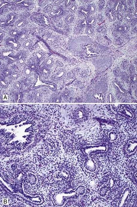
Fig 1 Development of lung. Pseudoglandular phase of fetal lung: low (A) and high (B) powers. Note undifferentiated mesenchyme and primitive tubular glands lined by nonciliated, glycogen-rich columnar cells with clear cytoplasm.
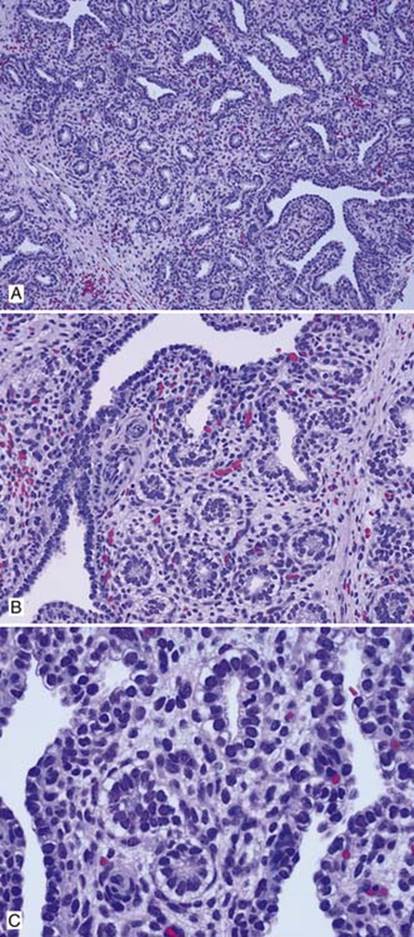
Fig 2 Development of lung. Canalicular phase of fetal lung: low (A), medium (B), and high (C) powers. Note the capillary network within the mesenchyme; some of the lining epithelium cells are flattened.
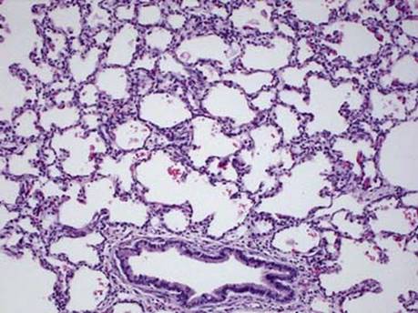
Fig 3 Development of lung. Terminal sac phase of lung development seen in this near term infant.
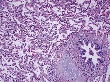
Fig 4 Development of lung. Alveolar phase of lung development in a 2½-year-old.