
Definition
• An alkylating agent used in combination with other chemotherapeutic agents to treat a variety of malignancies and in combination with corticosteroids to treat inflammatory diseases such as Wegener granulomatosis, which can cause lung toxicity
Clinical features
Epidemiology
• Pulmonary disease attributable to cyclophosphamide is rare
• Patients taking cyclophosphamide who present with lung disease are usually taking a combination of chemotherapeutic agents, which makes it difficult to definitively attribute the disease to cyclophosphamide alone
Presentation
• Early onset: fever, coughing, and dyspnea within the first 6 months of taking the drug
• Late onset: insidious onset of coughing and dyspnea with restrictive changes on pulmonary function tests, occurring after several years of therapy
Prognosis and treatment
• Early onset: resolution of symptoms and good prognosis with discontinuation of cyclophosphamide and treatment with corticosteroids
• Late onset: increased risk of progressive respiratory failure and death
Pathology
Histology
• A variety of histopathological patterns of lung injury can be seen, including type II pneumocytic hyperplasia, organizing pneumonia or organizing diffuse alveolar damage (DAD), interstitial fibrosis in a nonspecific interstitial pneumonia or usual interstitial pneumonia pattern, interstitial edema, alveolar hemorrhage, increased numbers of pigmented alveolar macrophages, and acute DAD with hyaline membranes
• Thickened pleura with fibrosis
Immunopathology/special stains
• Not contributory
Main differential diagnoses
• Other causes of organizing pneumonia, DAD, and interstitial fibrosis

Fig 1 Cyclophosphamide. In this patient receiving combination chemotherapy that included cyclophosphamide, there are prominent interstitial fibrosis, hyperplasia of type II pneumocytes, and increased numbers of pigmented alveolar macrophages.

Fig 2 Cyclophosphamide. Severe interstitial fibrosis is present in this patient with lung injury due to cyclophosphamide.
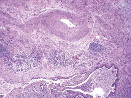
Fig 3 Cyclophosphamide. Other areas from the same patient as in Fig 2 show fibrosis of the bronchial wall and submucosal edema (lower left), as well as secondary pulmonary hypertensive changes in vessels.

Fig 4 Cyclophosphamide. Hyperplasia of type II pneumocytes with reactive atypia is seen at higher power.

Fig 5 Cyclophosphamide. Subepithelial fibrosis of an airway is seen here on H&E (A) and trichrome (B) stains.