
Definition
• Locally aggressive vascular tumor composed of spindled neoplastic endothelial cells associated with human herpesvirus (HHV)-8 infection
Clinical features
Epidemiology
• Occurs most often in AIDS patients and immunocompromised patients with solid organ transplant
Presentation
• Respiratory complaints include dyspnea, stridor, coughing, and hemoptysis
• Bronchoscopic examination may reveal nodular or flat bluish-red discolorations in the mucosa
• Chest x-ray films: bilateral interstitial infiltrates with ill-defined nodularity
Prognosis and treatment
• Poor prognosis: survival time approximately 2 years
• Surgical resection is usually not feasible due to tumor’s multicentricity
• Tumor responds relatively well to chemotherapy
Pathology
Gross
• Multiple ill-defined red-bluish lesions
Histology
• Proliferation of oval to spindled tumor cells with mild atypia; form slitlike spaces containing extravasated erythrocytes
• Irregular thin-walled blood vessels invading through the pulmonary parenchyma
• Tends to grow along preexisting intrapulmonary septae and concentrate around blood vessels and small airways
Immunopathology/special stains
• Tumor cells are positive for endothelial markers (CD31, CD34, and FLI1) and HHV-8
Ultrastructure
• Endothelial differentiation with basal lamina and Weibel-Palade bodies
Main differential diagnoses
• Angiosarcoma: a high-grade malignant vascular tumor comprising anastomosed irregular vascular channels lined by neoplastic endothelial cells and infiltrating the surrounding tissue. The tumor can be morphologically biphasic; composed of spindle and epithelioid zones
• Epithelioid hemangioma: well-formed blood vessels lined by plump, epithelioid endothelial cells, often associated with proliferation of eosinophils and lymphocytes
• Hemangioma: thin-walled blood vessels lined by bland, flat endothelial cells
• Kaposiform hemangioendothelioma: locally aggressive vascular neoplasm; typically occurs in infants and children; most frequently occurs in retroperitoneum and skin; characterized by predominant Kaposi sarcoma–like fascicular spindle cell proliferation; relatively well-formed vascular channels are present in peripheral of the tumor; no association with HIV or HHV-8 infection

Fig 1 Kaposi sarcoma. A, Oval to spindle tumor cells with mild atypia forming slitlike spaces with extravasated erythrocytes. B, Mitotic figures are identified.

Fig 2 Kaposi sarcoma. Transbronchial biopsy shows spindle cell neoplasm with slitlike spaces containing erythrocytes.

Fig 3 Kaposi sarcoma. Tumor cells show nuclear positivity for HHV-8 (immunohistochemical test).
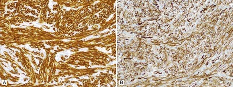
Fig 4 Kaposi sarcoma. Tumor cells are positive for CD34 (A) and CD31 (B).