
Definition
• PPHL is defined by the following parameters:
• Histological documentation of Hodgkin lymphoma
• Restriction to the lung parenchyma, with or without “minimal” hilar lymph node involvement
• Extrapulmonary disease not detected by imaging studies or bone marrow assessment
Pathogenesis
• Epstein-Barr virus (EBV) may be associated/found in some tumor cells
Clinical features
Epidemiology
• Primary extranodal Hodgkin lymphomas are rare, and only 8% of them originate in the lung
• On the other hand, secondary lung infiltration develops in approximately 40% of patients with Hodgkin lymphoma
• Slight female predominance
• Bimodal age distribution: third and seventh to eighth decades
Presentation
• Dry cough, dyspnea, chest discomfort, hemoptysis, wheezing, systemic symptoms (B symptoms), which divide each clinical stage of lymphoma into (A) absence of symptoms and (B) presence of symptoms; thus these are referred to as “B symptoms”
• Chest radiographs show discrete nodular opacities, which may be solitary or multiple and usually arise in the superior portions of the lung
• Secondary involvement by systemic Hodgkin lymphoma is characterized by a random miliary distribution lacking zonal predilection
Prognosis and treatment
• Clinical course is usually indolent, although aggressive variants have been reported
• Poor prognostic indicators include presence of B symptoms, age >60 years, multiple and/or bilateral nodules, penetration of the pleura, pleural effusions, and cavitary disease
• Treatment includes radiotherapy, combination chemotherapy, and combined modality therapy
Pathology
Gross
• PPHL tends to form fairly well-demarcated nodular masses, with the infiltrate spreading out along alveolar septa
Histology
• The diagnosis is based on the recognition of mononuclear Hodgkin and multinucleated Reed-Sternberg cells:
• Large cells with abundant, slightly basophilic cytoplasm
• Nuclei are large and round with prominent nuclear membranes, pale chromatin, and usually one prominent eosinophilic nucleolus
• Occasional “mummified” Hodgkin and Reed-Sternberg (HRS) cells have condensed cytoplasm and smudged, ill-defined nuclei
• The density of HRS cells varies from case to case, but they usually represent a small minority of the cellular infiltrate
• HRS cells reside in a mixed infiltrate composed of nonneoplastic lymphocytes, eosinophils, neutrophils, histiocytes, plasma cells, fibroblasts, and collagen fibers
• All histological subtypes of classic Hodgkin lymphoma have been described in PPHL
• Mixed cellularity and nodular sclerosing are the most frequent subtypes reported in the lung
• Central necrosis and cavitation is not unusual
• Adjacent nonneoplastic lung can demonstrate lymphoid hyperplasia, hypersensitivity, pneumonia-like changes, and fibrosis
• Most patients require wedge lung biopsy for diagnosis because of sampling or technical artifacts
Immunopathology/special stains
• HRS cells are characteristically positive for CD15 and CD30, demonstrating membranous and Golgi accentuation with both
• HRS cells are negative for CD45 (leukocyte common antigen) and EMA
• HRS cells are usually positive for PAX5 and negative for other B-cell markers (CD20, CD19, CD79a)
• The mixed inflammatory cells in the background are benign and demonstrate typical immunophenotypes
Main differential diagnoses
• Other lymphoid neoplasms:
• Non-Hodgkin lymphomas, particularly pleomorphic T-cell, large B-cell, and anaplastic large-cell lymphomas
• Lymphomatoid granulomatosis
• Nonlymphoid neoplasms:
• Large-cell carcinoma
• Melanoma
• Undifferentiated carcinoma
• Inflammatory myofibroblastic tumors
• Langerhans cell histiocytosis
• If central necrosis is present, consider granulomatosis with polyangiitis (Wegener), tuberculosis, and other infectious conditions
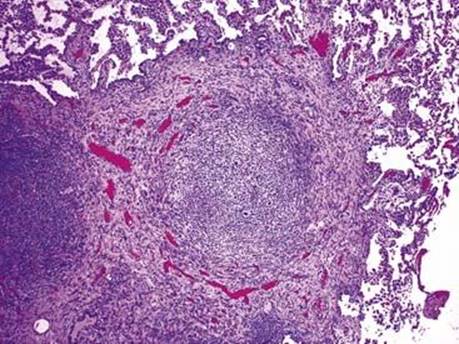
Fig 1 Primary pulmonary Hodgkin lymphoma. The tumor forms a fairly well-demarcated mass within the lung parenchyma.
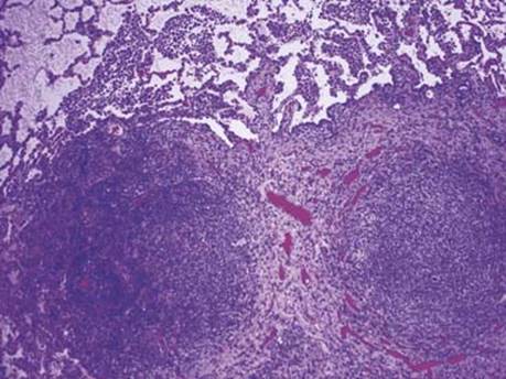
Fig 2 Primary pulmonary Hodgkin lymphoma. Fibrous collagen bands divide the mass into nodules.
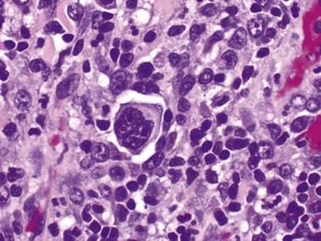
Fig 3 Primary pulmonary Hodgkin lymphoma. A multinucleated Reed-Sternberg cell is seen in a background of lymphocytes and histiocytes.
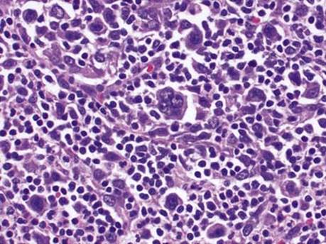
Fig 4 Primary pulmonary Hodgkin lymphoma. Numerous mononuclear Hodgkin cells are also present.
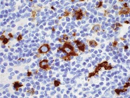
Fig 5 Primary pulmonary Hodgkin lymphoma. HRS cells demonstrate membranous and paranuclear dot-like staining with CD15.
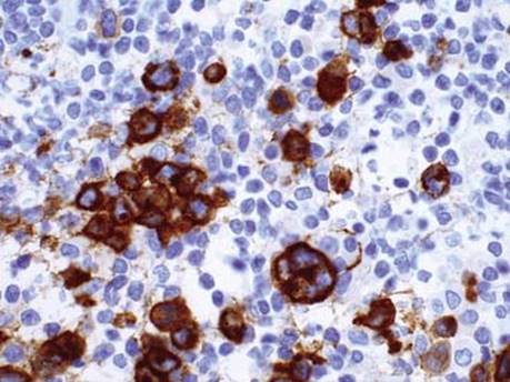
Fig 6 Primary pulmonary Hodgkin lymphoma. CD30 shows a similar membranous and paranuclear staining pattern, highlighting the presence of a large Reed-Sternberg cell and smaller Hodgkin cells.