
Definition
• Extraosseous plasmacytomas are localized plasma cell neoplasms that arise in tissue other than bone; primary pulmonary plasmacytomas (PPPs) involve the lower respiratory tract
• Complete skeletal radiographs show no other lesions; there are no clinical features of plasma cell myeloma (anemia, hypercalcemia or renal impairment) and no evidence of bone marrow plasmacytosis
Clinical features
Epidemiology
• Extraosseous plasmacytomas account for approximately 3% to 5% of all plasma cell neoplasms, and approximately 80% involve the upper respiratory tract, including the oropharynx, nasopharynx, sinuses, and larynx
• Pulmonary plasmacytomas are exceptionally rare, and the variance in the reported incidence of PPP (3%-6% of all extraosseous plasmacytomas) is wide
• Two thirds of patients with extraosseous plasmacytomas are male; however, PPP has no sex predilection
• Median age at diagnosis, approximately 55 years
Presentation
• Most commonly seen as a pulmonary nodule in the hilum; however, presentations as lobar consolidations and bilateral diffuse infiltrates have been reported
• Right and left lungs are equally affected, and the upper lobes are more commonly affected
• Patients may be asymptomatic or may present with coughing, dyspnea, shortness of breath, fever, or hemoptysis caused by bleeding from endobronchial extension
• A small portion of patients may have a small serum M-protein or Bence-Jones light chains in the urine. The presence of an associated M-protein is directly related to the tumor cell mass and may be measured to follow disease progression
• The typical radiographic appearance is that of a solitary mass or nodule, most frequently involving the hilar areas. Pleural lesions and peripheral masses may also occur
• No bone marrow involvement
Prognosis and treatment
• In general, patients with extramedullary plasmacytomas outlive those with multiple myeloma; reported survival time is up to 10 years or more
• Conversion to multiple myeloma is uncommon but is a definite risk, resulting in shortened survival
• The treatment for PPP is usually resection, but local radiation may also be utilized
Pathology
Histology
• Plasmacytomas exhibit discrete demarcation from the surrounding pulmonary parenchyma and consist of syncytia of mature plasma cells with no residual germinal centers. The numbers of intermingled lymphocytes are negligible
• Anaplastic and plasmablastic variants feature pleomorphic cells often with prominent nucleoli, multiple nuclei, or multilobated nuclei
• If present, amyloid deposits consist of pale, waxy, amorphous material
Immunopathology/special stains
• The neoplastic plasma cells usually have monotypic cytoplasmic immunoglobulin (Ig) and lack surface Ig. They also express CD79a, CD138, and CD38. There may be aberrant CD56 (up to 80% of cases), CD117, CD20, CD52, or CD10 expression
• CD19, CD20, and PAX5 are usually negative
Main differential diagnoses
• Inflammatory myofibroblastic tumor (inflammatory pseudotumor, plasma cell granuloma): characterized by hyalinized fibroconnective tissue, sheets of polytypic plasma cells together with plump fibroblasts, histiocytes, mast cells, and lymphocytes
• Lymphoplasmacytic lymphoma usually has a mixture of small lymphocytes mixed with plasma cells: CD45+, CD19+, scattered CD138+ cells, CD56–
• Nodal, extranodal, and splenic marginal zone lymphomas may have plasmacytoid differentiation, but small lymphocytes are usually present: CD45+, CD19+, scattered CD138+ cells, CD56–
• Plasmablastic lymphoma is usually seen in HIV+ or immunosuppressed patients and usually is seen in the oral cavity: EBV+ (60%)
• Melanoma or poorly differentiated carcinoma: CD45–, CD38–, CD138–, CD56–
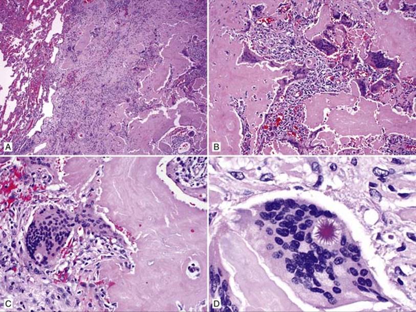
Fig 1 Primary pulmonary plasmacytoma. A, Low power shows well-demarcated tumor in the lung, which is composed of amorphous, eosinophilic material being engulfed by multinucleated giant cells (B). C and D, Asteroid bodies are present in some of the multinucleated giant cells.
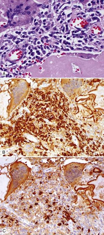
Fig 2 Primary pulmonary plasmacytoma. A, Sheets of plasma cells are present between amyloid deposits, which are predominantly lambda light chain positive (B) with only a few kappa light chain positive cells (C).
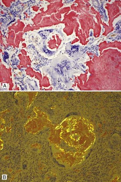
Fig 3 Primary pulmonary plasmacytoma. Amyloid is confirmed by Congo red positivity (A), which shows apple green birefringence under polarized light (B).