
Definition
• Multifocal nodular proliferation of benign-appearing smooth muscle tumors in women with previous or concomitant uterine leiomyomas
Clinical features
Epidemiology
• Rare lesion
• Affects women of reproductive age
• Could represent:
• True metastases from inadequately sampled uterine leiomyoma with sarcomatoid areas
• Embolization after surgical trauma during uterine dilatation and curettage, myomectomy, or hysterectomy
• Metaplastic transformation of pulmonary smooth muscle elements
• Development from circulating stem cells
Presentation
• History of previous or concurrent uterine leiomyomas
• Radiologic detection of multiple slow-growing lung nodules
• Either asymptomatic or could have coughing, asthma-like symptoms, hemoptysis, obstructive pneumonia
Prognosis and treatment
• Surgical enucleation of the nodules
• Favorable prognosis
• Hormonally dependent: grows during pregnancy and spontaneously regresses after pregnancy, oophorectomy, menopause, use of megestrol, selective estrogen modulators, aromatase-P450 inhibitors, and gonadotropin-releasing hormone agonists
Pathology
Gross
• Sharply circumscribed firm tan-gray masses with “whorled” appearance within lung parenchyma, with no specific pattern
• Nodule size ranges from 0.3 to 4 cm in greatest dimension
• Sometimes present as solitary nodule or multiple miliary nodules
Histology
• Homogeneous population of fusiform cells with fibrillary eosinophilic cytoplasm and blunt-ended nuclei with dispersed chromatin
• Low mitotic activity (less then 5 mitoses per 10 high-power fields [hpf])
• Lack of nuclear pleomorphism and atypia; absence of necrosis and pseudocyst formation
• Areas of fibrosis with entrapped alveolar and bronchial glandular spaces that may have metaplastic lining
Immunopathology/special stains
• ER/PR+, SMA+, desmin+, MSA+, calponin+, and caldesmon+, but S100–, CD56–, keratin–, and HMB45–
• Molecular evidence of clonal (neoplastic) nature
Main differential diagnoses
• Metastatic low-grade leiomyosarcoma: mitotically active
• Primary lung leiomyoma: solitary, distal part of the tracheobronchial tree
• Lymphangioleiomyomatosis (LAM): cyst formation, more epithelioid lesional cells, HMB45+, MiTF-1+
• Intravenous leiomyomatosis: thought to have similar pathogenesis, elongated branching lesions, with presence of lesions within pelvic veins
• Fibroleiomyomatous hamartoma: suggested to be synonymous to BML but without history of uterine leiomyomas
• Malignant peripheral nerve sheath tumor: muscle markers negative, at least focally S100+

Fig 1 Benign metastasizing leiomyoma. Large nodule of BML. Epithelial-lined clefts of bronchial and alveolar epithelium entrapped by fibrotic fusiform smooth muscles

Fig 2 Benign metastasizing leiomyoma. Plump spindle-shaped smooth muscle cells with abundant eosinophilic cytoplasm and blunt-ended nuclei. Note lack of atypia, nuclear pleomorphism, and mitotic figures.
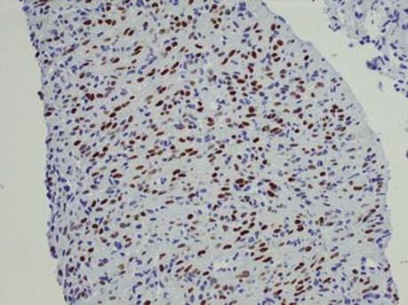
Fig 3 Benign metastasizing leiomyoma. Strong nuclear ER expression in BML; note not all cells are positive.
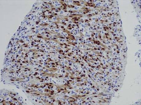
Fig 4 Benign metastasizing leiomyoma. Diffuse strong PR positivity in cigar-shaped smooth muscle cell nuclei of BML; note the nonspecific background staining of cytoplasm

Fig 5 Benign metastasizing leiomyoma. BML demonstrating strong immunoreactivity with smooth muscle actin.

Fig 6 Benign metastasizing leiomyoma. Muscle-specific actin strongly stains BML nodule.
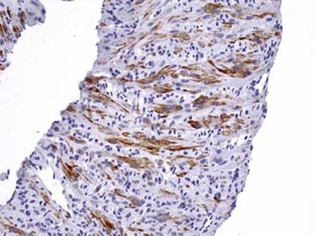
Fig 7 Benign metastasizing leiomyoma. Caldesmon expression in spindle cells of BML.
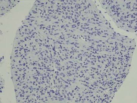
Fig 8 Benign metastasizing leiomyoma. HMB45 (shown) and S100 markers are uniformly negative in BML.