
Definition
• A group of benign or low-to-moderate-grade thymic neoplasms with thymic epithelial differentiation
Clinical features
Epidemiology
• Uncommon
• No gender predilection
• Usually occurs in older patients 50 to 60 years old; rarely in younger individuals
Presentation
• Anterosuperior mediastinal mass; rarely ectopic in posterior mediastinum, neck, pleura, or lung
• Local symptoms: chest pain, coughing, dyspnea, dysphagia, hoarseness, or superior vena cava syndrome
• Up to half of the tumors are associated with myasthenia gravis (MG); on the other hand, 20% to 30% of MG patients have thymoma
• Rarely associated with paraneoplastic syndromes (hypogammaglobulinemia and pure red cell aplasia) or connective tissue diseases (systemic lupus erythematosus and rheumatoid arthritis)
• May be asymptomatic and discovered incidentally
Prognosis and treatment
• For stage I and II tumors: complete removal of thymoma as well as thymus is recommended
• For higher stage tumor: surgical resection with chemotherapy or radiation therapy
• The prognosis depends on tumor stage, World Health Organization (WHO) histological subtype, and completeness of resection
• Types A and AB thymomas are considered to be clinically benign tumors with exceptionally rare recurrence and metastases
• Type B1: low grade malignant potential
• Types B2 and B3: moderate malignant potential
Pathology
Histology
• Broad lobulation is seen in all thymomas, and fibrous tissue separates the tumor into cellular lobules (organotypic architecture)
• All thymomas have varying amounts of neoplastic epithelial cells and reactive T cells that may be mature or immature
• In many there is perivascular clearing due to accumulation of fluid within which are found lymphocytes, red blood cells, and macrophages
• Although multiple histological classification schemes exist, the WHO scheme is currently the most widely accepted because it correlates with clinical behavior and outcome; used as follows:
• Conventional thymoma:
• Type A: neoplastic epithelial cells are spindle shaped
• Type B: neoplastic epithelial cells are round polygonal cells
– Type B1: with most amount of lymphocytes
– Type B2: with moderate amount of lymphocytes
– Type B3: lymphocyte poor
• Type AB (mixed spindle and epithelioid cells)
• Most common subtypes: B2 and AB
• Rare subtypes: B1 and A
• Nonconventional and rare thymomas:
• Micronodular thymoma
– Tumor consists of multiple discrete small nodules of epithelial cells separated by lymphocyte-rich stroma with prominent lymphoid follicles
– The epithelial cells are spindle to oval with few intraepithelial lymphocytes
– Organotypic architecture of conventional thymoma; however, the lymphocyte component (mature B cells) is different from that of conventional thymoma (immature T cells)
• Metaplastic thymoma
– Biphasic thymoma composed of anastomosing islands to broad trabeculae of epithelial cells intermingled with bland spindle cells
– Different from conventional thymoma: no association with MG; lack of lobulated growth pattern and perivascular spaces
• Microscopic thymoma
– Higher incidence rate in MG-associated thymus
– Multifocal microscopic (<1 mm) proliferation of epithelial cells
• Sclerosing thymoma
– Extremely rare
– An otherwise conventional thymoma with marked collagen-rich stroma without necrosis or hemorrhage
• Lipofibroadenoma
– Extremely rare
– Morphologically resembles fibroadenoma of the breast or thymolipoma
Staging of thymoma (Masaoka classification)
• Stage I: grossly and microscopically encapsulated (includes microscopic invasion into, but not through, the capsule)
• Stage II: tumor with transcapsular invasion
• IIa: microscopic capsular invasion
• IIb: macroscopic capsular invasion
• Stage III: invasion of neighboring organs (pericardium, lung, or great vessels)
• Stage IV: pleural or pericardial dissemination (IVa) or hematogenous /lymphatic dissemination (IVb)
Immunopathology/special stains
• Tumor epithelial cells:
• Positive for cytokeratins (e.g., AE1/AE3, CAM5.2, and CK7, except CK20)
• Frequently positive for p63 and CK5/6
• Usually negative for CD5 and CD70
Main differential diagnoses
• Lymphoma: usually positive for LCA and negative for cytokeratin
• Neuroendocrine carcinoma: neuroendocrine features with positive neuroendocrine markers
• Germ cell tumors: positive for OCT3/4 and PLAP
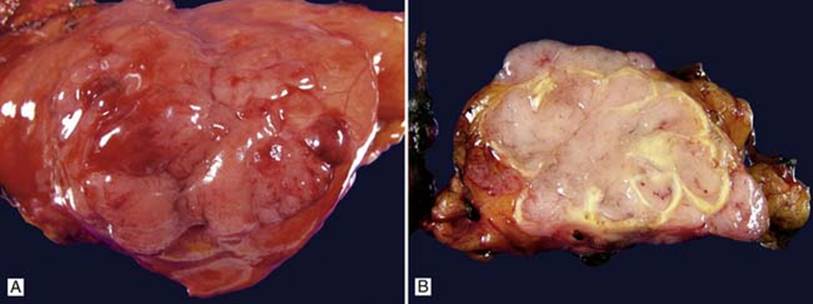
Fig 1 General features of thymoma. Gross photographs of a circumscribed thymoma: outside showing smooth surface and nodular appearance (A) and lobulated cut surface with fibrous septa (B).
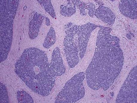
Fig 2 General features of thymoma. Low power of thymoma with lobulated cellular growth and intervening fibrous septa.
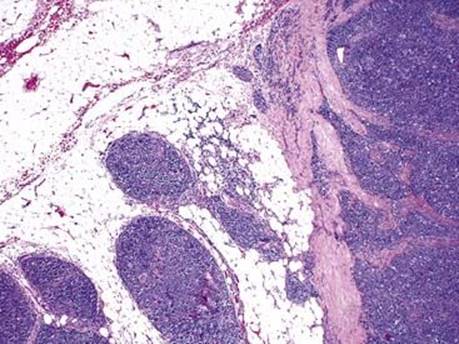
Fig 3 General features of thymoma. Capsular invasion.
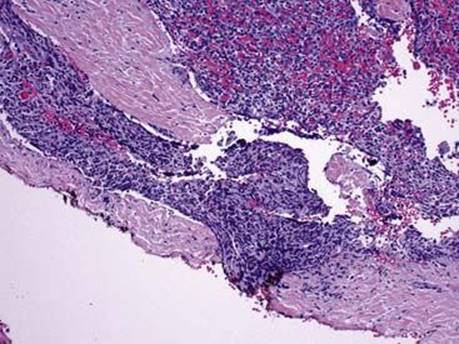
Fig 4 General features of thymoma. Margin positive.
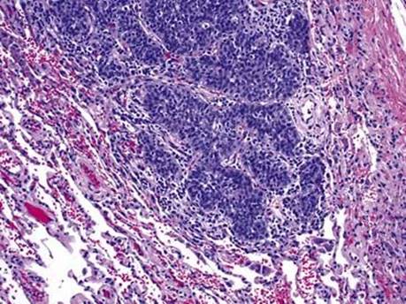
Fig 5 General features of thymoma. Lung invasion.