
Definition
• Inflammation due to infection (acute mediastinitis) or an excessive fibrotic reaction in the mediastinum (sclerosing mediastinitis) that results from a host reaction to a prior infection involving the mediastinal lymph nodes and may include granuloma formation (granulomatous mediastinitis)
Pathogenesis
• Acute mediastinitis is commonly due to infection after thoracic surgery
• Sclerosing mediastinitis commonly occurs after infection with Histoplasma capsulatum; less commonly due to tuberculosis, Aspergillus infection, mediastinal radiation, or idiopathic form
• H. capsulatum infection begins in the lungs and typically disseminates to the mediastinal lymph nodes before the fungus is contained by the immune system
• Fungal antigens cause a hypersensitivity reaction and fibroblast proliferation
• Granulomatous mediastinitis occurs when affected lymph nodes in sclerosing mediastinitis enlarge, coalesce, and form inflamed granulomas that extend into the fat
Clinical features
Epidemiology
• Less than 1% of patients with histoplasmosis develop sclerosing mediastinitis
• Can occur at any age
Presentation
• Acute mediastinitis presents with inflamed incision site, poor wound healing, and fever or other signs of infection
• Sclerosing mediastinitis presents with symptoms of compression
• Airway compression leading to obstructive pneumonia, atelectasis, shortness of breath
• Compression of the great vessels of the heart resulting in superior vena cava syndrome and pulmonary/vein obstruction and constrictive pericarditis
• Esophageal compression leading to dysphagia
• Hemoptysis caused by invasion of fibrous tissue into the bronchus
Prognosis and treatment
• Acute mediastinitis is treated with wound débridement and antibiotics
• Sclerosing mediastinitis is a variable and progressive disease with no effective medical therapy, but surgery can alleviate compression
Pathology
Gross
• Sclerosing mediastinitis
• Firm white tissue on cut section
• Clear-cut junction with adjacent adipose tissue can be seen
Histology
• Acute mediastinitis
• Granulation tissue with acute and chronic inflammation
• Sclerosing mediastinitis
• Dense fibrohyaline tissue that is relatively avascular and extremely paucicellular
• Necrotizing or nonnecrotizing granulomas can be present
Immunopathology/special stains
• GMS, PAS, and AFB stains to rule out infection
Main differential diagnoses
• Other fibromatoses

Fig 1 Mediastinitis. Débridement of tissues in a patient with acute mediastinitis after thoracotomy, showing extensive acute inflammation.
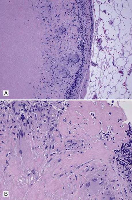
Fig 2 Mediastinitis. Low power (A) and high power (B) views of necrotizing granulomas with fibrosis in a mediastinal lymph node in a patient with histoplasmosis.