
Definition
• Thickening and stiffening of the mitral valve resulting in immobility of the valve and obstruction of blood flow from the left atrium to the left ventricle
Pathogenesis
• Tiny nodules form along lines of closure of the valve leaflets
• Deposition of fibrin thickens the leaflets, and the cusps lose their normal morphology
• Increasing fibrosis often with calcifications leads to fusion of leaflet commissures
Clinical features
Epidemiology
• The most common cause in developed and underdeveloped countries is rheumatic heart disease
• Congenital mitral valve stenosis is less common
Presentation
• Obstruction of blood flow causes increased pressure in the left atrium, pulmonary vasculature, and right side of the heart
• Most common symptom is dyspnea followed by fatigue
• Thromboembolism, hemoptysis, and right-sided heart failure also occur
• An opening snap of the mitral valve can be heard
• Commonly, a low-pitched diastolic rumble is heard best at the apex because there is also insufficiency of the valve
Prognosis and treatment
• Progressive disease that worsens quickly after onset of symptoms; mild symptoms to severe disability can occur in less than 10 years
• Death due to right-sided heart failure, pulmonary edema, and cerebral or pulmonary thromboembolism
• Symptoms are initially treated medically
• Mitral valve repair or replacement; replacement considered final option because of high perioperative mortality
Pathology
Gross
• Symmetric, narrow, oval-shaped orifice of valve (“fish mouth”)
• Doming of leaflets due to commissure fusion; leaflets thickened, retracted, and calcified
• Chordae fused and shortened
Histology
• Fibrosis, calcification with or without ossification, and neovascularization
• Chronic inflammatory infiltrate consisting of lymphocytes, monocytes, and mast cells
• Aschoff bodies are not seen
Immunopathology/special stains
• Not contributory
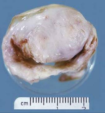
Fig 1 Mitral valve stenosis. Gross photograph of surgically resected thickened “fish mouth” valve.

Fig 2 Mitral valve stenosis. Medium-power view of fibrosis of mitral valve.
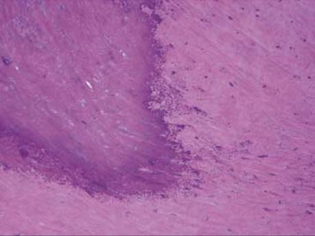
Fig 3 Mitral valve stenosis. Medium-power view of mitral valve calcification.