
Definition
• Rare primary malignant tumors of cardiac mesenchyme
Pathogenesis
• Studies of molecular pathogenesis of primary cardiac sarcomas are few because of the rarity of these entities
• Mutations in K-ras and p53 have been noted in some tumors (angiosarcoma and rhabdomyosarcoma)
• Nonspecific chromosomal aberrations have been detected in others
• Synovial sarcoma is unique in that it harbors the characteristic t(X;18), which results in fusion involving the SS18 and SSX genes. The transcript is thought to be implicated in carcinogenesis. See separate chapter for details
Clinical features
Epidemiology
• Cardiac sarcomas are rare; cardiac tumors in general account for about 0.2% of all neoplasms, with malignant tumors constituting about 25% of those; most of these are sarcomas
• The most common sarcomas are angiosarcoma, undifferentiated pleomorphic sarcoma (previously called malignant fibrous histiocytoma [MFH]), and fibrosarcoma
• Most sarcomas have a wide age range at presentation, and the average is about 40 years; there is no sex predilection
• The exception is rhabdomyosarcoma, which is frequently the embryonal type and affects children and young adults; the average age at presentation is 20 years
Presentation
• Presentation is variable depending on the location of the tumor
• Many patients present with dyspnea and orthopnea due to pulmonary venous congestion
• Other symptoms include conduction defects and arrhythmias, chest pain, and pericardial effusion. Congestive heart failure is also frequent
• The atrium is the more common location of sarcomas, especially on the left; angiosarcomas, however, are often seen in the right atrium; involvement of pericardium may result in a hemorrhagic pericardial effusion
• Systemic embolization is common
• The diagnosis can be facilitated by echocardiogram, followed by a metastatic work-up with MRI and/or CT and biopsy
Prognosis and treatment
• The mainstay of treatment is complete surgical excision, which may be unrealistic if the tumor extends to vital cardiac structures
• Many patients receive postsurgical chemotherapy, including those who underwent complete excision. Those with incomplete excision may also receive radiotherapy
• Chemotherapy and radiation therapy, however, are both toxic to the heart, limiting the efficacy of these modalities. These can be performed in a neoadjuvant setting in certain cases to decrease the tumor burden and facilitate subsequent surgical excision
• Prognosis is poor. Mean survival is 17 months with successful excision of the tumor, and 6 months with incomplete excision
Pathology
• Gross and microscopic morphology: because of the rarity of these entities, angiosarcoma (the most common sarcoma) and synovial sarcoma are discussed in detail in their respective chapters. Other cardiac sarcomas are briefly described below:
• MFH/undifferentiated pleomorphic sarcomas are soft, tan, polypoid masses, most often located in the left atrium. They are commonly multiple and frequently have areas of hemorrhage and necrosis. Microscopically, there is a proliferation of spindle to epithelioid cells, often in a storiform pattern. Marked pleomorphism and mitotic activity are easily recognized. There may be areas of osseous differentiation with variable giant cells, prompting the debate whether this subset should be classified as an extraskeletal osteosarcoma
• Fibrosarcomas grossly resemble MFH. However, the polypoid lesion is devoid of hemorrhage or necrosis. Histologically, they are characterized by spindle cells arranged in a herringbone pattern. Cases with prominent myxoid features may be diagnosed as myxosarcomas. As opposed to MFH, pleomorphism is not as apparent
• Rhabdomyosarcomas may resemble myxomas with a gelatinous appearance; they may also be soft and necrotic. Unlike most other sarcomas, they are often myocardial-based and may not form an intraluminal mass. On histological analysis, these tumors are almost exclusively of the embryonal type with classic rhabdomyoblasts featuring abundant eosinophilic cytoplasm
• Leiomyosarcomas are grossly firm, tan, sessile masses that often arise in the left atrium. Microscopically, there is a proliferation of cigar-shaped spindle cells that may exhibit perinuclear vacuoles or glycogen deposits in the cytoplasm, similar to lesions found elsewhere. Mitotic activity and necrosis are readily identifiable
Immunopathology/special stains
• Immunohistochemical analysis can be extremely helpful in the diagnosis of sarcomas
• Angiosarcomas: will immunostain for vascular markers such as CD31 and CD34. In epithelioid hemangioendotheliomas, keratins and epithelial membrane antigen may be positive
• Undifferentiated pleomorphic sarcomas: may be positive for vimentin and CD34; however, these are nonspecific, and definitive diagnosis relies on H&E morphology with obvious pleomorphism, mitoses, and necrosis
• Fibrosarcomas: positive for vimentin and variably positive with CD34; again, these are nonspecific, and diagnosis will rely on demonstration of low- to moderate-grade sarcomatous features without the pleomorphic features seen in undifferentiated pleomorphic sarcoma
• Rhabdomyosarcomas: will immunostain for muscle markers such as desmin and myogenin
• Leiomyosarcomas: immunostain for smooth muscle marker actin as well as vimentin and desmin. Leiomyomas are not known to occur in the heart
Main differential diagnoses
• A metastatic lesion from a sarcoma elsewhere in the body must be ruled out, as metastases are much more common than primary tumors of the heart, especially sarcomas
• Melanoma and mesothelioma: may histologically resemble sarcoma; immunostains (S100, HMB45, melan-A for melanoma; WT-1, calretinin for mesothelioma) and a proper clinical history will be helpful
• For differentiating primary sarcomas from each other, immunostains, as already listed, will be helpful as an adjunct to H&E morphology
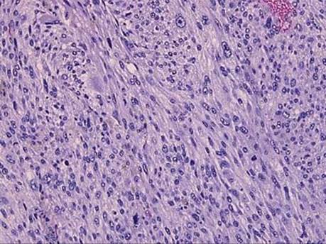
Fig 1 Rare cardiac sarcomas. Undifferentiated pleomorphic sarcoma: highly atypical spindled mesenchymal cells characterize this lesion, with easily identifiable mitotic figures. Other than vimentin, all markers of differentiation were negative.

Fig 2 Rare cardiac sarcomas. Rhabdomyosarcoma, solid variant: the relatively small tumor cells have high nuclear/cytoplasmic ratio and are arranged in sheets and clusters.

Fig 3 Rare cardiac sarcomas. Rhabdomyosarcoma, solid variant: myogenin is focally positive, indicating skeletal muscle differentiation.

Fig 4 Rare cardiac sarcomas. Leiomyosarcoma: the lesion is composed of spindle cells with pleomorphic nuclei, eosinophilic cytoplasm, and mitotic figures.

Fig 5 Rare cardiac sarcomas. Leiomyosarcoma: stain for SMA highlights smooth muscle differentiation.