
TABLE 1 Characteristic Features of Five Types of Congenital Pulmonary Airway Malformation

TABLE 2 Useful IHC Markers for Identifying Metastatic Pediatric Tumors

TABLE 3 Clinical Classification of Pulmonary Hypertension
|
Group I |
Pulmonary Arterial Hypertension |
|
Group II |
Pulmonary venous hypertension |
|
Group III |
Pulmonary hypertension associated with lung diseases or hypoxemia |
|
Group IV |
Pulmonary hypertension due to chronic thrombotic and/or embolic disease |
|
Group V |
Miscellaneous |
TABLE 4 Characteristic Features of Bacterial Pneumonias
|
HISTOLOGY |
ORGANISM AND SPECIAL STAINS |
|
|
Lobar and bronchopneumonia |
Vascular congestion, alveolar exudate with neutrophils, and bacterial colonies |
Gram-positive bacteria much more common than gram-negative bacteria |
|
Aspiration pneumonia |
Airway and alveolar neutrophils ± necrosis or abscess |
Superficial squames, vegetable material, or skeletal muscle are confirmatory; Gram stain |
|
Tuberculosis |
Necrotizing granulomatous lesions, often apical |
Acid-fast positive small beaded rods; organisms often present within the necrotic tissue |
|
Atypical mycobacterial infections |
Necrotizing granulomatous lesions; may have abundant neutrophils |
Acid-fast positive thin beaded rods; organisms mostly intracellular |
|
Actinomycosis |
Lung abscess with acute inflammation and necrosis; no granulomas |
GMS positive, gram-positive non–acid-fast long-branching filamentous rods; amphophilic crystalline sulfur granules with peripheral eosinophilic deposits (Splendore-Hoeppli phenomenon) in acutely inflamed areas |
|
Nocardiosis |
Lung abscess with necrosis and neutrophils surrounded by organizing pneumonia |
GMS positive, gram-positive, weakly acid-fast branching filamentous rods |
|
Legionnaires disease |
Airway and alveolar neutrophils and macrophages with prominent karyorrhexis, eosinophilic exudate, and fibrin, often with DAD |
Small gram-negative pleomorphic rods; best seen with silver impregnation stains; present within macrophages or extracellularly |
DAD, diffuse alveolar damage; GMS, Gomori methenamine silver.
TABLE 5 Characteristic Features of Viral Pneumonias

TABLE 6 Features of Fungal Pneumonias

TABLE 7 Histological Features of Mild, Moderate, and Severe Squamous Dysplasia, and Carcinoma in Situ
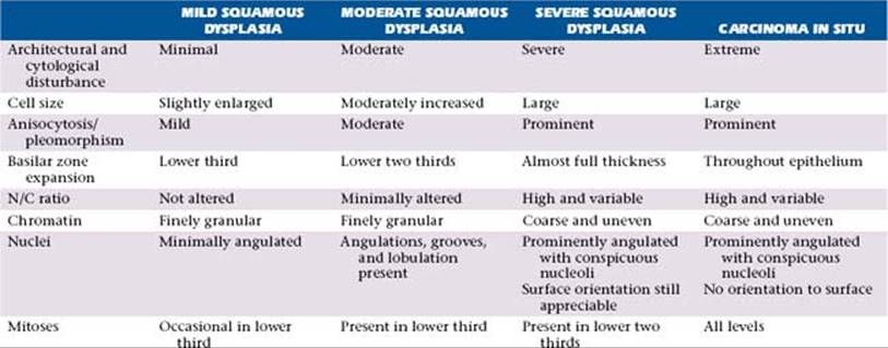
TABLE 8 Diagnostic Table for Prediction of Primary Site of Metastatic Carcinoma Using IHC Panel
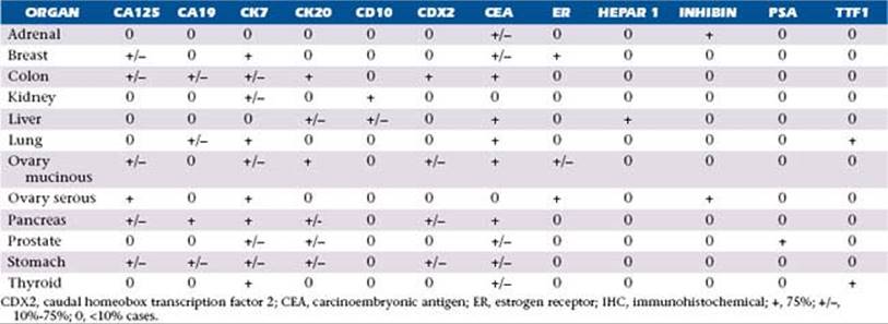
TABLE 9 Useful Positive IHC Markers for Metastatic Carcinomas from Breast and Female Genital Tract

TABLE 10 Useful Positive and Negative IHC Markers for GI Metastatic Carcinomas
|
ORGAN |
PANEL OF POSITIVE MARKERS |
NEGATIVE |
|
Colon |
CK20, CDX2, CEA, CA19, CA125, β-catenin |
CK7 |
|
Stomach |
CK7, CDX2, CEA, DPC4 |
|
|
Liver |
Hepar1, AFP, canalicular staining with CEA, CD10 and CD13, Arginase |
CK7, except fibrolamellar |
|
Pancreas |
CA19, CA125, CEA, CK7, CK17 |
DPC4 (SMAD4) |
|
Biliary tract |
CK7, CK20, CEA, villin |
AFP, alpha-fetoprotein; CDX2, caudal homeobox transcription factor 2; CEA, carcinoembryonic antigen; DPC4, deleted in pancreatic carcinoma, locus 4 tumor suppressor gene, also known as SMAD4; GI, gastrointestinal; IHC, immunohistochemical.
TABLE 11 Useful Positive and Negative IHC Markers for GU Metastatic Carcinomas
|
ORGAN, TUMOR |
PANEL OF POSITIVE MARKERS |
NEGATIVE |
|
Renal, clear cell |
Vimentin, CD10, CAIX, RCC, PAX2, EMA |
CK7 (or focal) |
|
Renal, papillary |
AMACR, CK7 |
|
|
Bladder |
p63, Uroplakin, CK20, Thrombomodulin, HMCK |
|
|
Prostate |
PSA, PAP |
CK7, CK20 |
|
Testis, seminoma |
PLAP, OCT4, CD117, D2-40, SALL4 |
AE1/AE3, CK7 |
|
Testis, EC |
PLAP, OCT4, CD30, AE1/AE3, SOX2, SALL4 |
|
|
Testis, YST |
PLAP, AFP, AE1/AE3, glypican-3, SALL4 |
CK7 |
|
Testis, Choriocarcinoma |
PLAP, hCG, AE1/AE3, SALL4 |
CK7 |
|
Adrenal |
Melan-A (clone A103), Inhibin, synaptophysin, Calretinin, Vimentin, SF1 |
CK, EMA/MUC1, S100, CD10, Hepar1 |
D2-40, podoplanin; EC, embryonal carcinomas; EMA, epithelial membrane antigen; GU, genitourinary; IHC, immunohistochemical; SALL4, sal-like protein 4; SOX2, SRY (sex-determining region Y)-box 2 protein; YST, yolk-sac tumors.
TABLE 12 Useful Positive and Negative IHC Markers for Head and Neck Metastatic Carcinomas
|
ORGAN, TUMOR TYPE |
PANEL OF POSITIVE MARKERS |
NEGATIVE |
|
Thyroid, papillary, and follicular |
TTF1, thyroglobulin, HBME, CK19, CK7 |
CK20 |
|
Thyroid, medullary |
TTF1, calcitonin, CEA, AE1/3, chromogranin |
Thyroglobulin |
|
Thyroid, anaplastic |
Pankeratin, vimentin, CEA |
TTF1, thyroglobulin |
|
Squamous cell carcinoma |
CK5/6, p63 |
TTF1, CK7/20, CAM5.2 |
|
Adenoid cystic carcinoma |
EMA, LMWK, SMA, calponin, p63, S100 |
TTF1 |
|
Acinic cell carcinoma |
Amylase, transferrin, lactoferrin, CEA |
TTF1 |
|
Parathyroid carcinoma |
PTH |
TTF1 |
CEA, carcinoembryonic antigen; EMA, epithelial membrane antigen; HBME, Hector Battifora mesothelial epitope; IHC, immunohistochemical; LMWK, low molecular weight keratin; PTH, parathyroid hormone; SMA, smooth muscle actin.
TABLE 13 Useful IHC Markers for Metastatic Sarcomas
