
Definition
• NSIP is a chronic idiopathic lung disease with interstitial widening that does not have features of usual interstitial pneumonia (UIP) and has a good prognosis
• Subtypes: cellular, fibrotic, and mixed
Clinical features
Epidemiology
• Second most common type of chronic interstitial pneumonia
• Usually affects people in their early to mid 50s; the cellular subtype is seen earlier in life. It can also develop in children or the elderly
• Slight predominance in women
• Most common interstitial pneumonia in patients with collagen vascular disease
Presentation
• Coughing and dyspnea occurring over a period of several months
• Systemic symptoms, such as fever, are occasionally present
Prognosis and treatment
• In general much better prognosis than UIP
• Nearly all patients with cellular NSIP are alive at 10 years
• Only one third of the patients with fibrotic NSIP are alive at 10 years
• Mixed pattern has a better prognosis than fibrotic variant but not as favorable as cellular
• Corticosteroids are the mainstay of therapy
Pathology
Histology
• Cellular NSIP:
• Uniform in intensity and time, mild diffuse or patchy, interstitial lymphoplasmacytic infiltrate with minimal fibrosis and preservation of lung architecture
• Prominent type II pneumocyte hyperplasia
• Focal organizing pneumonia, lymphoid aggregates, and alveolar macrophages may be present
• Lack of dense fibrosis, honeycombing, granulomas, eosinophils, neutrophils, organisms, hyaline membranes, and necrosis
• Fibrotic NSIP:
• Prominent interstitial fibrosis causing uniform thickening of alveolar walls with preservation of lung architecture and mild lymphocytic inflammation
• Fibroblastic foci are inconspicuous or insignificant in number and lack temporal heterogeneity and honeycombing of UIP
• Organizing pneumonia, lymphoid aggregates, alveolar macrophages, bronchial metaplasia, and metaplastic calcifications/bone formation may be present
• Inconspicuous to rare granulomas and no eosinophils or organisms
• Mixed NSIP:
• Has both inflammation and fibrosis but no fibroblastic foci or honeycombing
Immunopathology/special stains
• Not contributory
Main differential diagnoses
• UIP: patchy fibrosis that distorts/destroys the lung architecture together with fibroblastic foci are the main differentiating features
• Hypersensitivity pneumonitis: poorly formed granulomas and mixed inflammatory infiltrates including eosinophils are characteristic
• Lymphocytic interstitial pneumonia: once low-grade lymphomas are excluded, almost all cases are associated with HIV infection or other immunodeficiency diseases; much more inflammation than NSIP
• Organizing pneumonia: intraalveolar nodules of fibrosis (Masson bodies)
• Diffuse alveolar damage: organizing hyaline membranes

Fig 1 Nonspecific interstitial pneumonia. Cellular variant with uniform, diffuse interstitial lymphoplasmacytic infiltrate, minimal fibrosis, and preservation of lung architecture is seen in this 14-year-old female with juvenile rheumatoid arthritis.

Fig 2 Nonspecific interstitial pneumonia. High-power image of NSIP, cellular variant (same case as in Fig 1).

Fig 3 Nonspecific interstitial pneumonia. Cellular variant with multiple lymphoid aggregates (same case as in Fig 1).

Fig 4 Nonspecific interstitial pneumonia. Fibrotic variant with uniform interstitial fibrosis and thickening of the alveolar septae, low power.

Fig 5 Nonspecific interstitial pneumonia. Fibrotic variant, high power.
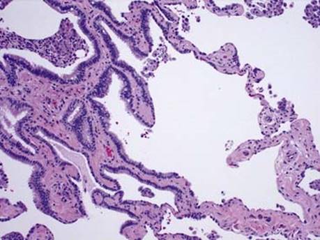
Fig 6 Nonspecific interstitial pneumonia. Fibrotic variant with area of bronchiolar metaplasia. Note absence of honeycombing and fibroblastic foci.
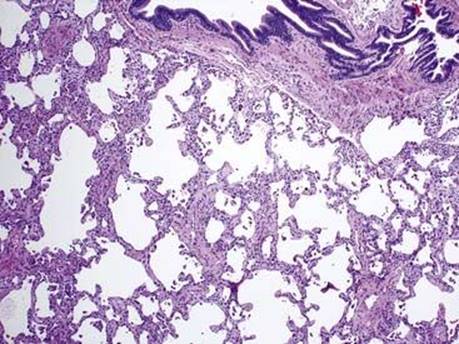
Fig 7 Nonspecific interstitial pneumonia, mixed variant. Both mild chronic inflammation and fibrosis are present in the alveolar septae, low power.

Fig 8 Nonspecific interstitial pneumonia. Mixed variant. High power of Fig 7.