
Definition
• A fibrosing inflammatory disease associated with an infiltration of IgG4-positive plasma cells
Clinical features
Epidemiology
• Occurs mainly in adults; male predominance
Presentation
• Patients may be asymptomatic or present with coughing, dyspnea, hemoptysis, or pleural effusion
• Imaging may show ground-glass opacities, lung nodules, peribronchial consolidation, or hilar lymphadenopathy
• Elevated serum levels of IgG4
• Patients frequently have IgG4-related disease in other organs
Prognosis and treatment
• This is a benign condition that responds well to steroids and immunosuppression
Pathology
Gross
• Solid nodular type: distinct gray nodules, more often located peripherally
• Alveolar interstitial type: firm parenchyma and thickened pleura
• Bronchovascular type: irregular areas of fibrosis
Histology
• Inflammatory infiltrate consisting of lymphocytes, plasma cells, and eosinophils
• Lymphoid follicles are present in approximately 50% of cases
• Veins and arteries are commonly involved by a subendothelial lymphoplasmacytic infiltrate that obliterates the lumen
• If involved, the pleura may demonstrate fibrous thickening, fibrinous exudate, and chronic inflammation
• There are several histological patterns:
• Solid nodular (also called inflammatory pseudotumor with IgG4+ plasma cells):
– Tumor-like lesion with sclerosis and lymphoplasmacytic infiltrate
– Surrounding alveoli infiltrated by lymphocytes and plasma cells
– Inflammation and sclerosis of the bronchial wall and bronchial glands
• Bronchovascular:
– Lymphoplasmacytic infiltrate surrounding the bronchovascular bundles and within the alveolar interstitium, interlobular septa, and pleura
– Lymphatics may be filled with histiocytes demonstrating emperipolesis of lymphocytes
• Alveolar interstitial:
– Lymphoplasmacytic infiltrate is present in the alveolar interstitium
– Nonspecific interstitial pneumonia (NSIP) pattern
Immunopathology/special stains
• Increased IgG4+ plasma cells (>50 per high-power field with IgG4/IgG ratio >40%); however, criteria vary in the literature
Main differential diagnoses
• Multicentric Castleman disease
• Connective tissue disease–related lung diseases (e.g., NSIP)
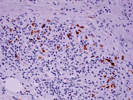
Fig 1 IgG4-related sclerosing disease. Immunohistochemical stain for IgG4 shows many positive plasma cells.

Fig 2 IgG4-related sclerosing disease. NSIP-like pattern of IgG4-related lung disease with varying amounts of lymphoplasmacytic infiltrates in the interstitium (A-C) and airway (D). Note that this case has very little fibrosis.