
Definition
• A progressive sclerosing autoimmune disease with frequent lung involvement
Clinical features
Epidemiology
• Almost half of patients with scleroderma develop a fibrosing lung disease
• About a quarter of scleroderma patients develop pulmonary hypertension
Presentation
• Lung disease almost always presents in patients with known scleroderma, and only rarely precedes a diagnosis of scleroderma
• Dyspnea on exertion and dry coughing
• Reticulonodular pattern and ground-glass opacities on chest CT involving the lower lobes
Prognosis and treatment
• Patients with scleroderma and lung disease have a worse prognosis than those without lung disease and have an increased incidence of primary lung adenocarcinoma
• Lung disease and pulmonary hypertension are major causes of death in patients with scleroderma
• Immunosuppressive therapy may stabilize or improve lung function and CT changes
Pathology
Histology
• Interstitial fibrosis predominant in the lower lobes with nonspecific interstitial pneumonia (NSIP) and usual interstitial pneumonia (UIP) patterns
• Early disease has preserved underlying lung architecture with diffuse interstitial fibrosis, in a fibrotic NSIP pattern
• Later disease has more florid fibrosis with loss of lung architecture, resembling UIP pattern, but usually without fibroblastic foci
• Other findings may include chronic aspiration changes, organizing pneumonia, alveolar hemorrhage, adenocarcinoma, and, rarely, diffuse alveolar damage
• Pleural fibrosis and adhesions
• Secondary pulmonary hypertensive changes characterized by medial and intimal hypertrophy of pulmonary arteries and arterioles; also mucinous degeneration of the media, elastic fiber disruption, and concentric intimal fibrosis may be seen
Immunopathology/special stains
• Not contributory
Main differential diagnoses
• Other causes of NSIP or UIP pattern interstitial lung disease

Fig 1 Scleroderma. Combined features of NSIP (left) and UIP (right) patterns of interstitial lung disease are present in this patient with scleroderma.

Fig 2 Scleroderma. Florid interstitial fibrosis with bronchiolar metaplasia and mucostasis without obvious fibroblastic foci are seen in this section of lung from a patient with scleroderma.

Fig 3 Scleroderma. Alveolar foamy macrophages consistent with postobstructive pneumonitis are seen at high power.
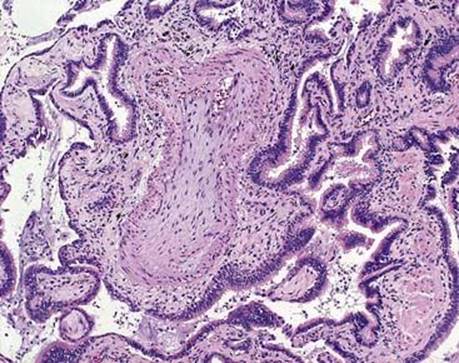
Fig 4 Scleroderma. Severe secondary pulmonary hypertensive change with myxoid features is seen in the same patient as in Figs 2 and 3.

Fig 5 Scleroderma. Pleural and subpleural pulmonary fibrosis is seen here in an area of early UIP. Note fibroblastic focus.