
Definition
• Rare chronic progressive disease characterized by pulmonary cyst formation associated with proliferation of abnormal smooth muscle cells
Pathogenesis
• Experimental data demonstrate loss of heterozygosity of chromosomes 9p and 16p, and mutation of the tuberous sclerosis complex gene (TSC)
Clinical features
Epidemiology
• Affects females of child-bearing age
• Some cases are associated with renal angiomyolipoma and, rarely, with tuberous sclerosis
• Four cases have been reported in males
Presentation
• Dyspnea with pneumothorax or emphysema with no smoking history
• Worsened by pregnancy or menstruation
• Clinical improvement postmenopause
Prognosis and treatment
• Progressive course, prognosis poor, death due to respiratory failure or cor pulmonale
• Treatment is antiestrogen therapy (e.g., tamoxifen), progesterone, and oophorectomy, with variable response
• Lung transplantation as last resort
Pathology
Histology
• Thin-walled cysts with patchy, nodular, variably cellular proliferation of bland smooth muscle cells around airways, lymphatics, and blood vessels
• Smooth muscle cells are fusiform, plump, have a slightly increased nuclear/cytoplasmic ratio; optically clear cytoplasm; intracytoplasmic glycogen
• Intraalveolar hemosiderin-laden macrophages are common
Immunopathology/special stains
• Lesional smooth muscle cells are positive for
• HMB45
• Desmin
• Smooth muscle actin (SMA)
• Estrogen receptor (ER)
• Progesterone receptor (PR)
• D2-40
• Melan-A/MART-1
Main differential diagnoses
• Alveolar duct smooth muscle hyperplasia: smooth muscle cells are HMB45, Melan-A/MART-1 negative
• Pulmonary Langerhans cell histiocytosis: nodular aggregates of Langerhans cells (CD1a, S100-positive) with bronchiolocentric distribution and prominent eosinophils
• Metastatic low-grade sarcomas: distinct nodule(s) of spindle cells that are HMB45, Melan-A/MART-1 negative
• Benign metastasizing leiomyoma: nodule with smooth muscle cells that are HMB45, Melan-A/MART-1 negative
• Emphysema: distended alveolar spaces with no nodule formation

Fig 1 Lymphangioleiomyomatosis. Low-power view shows peribronchial and perivascular expansion and patchy thickening of alveolar walls.

Fig 2 Lymphangioleiomyomatosis. Medium-power view shows nodular proliferation of smooth muscle cells and cyst formation.
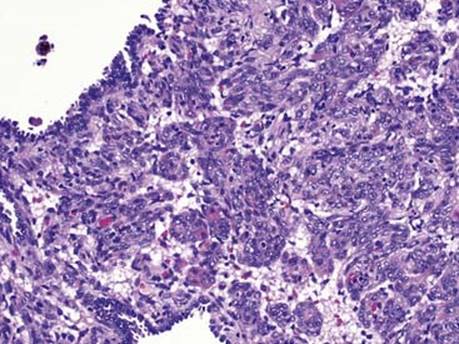
Fig 3 Lymphangioleiomyomatosis. High-power view shows a nodular proliferation of plump fusiform smooth muscle cells with a slightly increased nuclear cytoplasmic ratio.

Fig 4 Lymphangioleiomyomatosis. Lesional cells are positive for HMB45.
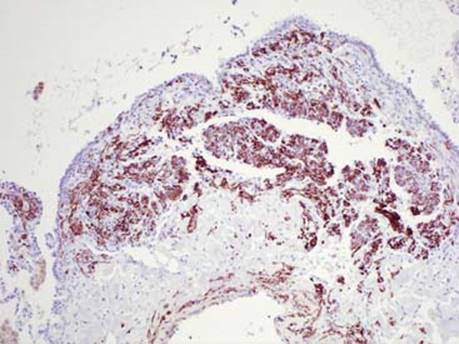
Fig 5 Lymphangioleiomyomatosis. Lesional cells are positive for desmin.

Fig 6 Lymphangioleiomyomatosis. Lesional cells are positive for PR.
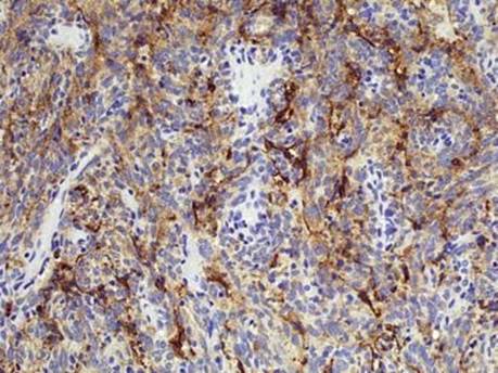
Fig 7 Lymphangioleiomyomatosis. Lesional cells are positive for D2-40.
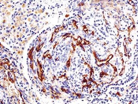
Fig 8 Lymphangioleiomyomatosis. Lymphatic channels within the lesion are highlighted by D2-40.