
Definition
• Severe form of sickle cell crisis manifesting as a new pulmonary infiltrate involving at least one complete lung segment consistent with alveolar consolidation accompanied by chest pain, fever (>38.5° C), tachypnea, and wheezing, or coughing
Pathogenesis
• ACS is a clinical diagnosis, the etiology of which is often unknown at the time of diagnosis
• Possible precipitating factors:
• Infections: atypical bacteria and viruses (e.g., Chlamydia pneumoniae, Mycoplasma pneumoniae, respiratory syncytial virus, and community acquired/encapsulated bacteria)
• Bone marrow embolism (usually in patients older than 20 years of age)
• Vascular risk factors:
• Hypoxia-vasoconstriction cycle:
– Pulmonary vessels are unique because they constrict in hypoxia, which amplifies this cycle
• Slow transit of abnormally adherent red blood cells (RBCs) through small vessels
• Other factors: sequestration of sickled cells; shunting and V-Q mismatch; acute hemolysis and hypercoagulable state; fat emboli from necrotic marrow; inflammation; reperfusion injury; unstable hemodynamics
Clinical features
Epidemiology
• Approximately 40% of sickle cell patients experience at least one episode of ACS
• Leading cause of intensive care unit admission and premature death in sickle patients
• Incidence is higher in children than in adults
• All sickle patients with fever, irrespective of respiratory symptoms, should have a chest radiograph done to rule out ACS (early detection improves outcome)
Presentation
• ACS usually presents with fever, chest pain, coughing/wheezing, acute drop in hemoglobin level and hypoxemia
• Acute vasoocclusive pain crisis (VOC) of sickle disease is generally self-limiting, but 20% will progress into ACS in the first 3 days of hospitalization
• Predictive factors for developing ACS from VOC:
• Leukocytosis
• Higher steady-state total hemoglobin (Hb) (risk for hyperviscosity)
• Lower steady-state HbF/HbA2 (i.e., higher HbS)
• Hypoventilation due to asthma (in children), anesthesia/surgery, rib infarcts, avascular necrosis of the hip, pregnancy, narcotic overuse, previous pulmonary events
• Secretory phospholipase A2 rise precedes radiographic changes and can be used as a predictive marker
Prognosis and treatment
• Risk factors for respiratory failure: age older than 20 years, multilobar lung involvement, cardiac disease, platelets <200 K/μL
• Early treatment is critical because full-blown ACS is difficult to reverse, even with treatment
• Severe ACS may progress to acute respiratory distress syndrome (ARDS)
• Mortality rate in ACS is 9% in adults and 1% in children
• Causes of death include bronchopneumonia and pulmonary embolism
• Repeated ACS leads to pulmonary hypertension: poor prognosis (mortality, 40%)
• Treatment includes oxygen, bronchodilators, analgesics, broad-spectrum antibiotics; avoid overhydration
• Transfusion needs to be judicious (can prevent ARDS if timely)
• Target: make HbS <30%, Hct <30%
• Simple transfusion (has risk of hyperviscosity and iron and volume overloading)
• Exchange transfusion (avoids iron and volume overloading) indicated in rapid progression, severe hypoxia, multilobar disease, and mechanical ventilation
• Refractory cases: aggressive ventilator support, corticosteroids, nitric oxide
• Bone marrow transplant can be curative of HbSS, but irreversible lung injuries persist
Pathology
• No biopsy is generally done during ACS, and the only specimen available may be autopsy, if fatal
• If bronchioalveolar lavage (BAL) is available, lipid-laden macrophages may suggest fat embolism
Gross
• Dark red, heavy, and edematous lungs
• Patchy focal scars
• Thrombi/emboli rarely seen grossly (too small in most cases)
Histology
• Capillaries are distended by sickled RBCs
• Edema fluid, inflammatory cells, and heart failure cells in alveolar spaces, sometimes hemorrhage (usually scant)
• Characteristic multifocal microscopic foci of alveolar wall necrosis with focal inflammation, small amount of fibrinous exudate, and hardly any hemorrhage
• Variable amount of bronchopneumonia, sometimes extensive and necrotizing
• Thromboembolism:
• More common as age increases
• May be small and peripheral or massive and central (especially in pregnancy)
• Recent or organized or recanalized
• Bone marrow emboli:
• Vascular occlusion by fragments of necrotic bone marrow
• Lipid-laden macrophages in older emboli
• Pulmonary wedge infarction (generally after massive thromboemboli)
• Multiple infarcts without vasoocclusion have been reported (vasospastic)
• With ARDS: interstitial edema and hyaline membranes
• Aforementioned acute changes may be superimposed on a background of “sickle cell chronic lung disease” pathology:
• Pulmonary hypertension (PH)
– 20% to 40% of all sickle patients have PH (by time of death, >75%)
– Children and adolescent sickle patients develop PH due to recurrent ACS
– Sickle-associated pulmonary hypertension histology:
– Widespread thrombosis of small arteries (in situ)
– Recanalized arteries
– Eccentric and concentric intimal fibrosis of small and medium arteries
– Subintimal fibrosis may be present
– Fibroelastic degeneration of small arteries, arterioles, and venules
– Bronchopulmonary anastomoses
– Severe cases may be associated with:
° Extensive recanalized thrombi resembling plexiform lesions
° Dilatation of lesions of muscular arteries distal to their origin
° Focal aneurysms
° No angiomatoid lesion or fibrinoid necrosis
• Cor pulmonale
• Healed wedge infarcts, patchy fibrosis (often perivascular)
• Fibrous pleural adhesions: scars from past pleural exudates during episodes of pneumonia and/or pulmonary infarction
• Hemosiderin-laden macrophages
Immunopathology/special stains
• Oil red O for fat embolism (can also be done on BAL/induced sputum/frozen section)
• Homogenous red stain for fat embolism (soluble in organic solvents)
• Must be distinguished from granular lipochrome pigment frequently
• Elastic staining for pulmonary hypertension
• Prussian blue for secondary hemochromatosis
Main differential diagnoses
• Vasoocclusive crisis without ACS (must obtain radiograph)
• Acute exacerbation of asthma
• Continuation of preexisting pneumonia
• Rib-cage infarction/fracture
• Myocardial infarction
• Pulmonary embolism
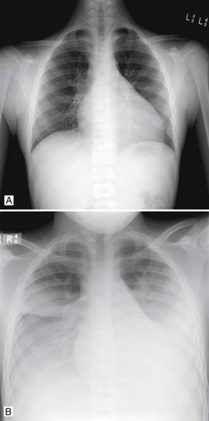
Fig 1 Acute chest syndrome of sickle cell disease. Chest radiographs from a 10-year-old with sickle cell disease: during vasoocclusive crisis without ACS (A) and during ACS (B). Note presence of bilateral areas of consolidation.

Fig 2 Acute chest syndrome of sickle cell disease. A 36-year-old patient with sickle cell disease and ACS: A, gross photograph of cut surface of the lung at autopsy with thromboembolus in pulmonary artery branch in lower left; B, sickled RBCs are present within the thromboembolus, which is attached to the pulmonary arterial wall seen in the lower right.

Fig 3 Acute chest syndrome of sickle cell disease. This small vessel is occluded by a thrombus.
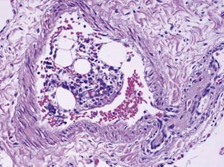
Fig 4 Acute chest syndrome of sickle cell disease. Bone marrow embolus in a small pulmonary artery.

Fig 5 Acute chest syndrome of sickle cell disease. Microscopic foci of alveolar necrosis are seen in this patient with ACS.
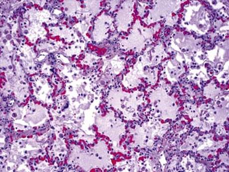
Fig 6 Acute chest syndrome of sickle cell disease. Pulmonary edema and congested capillaries. Note two darkly staining megakaryocytes in the left half.

Fig 7 Acute chest syndrome of sickle cell disease. Recent and old hemorrhage with interstitial fibrosis.

Fig 8 Acute chest syndrome of sickle cell disease. Sickled RBCs are distending the capillaries. Note hemosiderin-laden macrophages in the alveoli.

Fig 9 Acute chest syndrome of sickle cell disease. Organized thromboembolus in large pulmonary artery with adjacent old infarct.

Fig 10 Acute chest syndrome of sickle cell disease. Organized thromboembolus in small pulmonary artery resembling plexiform lesion.

Fig 11 Acute chest syndrome of sickle cell disease. Eccentric intimal fibrosis in medium-sized pulmonary artery branch is seen in this patient with ACS superimposed on sickle cell chronic lung disease.

Fig 12 Acute chest syndrome of sickle cell disease. Concentric intimal fibrosis and medial hypertrophy of small pulmonary artery is present in this patient with ACS superimposed on sickle cell chronic lung disease..