
Definition
• Pulmonary embolism (PE) refers to a detached intravascular solid, liquid, or gaseous mass that is carried by the blood to the lung from a distant site of origin to the pulmonary vasculature
• Pulmonary infarction refers to ischemic necrosis of the lung parenchyma. It is most often secondary to PE
Pathogenesis
• Most pulmonary emboli (about 95%) originate from deep vein thrombosis (DVT) in the legs. However, less than one half of patients with DVT will have a PE
• Nonthrombotic causes of emboli include tissues (brain, bone marrow, fat, tumor, trophoblast), amniotic fluid, air, and foreign bodies (silicon, tablet fillers, angiographic procedure-induced, parasites)
• Pulmonary infarction occurs secondary to emboli in the peripheral pulmonary circulation; large saddle emboli cause death before infarction
• Underlying heart failure and lung cancer can predispose to infarction. Pulmonary infarcts in young individuals should prompt investigation for hypercoagulable states and sickle cell anemia
• Invasive fungal pneumonia can cause hemorrhagic infarcts
Clinical features
Epidemiology
• PE has an approximate incidence of two to four per 1000 hospitalized patients and causes 200,000 deaths per year in the United States
• It is estimated that 600,000 persons each year in the United States experience a PE, resulting in approximately 300,000 hospital admissions
• History of a previous PE significantly increases the risk for a second PE
• PE does not always result in infarction. In an autopsy series, lung infarcts were seen in 20% of cases, while PE was present in 69% of cases
Presentation
• Most PEs are clinically silent
• Clinical manifestations of acute PE range from mild dyspnea to hemodynamic collapse
• Large PEs that occlude the main pulmonary trunk are seen early with symptoms mimicking acute myocardial infarction; sudden death or cardiovascular collapse can occur when 60% of the pulmonary circulation is obstructed
• Obstruction of smaller vessels causes hemorrhagic (“red”) infarcts due to the lung’s dual arterial blood supply (pulmonary and bronchial vessels)
• Peripheral infarcts cause pleuritic pain
Prognosis and treatment
• The case fatality rate for acute PE ranges from less than 1% to 60% (the high end is seen in patients who are hemodynamically unstable)
• Acute PE requires initial short-term therapy with a rapid-onset anticoagulant, followed by therapy with a vitamin K antagonist for at least 3 months. More extended therapy is required in patients at high risk of recurrence
• Hemodynamically unstable patients are candidates for more aggressive treatment, such as pharmacological or mechanical thrombolysis
• Vena cava filters are reserved for patients with contraindications to anticoagulant therapy
• The risk of recurrent PE is less than 1% per year while patients are receiving anticoagulant therapy, but the risk is 2% to 10% per year after the discontinuation of such therapy. Risk factors for recurrence include male sex, advanced age, malignancies, and idiopathic or unprovoked PE (i.e., occurring in the absence of any identifiable risk factor for venous thromboembolism)
• The incidence of chronic thromboembolic pulmonary hypertension 2 years after an acute PE ranges from 0.8% to 3.8%
Pathology
Gross
• A fatal embolus is usually situated at the bifurcation of the pulmonary trunk (saddle embolus) or in a main pulmonary artery, as a deep red, firm, and coiled clot. It represents a cast, usually of a deep leg vein, unlike a postmortem clot, which is a cast of the artery in which it is found
• Thromboemboli are firm and friable rather than being soft and gelatinous (postmortem clots). On cross-sectioning, true antemortem clots and emboli show alternating darker and lighter striations (lines of Zahn) representing sequential erythrocyte-rich and fibrin/platelet-rich layers. Postmortem clots are caused by the passive settling of erythrocytes and aggregation of leukocytes and have a biphasic red and yellow (“chicken fat”) appearance
• A lung infarct appears as a pleural-based hemorrhagic zone of parenchymal induration, with an “apex” pointing toward the hilum. An occluded vessel may be seen proximal to the infarct. Peripheral emboli can result in more rounded infarcts
• Organizing infarcts are yellow-tan and firm; superimposed pneumonia or abscess formation can cause cavitation
• Old organized infarcts may appear as subpleural scars with overlying pleural fibrosis and adhesions
Histology
• Antemortem clots show layering of erythrocytes and fibrin. The erythrocytes often appear “hyalinized.” Entrapped leukocytes show pyknosis and karyorrhexis. A thromboembolus can itself activate the coagulation cascade, and the thrombus can propagate within the affected vessel; alternatively a thromboembolus can lyse or organize
• If thrombolysis does not occur within 1 or 2 days, the thromboembolus gets organized by migrating fibroblasts and myofibroblasts. In larger arteries, these migrating cells combine with granulation tissue to cause contraction of the clot to one side, leading to an eccentric thickening of the affected vessel. In smaller vessels, organization may alternatively result in recanalization. These recanalized vessels may be fed by either pulmonary or bronchial arteries
• Various phases of organization have been proposed to help “date” the thrombus; these are, however, only approximations, are often imprecise, and are difficult to use in routine practice
• Pulmonary arteries may show nonspecific changes at sites of impaction of emboli. These include congestion of adventitial vessels, perivascular hemorrhage, and acute inflammation of the arterial wall
• Incipient lung infarcts (“preinfarcts”) show capillary congestion and acute alveolar hemorrhage, of which the latter is probably derived from the still patent bronchial circulation
• Progression to infarction is heralded by loss of alveolar nuclei and acute inflammation
• The overlying visceral pleura shows fibrinous pleuritis
• Organization of an infarct proceeds from the periphery to the center. Squamous metaplasia may occur within the organizing infarct
Immunopathology/special stains
• Oil red O or Sudan black stains on frozen sections help highlight fat emboli
• Elastic stains on organized infarcts show collapsed, entangled elastic fibers within a collagenous stroma
• Immunostains are not contributory
Main differential diagnoses
• Plexiform lesions of primary pulmonary arterial hypertension (PAH): disordered slitlike vascular spaces with proliferating endothelial cells that may obliterate the lumen; recanalized vessels show sharply demarcated, “punched-out” intraluminal spaces, with muscularized walls around each space

Fig 1 Pulmonary embolism and infarction. A, This example of an antemortem thrombus lodged in a pulmonary artery shows alternating lines of Zahn. B, In contrast, this postmortem clot does not show any organization.

Fig 2 Pulmonary embolism and infarction. Vascular occlusion results in a hemorrhagic infarct. The occluded vessel can be seen in the lower center at the margin of infarcted and viable lung.

Fig 3 Pulmonary embolism and infarction. A, An embolus often gets organized by granulation tissue that contracts and pulls the embolus eccentrically. B, After complete organization, the only evidence of the embolus may be an eccentric thickening of the artery, as seen in this trichrome stain. C, If the embolus cannot be cleared in the above manner, it undergoes organization with recanalization.
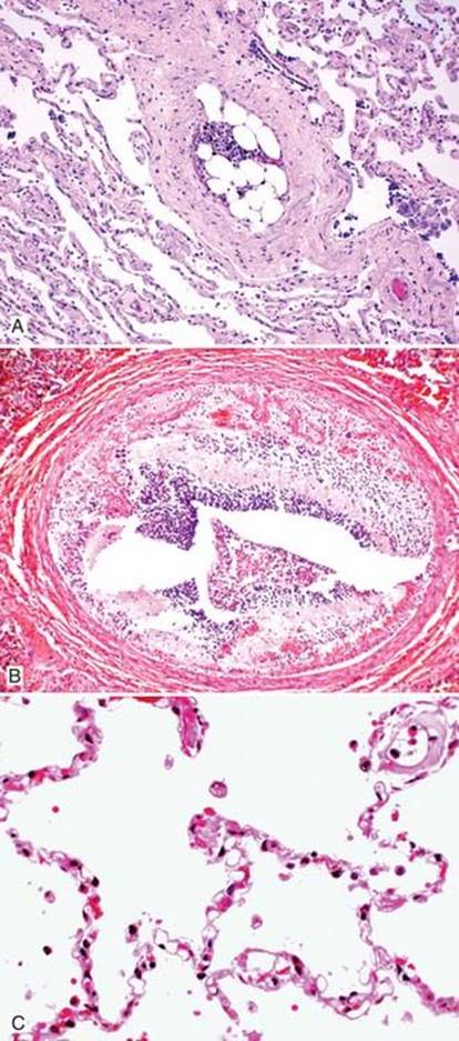
Fig 4 Pulmonary embolism and infarction. Emboli in the pulmonary vasculature can have other systemic sources such as bone marrow (A) and brain (B). Fat emboli (C) appear as distended rigid vacuoles within the pulmonary microcirculation and can be highlighted by stains for fat on frozen sections (not shown).

Fig 5 A and B, Pulmonary embolism and infarction. Septic emboli refer to embolic material that contains microorganisms. They are usually rich in neutrophils and contain bacteria or fungi. This example is from a patient with fungal sepsis. The fungi in the circulation are often derived from a focus of invasive fungal pneumonia elsewhere in the lung. Note the hemorrhagic infarct in the adjacent lung.

Fig 6 Pulmonary embolism and infarction. An example of dirofilariasis. The adult parasites (Dirofilaria immitis) reside in the pulmonary vasculature and can cause lung infarcts (trichrome stain). In heavy infections, the parasite may migrate to the right heart.
(Case courtesy of Dr. Dani S. Zander, Hershey, Pa.)

Fig 7 Pulmonary embolism and infarction. Extraneous elements may also be the source of pulmonary emboli. A, This patient underwent a vascular interventional procedure in the thorax and was seen later with Gelfoam emboli. B, Although the microcrystalline cellulose in this pulmonary vasculature is also derived from medication, the injury here is self-inflicted from IV drug abuse rather than iatrogenic. C,Cellulose elicits a giant cell response and can be highlighted with a GMS stain.