
Definition
• Pulmonary arterial hypertension (class I) includes pulmonary hypertension of variable causes that directly affects the vessels
Clinical features
Epidemiology
• Idiopathic PAH is more frequent in females than in males
• PAH may also be familial or secondary to collagen vascular diseases, congenital systemic to pulmonary shunts, portal hypertension, HIV infection, drugs and toxins (anorexigens, rapeseed oil, cocaine, methamphetamines), and other rare disorders
Presentation
• Gradual onset of fatigue and dyspnea, especially with exercise
• Edema, angina, and syncope are possible with more severe disease
Prognosis and treatment
• Poor prognosis: lung transplantation is the only curative therapy
• In congenital systemic to pulmonary shunts, early changes of pulmonary hypertension are reversible after correction of the shunt
• Clinical improvement may be seen with pulmonary vasodilators, including continuous intravenous infusion of prostacyclin analogues, phosphodiesterase type 5 inhibitors, and endothelin receptor blockers
Pathology
Histology
• Medial hypertrophy and concentric or eccentric intimal fibrosis of the pulmonary arteries are seen in PAH as well as in other forms of pulmonary hypertension
• Smooth muscle may extend into the distal arterioles
• Plexiform lesions are found mainly in PAH, are usually immediately distal to an arterial branch point, and are composed of glomeruloid tufts of endothelial cells within the arterial lumen or extending through the arterial wall
• Platelet thrombi are often seen within plexiform lesions
• Dilatation lesions form downstream of plexiform lesions and are composed of dilated, thin walled vessels
• Necrotizing arteritis can be present
• Plexiform lesions are less frequent in scleroderma-associated PAH
• Recanalizing thrombi may be seen in PAH as well as in pulmonary hypertension due to chronic thromboembolic disease
• In cases of congenital cardiac malformation with left to right shunts, the Heath-Edwards grading system can be used to divide potentially reversible (grades I-III: muscular hypertrophy and intimal fibrosis) from irreversible changes (grades IV-VI: plexiform and dilatation lesions and necrotizing arteritis)
Immunopathology/special stains
• Elastic and trichrome stains highlight the medial and intimal changes and demonstrate disruption of the elastic lamina in plexiform lesions
• Plexiform lesions may be highlighted by immunohistochemical analysis for endothelial markers, including CD34, CD31 and von Willebrand factor
• Extension of smooth muscle into the arterioles can be demonstrated with smooth muscle actin
Main differential diagnoses
• Chronic thromboembolic pulmonary hypertension: unlike plexiform lesions, recanalizing thrombi have “colander-like” new channel spaces, are less cellular, preserve the elastic laminas, and do not distend or invade through the arterial wall
• Pulmonary venoocclusive disease: prominent involvement of septal veins and preseptal venules
• Pulmonary capillary hemangiomatosis: angiomatous proliferation of capillaries widening the alveolar septa
• Pulmonary hypertension secondary to chronic lung diseases or left-sided heart failure: requires identification of other lung or heart disease
• Systemic forms of necrotizing vasculitis: no medial hypertrophy or intimal fibrosis

Fig 1 Pulmonary arterial hypertension. Concentric intimal fibrosis.

Fig 2 Pulmonary arterial hypertension. Concentric intimal fibrosis, elastic stain.

Fig 3 Pulmonary arterial hypertension. Concentric intimal fibrosis, trichrome stain.

Fig 4 Pulmonary arterial hypertension. Eccentric intimal fibrosis.

Fig 5 Pulmonary arterial hypertension. Eccentric intimal fibrosis, elastic stain.

Fig 6 Pulmonary arterial hypertension. Extension of smooth muscle into distal arterioles.

Fig 7 Pulmonary arterial hypertension. Smooth muscle actin stain highlights extension of smooth muscle into distal arterioles.

Fig 8 Pulmonary arterial hypertension. Plexiform lesions (arrows) downstream of a branch point with associated dilatation of vessels (arrowheads).
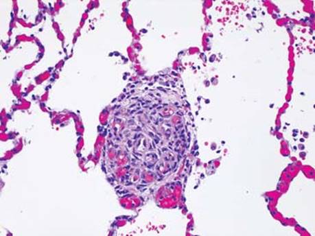
Fig 9 Pulmonary arterial hypertension. Plexiform lesions are composed of channels formed by cellular clusters of endothelial cells within the artery that extends out into the parenchyma.

Fig 10 Pulmonary arterial hypertension. Plexiform lesion with associated dilations (arrowhead).

Fig 11 Pulmonary arterial hypertension. The elastic lamina is destroyed in plexiform lesions (elastic stain).

Fig 12 Pulmonary arterial hypertension. Arteritis with eosinophils and neutrophils in the vessel wall.