
Definition
• A rare clinical entity characterized by diffuse alveolar hemorrhage and iron deficiency anemia
Pathogenesis
• Thought to be immune-mediated because of its excellent response to immunosuppression; however, no specific antibody has been identified
Clinical features
Epidemiology
• True incidence and prevalence of IPH is unknown. The estimated incidence among children is 0.21/100,000 per year
• In childhood, IPH occurs with equal frequency in males and females
• Extremely rare in adulthood, but when it occurs, IPH is more frequent in men
Presentation
• Symptoms often begin in the first decade. Most cases are recognized before age 30
• Presentation in children is with recurrent episodes of dyspnea, coughing, hemoptysis, and failure to thrive
• Adults frequently present with exertional dyspnea and fatigue due to intrapulmonary bleeding and iron deficiency anemia
Prognosis and treatment
• Prognosis of IPH is difficult to assess because of the lack of large and comparable patient series and inadequate follow-up
• Average survival from beginning of symptoms to death was around 2.5 years, with a wide time-range
• Treatment with systemic corticosteroids in both pediatric and adult patients has greatly improved outcomes
• Children and adolescents usually carry a poor prognosis. Adults have less pronounced symptoms and a more favorable prognosis
Pathology
Gross
• In advanced cases of IPH, lungs have a striking brown appearance due to siderosis, with varying degree of pulmonary fibrosis
Histology
• Alveoli may contain numerous hemosiderin-laden macrophages and erythrocytes
• Hemosiderin deposition in the vascular wall and supporting connective tissue is characteristic
• Alveolar capillaries are dilated and tortuous and may protrude into the alveolar lumen
• The alveolar walls are thickened, and type II pneumocytes appear enlarged and hyperplastic
• Localized interstitial fibrosis with formation of collagen is a prominent feature in long-standing disease
Main differential diagnoses
• Goodpasture syndrome: anti–glomerular basement membrane antibodies
• Infections

Fig 1 Idiopathic pulmonary hemosiderosis. Lung biopsy from a 2-year-old shows intraalveolar hemorrhage and hemosiderin-laden macrophages.

Fig 2 Idiopathic pulmonary hemosiderosis. This lung biopsy from a 3-year-old shows recent and old hemorrhages. In addition, there is interstitial fibrosis.
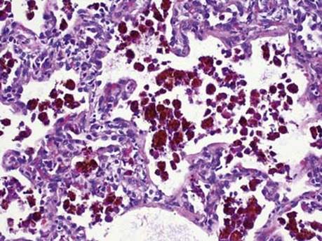
Fig 3 Idiopathic pulmonary hemosiderosis. This lung biopsy from another 2-year-old shows only numerous intraalveolar hemosiderin-laden macrophages without recent hemorrhage.

Fig 4 Idiopathic pulmonary hemosiderosis. In this lung biopsy, there are minimal recent and old hemorrhages; in addition, chronic inflammation and focal fibrosis illustrates the variable histological picture seen in IPH.

Fig 5 Idiopathic pulmonary hemosiderosis. Iron stain (Prussian blue) highlights the intraalveolar hemosiderin-laden macrophages as well as iron deposition within connective tissue.