
Definition
• A localized suppurative necrosis of lung tissue and cavitation containing necrotic fluid and/or debris, usually as a result of microbial infection
Pathogenesis
• Infection leading to a lung abscess can be a result of
• Aspiration of oropharyngeal or gastric secretion (most commonly due to altered consciousness secondary to alcohol consumption) or aspiration of infected materials (in oropharyngeal surgery)
• Primary bacterial infection of the lung (or after rupture of an extrapulmonary abscess into the lung)
• Direct traumatic puncture of the lung
• Septic embolism (bacterial endocarditis)
• Postobstructive pulmonary infection due to primary or metastatic tumors
• Continuous hypoperfusion of pulmonary tissue (such as in cyanotic heart disorders) may predispose to chronic infection and cavitation
• Localized pneumonitis progresses to a large area of exudate, blood, and necrotic lung tissue within 24 to 48 hours (pyogenic pneumonitis) that undergoes liquefactive necrosis producing an acute abscess. As the liquefied necrotic material empties through a draining bronchus, a necrotic cavity containing an air–fluid level is created
• About 60% of cases are associated with anaerobic organisms present in the oropharyngeal tract (heavily concentrated in areas of gingival disease). Both gram-positive and gram-negative agents, as well as multiorgan infections, have been found to be causative
Clinical features
Presentation
• Patients present with cough productive of copious amounts of foul-smelling sputum (approximately 70%) or hemoptysis (approximately 30%) or both
• Fever, shivering, night sweats, chest pain, shortness of breath, lethargy, with or without clubbing, are common
• Patients with necrotizing staphylococcal or gram-negative infections can be acutely ill
• Aspiration abscesses are more common on the right side (more vertical right bronchus)
• Abscesses secondary to pneumonia or bronchiectasis are usually, multiple, basal, and diffusely scattered
• Abscesses contain a variable mixture of pus and air, depending on the available drainage (air–fluid levels on chest radiography)
Prognosis and treatment
• Diagnosis can be made by use of a detailed history and physical examination findings with adjunct diagnostic studies such as sputum culture and staining, chest radiography (X-ray and CT scanning) and fiberoptic bronchoscopy
• Treatment includes prolonged broad-spectrum antibiotic therapy; drainage (physical therapy) with surgical options is reserved for cases with no response to medical management or for treating complications such as empyema or bronchopleural fistula
• Most cases respond to antibiotics; the prognosis is excellent unless there is a debilitating underlying condition
• Mortality rate from lung abscess alone is around 5% and is showing improvement
Pathology
• Lesions may vary in size from a few millimeters to large cavities measuring 5 to 6 cm
• Continued infection can be seen grossly as large, green, multiloculated lesions with poorly demarcated margins (gangrene of lung)
• Histologically, lung abscesses reveal suppurative destruction of lung parenchyma with central cavitation
• Chronic abscesses often develop a fibrotic wall due to reactive fibroblast proliferation
Complications
• Empyema
• Hemorrhage
• Septic embolism
• Secondary amyloidosis
• Bronchopleural fistula
Main differential diagnoses
• Pulmonary embolism
• Cavitary infarction
• Wegener granulomatosis
• Primary or metastatic neoplasms
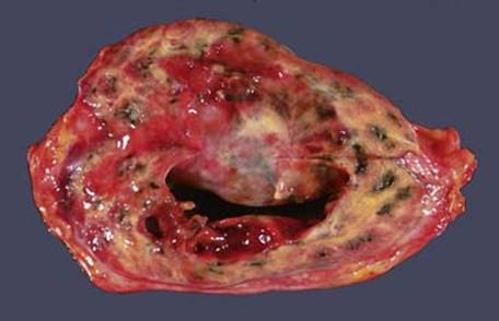
Fig 1 Lung abscess. Cut section of lobectomy specimen shows walled-off abscess. Note extensive fibrosis in adjacent lung.
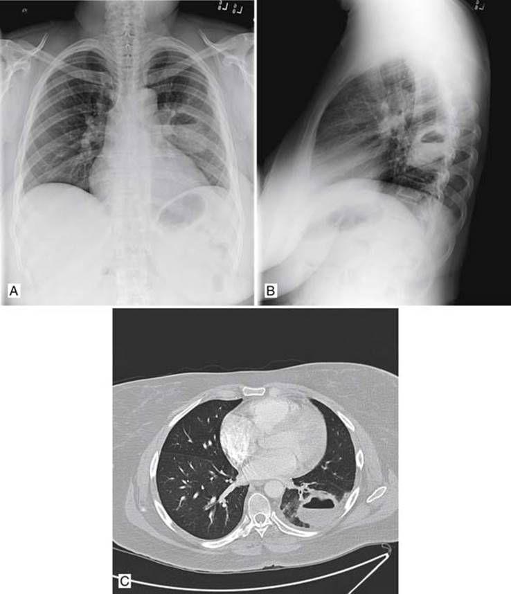
Fig 2 Lung abscess. Radiological images reveal diagnostic appearance of lung abscess with air–fluid level. Chest x-ray film PA view (A), lateral view (B), and CT (C).
(Courtesy of Dr. Christopher M. Strauss, Department of Radiology, University of Chicago, Ill.)
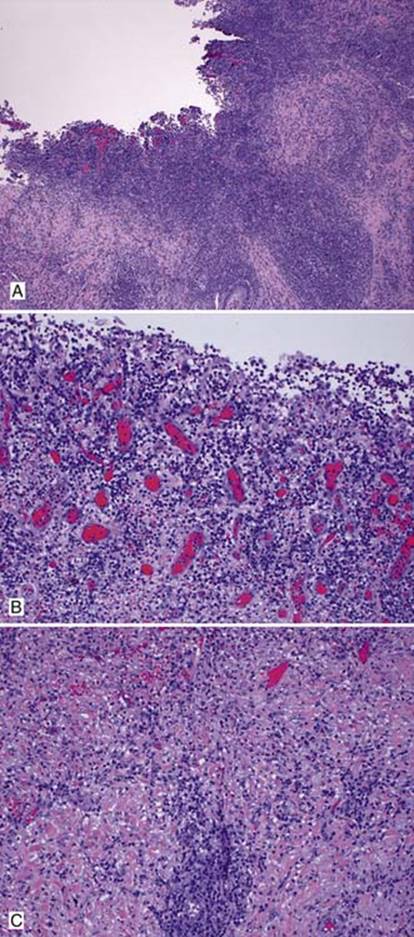
Fig 3 Lung abscess. A, At low power, a well-defined cavity is apparent, the wall of which is composed of inflammatory cells and fibrosis. B, The abscess wall has granulation tissue and chronic inflammation. C, Further away from the cavity, there is dense fibrosis with chronic inflammation.