
Definition
• Ubiquitous human pathogen and one of the most common opportunistic viral infections causing pneumonia
Clinical features
Epidemiology
• Large, double-stranded DNA virus
• 50% to 90% of the North American adult population is infected and harbors the virus for life
• Latent virus hosted by endothelial cells, monocytes, macrophages, neutrophils, and renal and pulmonary epithelial cells
• The majority of cases are unapparent and asymptomatic
• Disseminated form commonly seen in immunocompromised patients
Presentation
• Immunocompetent children and adults: self-limited mononucleosis-like infection, rarely manifesting as pneumonia
• Immunocompromised (AIDS, transplant, neonatal) patients: fever, cough, rales, and hypoxemia; radiographic bilateral nodular, reticular, interstitial or ground-glass opacities, consolidation, and pleural effusions. Can become disseminated with thrombocytopenia, hepatitis, adrenalitis, and encephalitis
Prognosis and treatment
• Ganciclovir, foscarnet, and intravenous CMV immune globulin
• Severe hypoxemia correlates with poor outcome
• Number of inclusions and cytomegaly increase with disease progression
• CMV pneumonia may be fatal, especially when complicated by coinfections and other morbidities
Pathology
Gross
• Scattered nodular foci of hemorrhage and necrosis (nodules 1-3 cm), or
• Heavy, diffusely consolidated lungs, or
• Single pulmonary nodule (rare)
Histology
• Four main patterns of tissue response:
• Miliary necroinflammatory lesions (most common; result of hematogenous spreading): multicentric small nodules with central hemorrhage and fibrin surrounded by necrotic alveolar walls with inflammatory cells, zonal necrosis, and neutrophilic necrosis
• Diffuse alveolar damage (DAD) or hemorrhagic pneumonia
• Minimal nonspecific inflammation or solitary nodule
• Diffuse interstitial pneumonitis with varying amounts of pneumocyte hyperplasia and lymphocytic and polymorphonuclear cell infiltrates
• Cytopathic effects:
• Cytomegaly (25-40 μm): affecting epithelial cells, endothelial cells, fibroblasts, and macrophages
• Nuclear inclusion (20 μm): single, basophilic, round to oval, with a peripheral halo and nuclear membrane accentuation (“owl’s eye” Cowdry type A cell); positive with Feulgen stain
• Cytoplasmic inclusions (1-3 μm): granular basophilic or amphophilic bodies positive with GMS and PAS stains
Immunopathology/special stains
• Immunohistochemical (IHC) analysis is very sensitive and specific, especially at early stages
• IHC is superior to fluorescence in situ hybridization, culture, and polymerase chain reaction
Main differential diagnoses
• Herpesvirus: eosinophilic ground-glass nuclear inclusions without cytomegaly
• Adenovirus: “smudgy” appearance of nuclear inclusions, obscuring nuclear membrane
• Measles: giant cell interstitial pneumonia with multiple, irregular cytoplasmic and eosinophilic nuclear inclusions
• Toxoplasmosis: 2-4 μm crescent-shaped free and intracellular tachyzoites, or true cysts with round to oval basophilic PAS-positive bradyzoites
• DAD and interstitial pneumonitis of other causes

Fig 1 Cytomegalovirus pneumonia. CMV infection with classic “owl’s eye” nuclear inclusions present in this case of DAD in organizing phase.

Fig 2 Cytomegalovirus pneumonia. The cytopathic effects of CMV include cytomegaly and a single nuclear inclusion commonly associated with multiple small basophilic cytoplasmic inclusions.
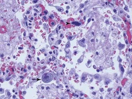
Fig 3 Cytomegalovirus pneumonia. CMV nuclear inclusion (arrow) can be confused with multilobated dense nuclei of megakaryocytes (arrowhead), which normally circulate in lungs, get “stuck” within the alveolar wall capillaries, and are more abundant after resuscitation.

Fig 4 Cytomegalovirus pneumonia. Dense inflammatory infiltrate masking several CMV-infected endothelial cells (arrows) in the submucosa of bronchial wall.
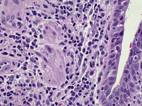
Fig 5 Cytomegalovirus pneumonia. CMV has a broad cellular tropism infecting virtually any cell, including smooth muscle, as shown in this example of miliary CMV pneumonia.

Fig 6 Cytomegalovirus pneumonia. Immunohistochemical staining is very useful in early CMV infection, highlighting cells without obvious cytomegaly or cytopathic effects.

Fig 7 Cytomegalovirus pneumonia. IHC with CMV antibody is very sensitive and easy to interpret. Neutrophils in this picture stain nonspecifically and should be disregarded.