
Christian A. Tomaszewski
EPIDEMIOLOGY
 Carbon monoxide (CO) is probably the most common cause of fatal poisoning, both intentionally and accidentally, with peak incidence in the fall and winter.
Potential sources of CO include incomplete combustion from such things as kerosene heaters, gas furnaces, wood burning stoves, charcoal grills, forklifts, generators, and automobiles. With inadequate ventilation any gasoline or natural gas-powered machinery can contribute to a high ambient CO.
Carbon monoxide contributes to fire deaths through smoke inhalation.
Methylene chloride can cause delayed, up to 8 hours post exposure, elevations of carboxyhemogoblin (COHb) because it is metabolized in the liver to CO.
PATHOPHYSIOLOGY
CO is an odorless, colorless gas. Ambient levels are usually around 10 ppm.
The permissible level of CO is 50 ppm averaged over an 8 hour shift (OSHA). Toxicity is usually seen at 100 ppm.
CO has an affinity for hemoglobin approximately 200 times that of oxygen. The half-life of COHb on room air is 250 to 320 minutes, reducing to about 85 minutes on 100% oxygen. The half-life of COHb from methylene chloride is longer and approaches 13 hours.
COHb cannot carry oxygen and it shifts the oxygen dissociation curve to the left for the remaining oxyhemo-globin, thereby providing less oxygen release to cells.
COHb does not account for all of the toxicity associated with CO poisoning. Ten percent to 15% of CO is dissolved in plasma. It binds to both myoglobin and cytochrome oxidase, interfering with cellular respiration in the heart and brain. CO also causes endothe-lial dysfunction and indirect vasodilation from nitric oxide release.
Delayed effects of CO include ischemic-reperfusion injury with attraction of neutrophils that trigger an inflammatory cascade with neuronal cell loss, particularly in the basal ganglia.
CLINICAL FEATURES
Clinical features of CO are varied, but commonly relate to hypoxic effects on the neurological and cardiovascular systems (Table 129-1). Symptoms range from “flu-like” symptoms, such as headache, dizziness, nausea, and vomiting, to coma. Older patients may present with syncope or cardiac ischemia.
A history of exposure to gas heat or smoke inhalation, or multiple victims with altered mental status, acidosis, or coma, should alert one to the possibility of CO poisoning.
Physical examination may reveal tachycardia, tachyp-nea, or hypotension. The hallmark neurological manifestations of CO poisoning beyond headache include confusion, irritability, seizures, focal neurological deficits, or coma. The “classic finding” of cherry red lips is rarely seen in living patients.
Patients with significant poisoning from acute or even chronic exposure may go on to experience long-term neuropsychiatric problems including memory loss and inability to concentrate.
TABLE 129-1 Signs and Symptoms of Acute Carbon Monoxide Poisoning
Headache
Visual disturbances
Vomiting
Confusion
Ataxia
Dyspnea/tachypnea
Seizure
ECG changes/dysrhythmias
Syncope
Retinal hemorrhage
Chest pain
Bullous skin lesions
Focal neurologic deficit
DIAGNOSIS AND DIFFERENTIAL
A venous, or arterial, blood sample for measurement of COHb on co-oximetry is the most reliable test to diagnose carbon monoxide poisoning. The use of bedside pulse co-oximetry in the ED to screen for CO exposure is still under investigation.
Although COHb levels confirm exposure, they do not necessarily correlate with symptoms or prognosis. CO poisoning is usually detected by measuring COHb levels. Normal COHb levels are around 1% but may be as high as 3% in nonsmokers and 10% in smokers. Higher levels are suggestive of CO exposure.
Standard pulse oximetry is unreliable in the presence of increasing COHb as oxygen saturation readings will be artificially high.
Additional lab abnormalities seen in symptomatic patients may include elevated anion gap metabolic acidosis, creatinine phosphokinase, or lactate. The latter may be indicative of concomitant cyanide poisoning in smoke inhalation victims.
Cardiac toxicity may be manifested by elevated tro-ponin or signs of ischemia on ECG.
Radiographic imaging is more useful for exploring alternate diagnoses. Early CT scanning of the brain is not very sensitive, but may show globus pallidus lesions in severely poisoned patients.
The differential diagnosis for CO poisoning is wide due to the nonspecific nature of the symptoms and includes flu-like illness, gastroenteritis, exposure to other toxins, and infectious causes of mental status changes. Cardiovascular compromise after poisoning may represent a concomitant myocardial infarction.
EMERGENCY DEPARTMENT CARE AND DISPOSITION
Remove patients from the source of exposure and address airway, breathing, and circulation.
Begin treatment in all patients suspected of CO poisoning with the highest concentration of supplemental oxygen available (eg, 100% oxygen via face-mask with reservoir) and continue until the patient is asymptomatic. Provide continuous monitoring of vital signs, heart rate, and rhythm. Establish IV access for seriously poisoned patients.
Guidelines for hyperbaric oxygen therapy (HBO) in patients with severe poisoning are the same in adults and children (Table 129-2). The threshold COHb for initiating HBO in pregnant patients is lower because of concerns for the fetus. Consult with a hyperbaric specialist.
Patients should have a secure airway and stable hemo-dynamics before transport and treatment with HBO as access may be limited en route and in the chamber.
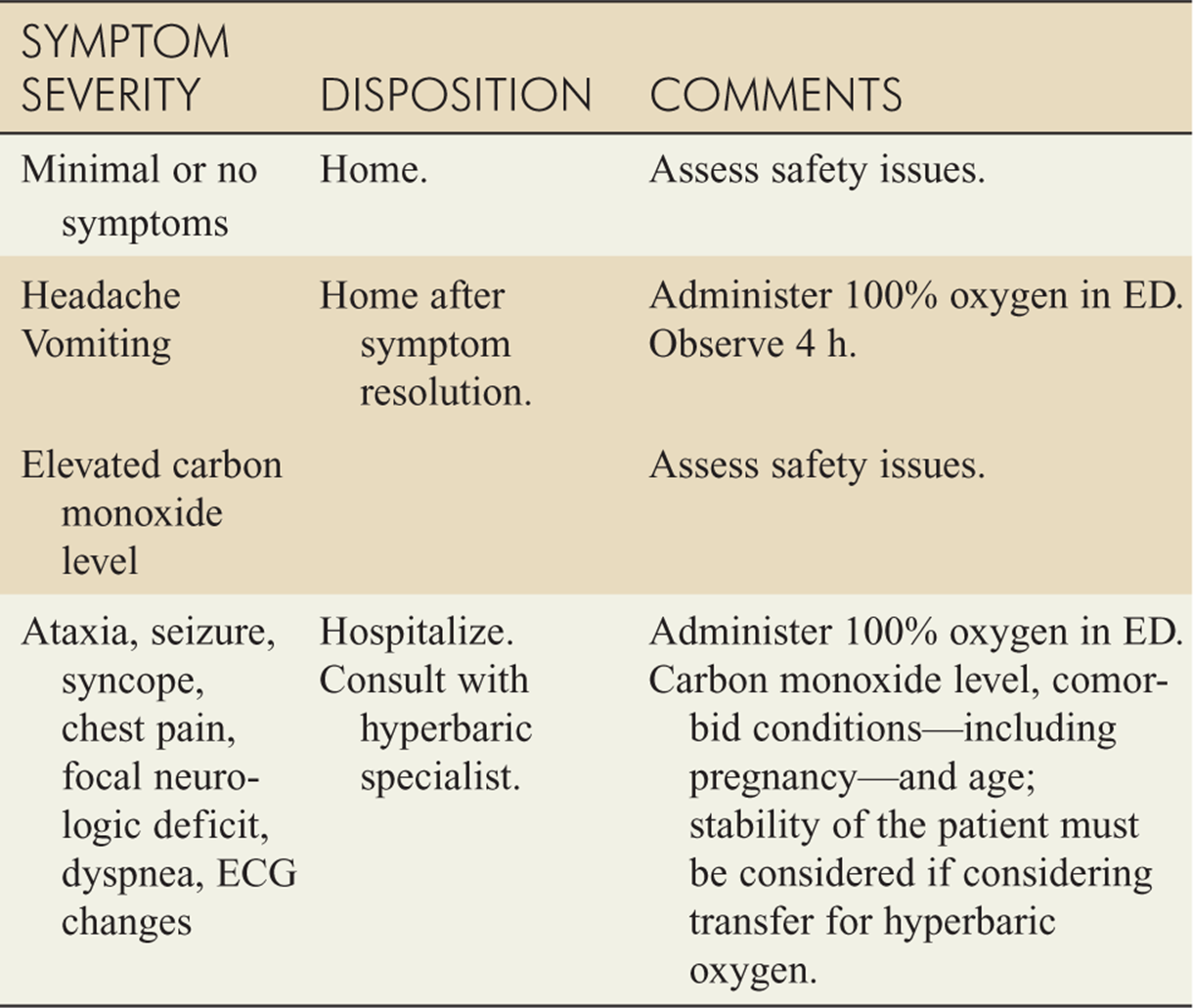
Guidelines for disposition of CO victims are based on severity of presentation (Table 129-3).
Symptomatic patients that do not require HBO treatment can be observed for approximately 4 hours while being treated with oxygen. If symptoms resolve and their neurological examination is normal, they can be discharged provided the home or work environment is no longer a source of carbon monoxide exposure.
TABLE 129-2 Commonly Utilized Indications for Referral for Hyperbaric Oxygen Treatment
Syncope
Confusion/altered mental status
Seizure
Coma
Focal neurologic deficit
Pregnancy with carboxyhemoglobin level >15%
Blood level >25%
Evidence of acute myocardial ischemia
TABLE 129-3 Disposition Considerations
For further reading in Tintinalli’s Emergency Medicine: A Comprehensive Study Guide, 7th ed., see Chapter 217, “Carbon Monoxide Poisoning,” by Gerald Maloney.