
Robert Jones
ECTOPIC PREGNANCY
EPIDEMIOLOGY
 Ectopic pregnancy (EP) occurs in 2% of all pregnancies and is the leading cause of maternal death in the first trimester.
Twenty percent of EPs are ruptured at the time of presentation.
Major risk factors include history of pelvic inflammatory disease; surgical procedures on the fallopian tubes, including tubal ligation; previous EP; diethyl-stilbestrol exposure; intrauterine device use; and assisted reproduction techniques.
More than 50% of cases of EP occur in patients without recognized risk factors.
This diagnosis must be considered in every woman of childbearing age presenting with abdominal pain.
PATHOPHYSIOLOGY
EP is postulated to be caused by (1) mechanical or anatomic alterations in the tubal transport mechanism, or (2) functional/hormonal factors that alter the fertilized ovum.
Tubal rupture is thought to be spontaneous, but trauma associated with coitus or a bimanual examination may precipitate tubal rupture. Tubal rupture may occur in the early weeks of an EP or as late as 16 weeks estimated gestational age.
CLINICAL FEATURES
The classic triad of abdominal pain, vaginal bleeding, and amenorrhea used to describe EP may be present, but many cases occur with more subtle findings.
Presenting signs and symptoms may be different in ruptured versus non-ruptured EP. Ninety percent of women with EP complain of abdominal pain; 80% have vaginal bleeding; and only 70% give a history of amenorrhea.
The pain may be sudden, lateralized, and extreme, or it may be relatively minor and diffuse.
The presence of hemoperitoneum causing diaphragmatic irritation may cause pain to be referred to the shoulder or upper abdomen.
Vaginal bleeding is usually light; heavy bleeding is more commonly seen with threatened abortion or other complications of pregnancy.
Presenting vital signs may be entirely normal even with a ruptured EP, or may indicate advanced hemor-rhagic shock.
There is poor correlation with the volume of hemoperitoneum and vital signs in EP. Relative bradycardia may be present even in cases with rupture and intra-peritoneal hemorrhage.
Physical examination findings are highly variable. The abdominal examination may show signs of localizing or diffuse tenderness with or without peritoneal signs.
The pelvic examination findings may be normal, but more often reveal cervical motion tenderness, adnexal tenderness with or without a mass, and possibly an enlarged uterus.
Fetal heart tones can be auscultated in advanced cases of EP. The presence of fetal heart tones cannot be used to rule out the presence of EP.
DIAGNOSIS AND DIFFERENTIAL
Urine pregnancy testing (for urinary beta-human chorionic gonadotropin [β-hCG]) should be performed immediately.
Dilute urine may result in a false-negative result; serum testing will give a more definitive result in such situations.
The primary goal of ultrasound in suspected EP is to determine if an intrauterine pregnancy (IUP) is present (Fig. 61-1). The presence of a heterotopic pregnancy should be strongly considered in patients who have undergone assisted reproduction.
Transvaginal ultrasound is the test of choice to identify EP and should be performed in cases where transabdominal ultrasound is nondiagnostic.
Definitive findings of an EP include the presence of an empty uterus along with visualization of extrauter-ine cardiac activity (Fig. 61-2).
Findings suggestive of an EP include the presence of an empty uterus and any of the following: adnexal mass (other than a simple cyst) with or without free fluid in the abdomen or moderate/large amount of free pelvic fluid (Fig. 61-3).
A normal pelvic ultrasound should be considered to be an indeterminate test and should be interpreted in light of the β-hCG levels.
A β-hCG level above the discriminatory zone (1500 mIU/mL) and an empty uterus suggests EP.
A β-hCG level below the discriminatory zone (1500 mIU/mL) and an empty uterus is indeterminate.
A β-hCG level should never be used to determine the need for performing a pelvic ultrasound in the symptomatic patient.
Failure of the β-hCG level to increase by 53% in 2 days is suggestive of EP or an abnormal IUP. However, an increase of 53% in 2 days does not rule out EP.
Differential diagnosis in the patient presenting with abdominal pain, vaginal bleeding, and early pregnancy includes threatened, incomplete, or missed abortion; recent elective abortion; or endometritis.
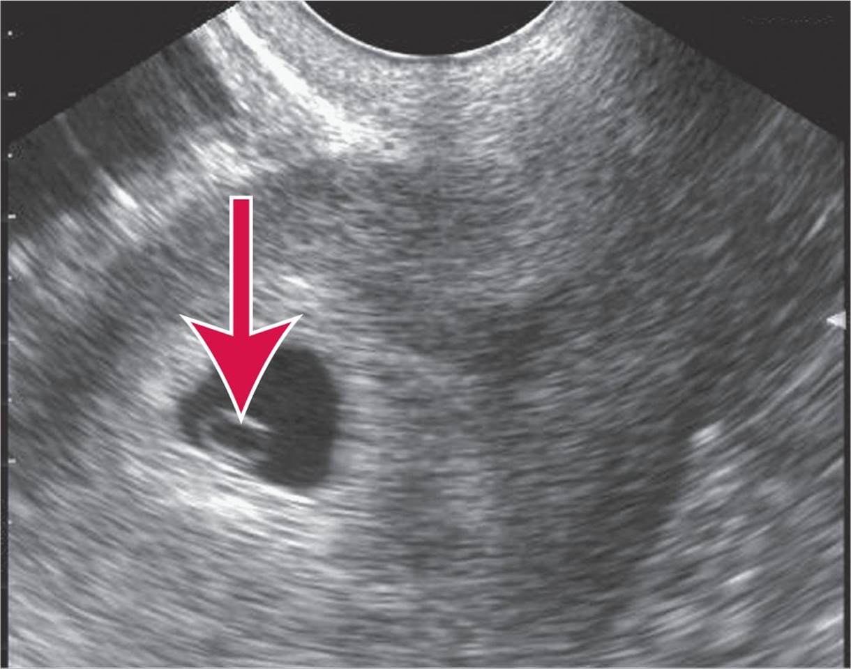
FIG. 61–1. Yolk sac {arrow) seen within an intrauterine gestational sac consistent with an early IUP. (Reproduced with permission from Ma OJ, Mateer JR, Blaivas M: Emergency Ultrasound, 2nd ed. © 2008, McGraw-Hill Inc., New York.)
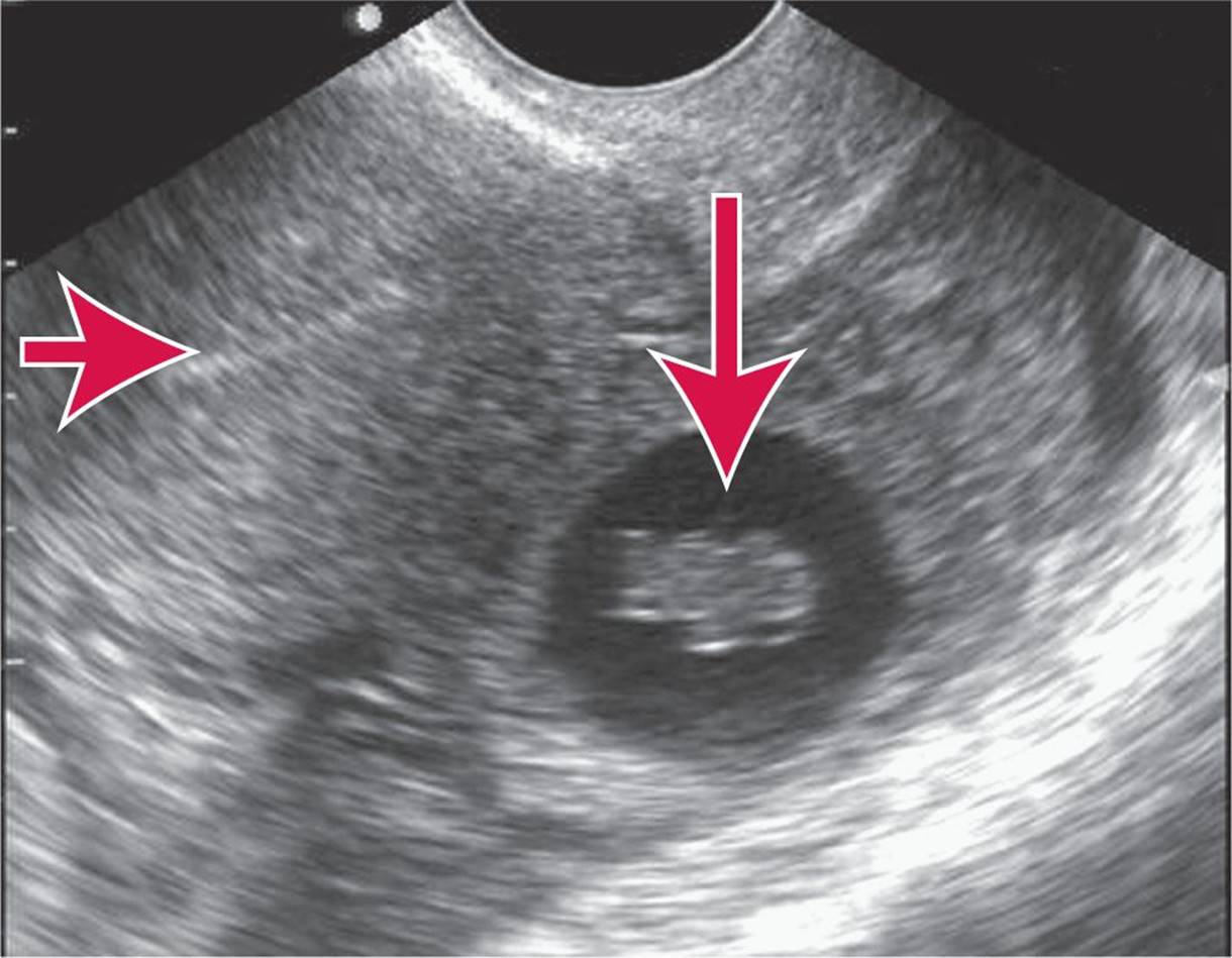
FIG. 61–2. Ectopic pregnancy. A living embryo in the adnexa and empty uterus is seen in this ectopic pregnancy (endome-trial echo is visible in the left upper portion of the image). Embryonic cardiac activity was present on real-time imaging. Transvaginal image. Horizontal arrow points to empty uterus (uterine stripe) and vertical arrow to ectopic pregnancy in the adnexa. (Reproduced with permission from Ma OJ, Mateer JR, Blaivas M: Emergency Ultrasound, 2nd ed. © 2008, McGraw-Hill Inc., New York.)
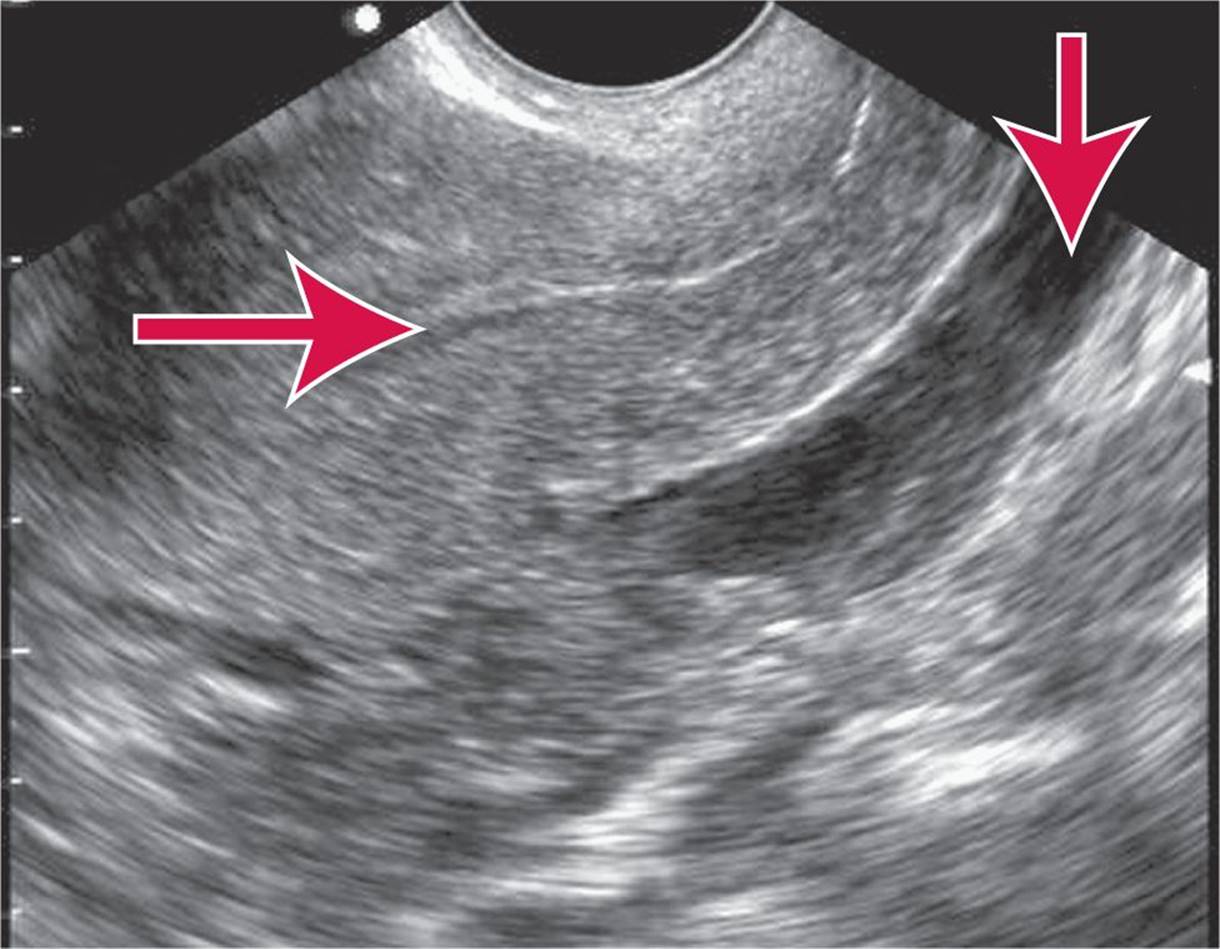
FIG. 61–3. Ectopic pregnancy: empty uterus and free fluid in the posterior cul-de-sac. Transvaginal sagittal image. Horizontal arrow points to empty uterus (uterine stripe), and vertical arrow points to fluid in the cul-de-sac. (Reproduced with permission from Ma OJ, Mateer JR, Blaivas M: Emergency Ultrasound, 2nd ed. © 2008, McGraw-Hill Inc., New York.)
EMERGENCY DEPARTMENT CARE AND DISPOSITION
Treatment of patients with suspected EP is dependent on the patient’s vital signs, physical signs, and symptoms. Close communication with the obstetric-gynecologic consultant is essential.
For unstable patients, start two large-bore intravenous lines for rapid infusion of crystalloid and/or packed red blood cells to maintain blood pressure.
Perform a bedside urine pregnancy test.
Perform a rapid bedside, transabdominal ultrasound in the unstable patient after ABCs have been addressed.
Notify an obstetric-gynecologic consultant immediately for the unstable patient, even before laboratory and diagnostic tests are complete.
Draw blood for complete blood count (CBC), blood typing, and Rh (rhesus factor) determination (or crossmatching for the unstable patients), quantitative β-hCG determination (if indicated), and serum electrolyte determination as required.
If the patient is stable, proceed with diagnostic workup, including transabdominal with or without transvaginal ultrasound. In reliable patients with indeterminate ultrasound results and a β-hCG level below 1000 mIU/mL, discharge with ectopic precautions and arranged follow-up in 2 days for repeat β-hCG determination and obstetric-gynecologic reevaluation is appropriate.
Definitive treatment, as determined by the obstetric-gynecologic consultant, may include laparoscopy, dilatation and curettage (D&C), or medical management with methotrexate.
EMERGENCIES DURING PREGNANCY AND THE POSTPARTUM PERIOD
The leading causes of maternal death are pulmonary embolus (see Chapter 27), EP (see Chapter 60), hypertensive disorders of pregnancy, hemorrhage, and infection.
Risk increases with increased maternal age, increased birth order, lack of prenatal care, unmarried status, and minority race.
EMERGENCIES IN THE FIRST 20 WEEKS
THREATENED ABORTION AND ABORTION
EPIDEMIOLOGY
Twenty to 40% of pregnancies abort spontaneously.
PATHOPHYSIOLOGY
Chromosomal abnormalities account for most fetal wastage. Risk increases with increasing maternal age and concurrent medical disorders, previous abortion, infections, and anatomic abnormalities.
CLINICAL FEATURES
Threatened abortion is defined as vaginal bleeding with a closed cervical os and benign physical examination.
Inevitable abortion will occur with vaginal bleeding and dilatation of the cervix.
Incomplete abortion is defined as partial passage of the conceptus and is more likely between 6 and 14 weeks of pregnancy.
Complete abortion is passage of all fetal tissue before 20 weeks’ gestation.
Missed abortion is fetal death at less than 20 weeks’ gestation without passage of fetal tissue for 4 weeks after fetal death.
Septic abortion implies evidence of infection during any stage of abortion, such as pelvic pain, fever, cervical motion or uterine tenderness, and purulent or foul-smelling discharge.
DIAGNOSIS AND DIFFERENTIAL
The differential diagnosis includes EP and gestational trophoblastic disease (GTD). GTD is aneoplastic disease of trophoblastic tissue, and is distinguished from threatened abortion by ultrasound. These patients will have an abnormally large uterus and an abnormally high β-hCG level. It may be noninvasive (hydatidiform mole) or invasive (choriocarcinoma).
EMERGENCY DEPARTMENT CARE AND DISPOSITION
Manage hemodynamic instability. Consult a gynecologist emergently in the unstable patient. Perform a pelvic examination, and obtain a CBC, blood typing and Rh factor determination, quantitative β-hCG, and urinalysis. Rh-negative women should receive Rh (D) immune globulin 300 micrograms IM.
Pelvic ultrasound should reveal a gestational sac in a normal pregnancy with a β-hCG >1500 mIU/Ml, but an IUP cannot be definitively diagnosed until a yolk sac or embryo is visualized. Absence of a gestational sac with a β-hCG >1500 mIU/mL suggests complete abortion or EP. An excessively large uterus in which the placenta has many lucent areas interspersed with brighter areas is seen in GTD.
Incomplete abortion or GTD requires D&C. GTD patients must receive close follow-up until quantitative β-hCG has returned to zero. Failure of the β-hCG to return to normal could indicate choriocarcinoma.
Septic abortion requires gynecologic consultation and broad-spectrum antibiotics such as ampicillin sul-bactam 3.0 grams IV or clindamycin 600 milligrams plus gentamicin 1 to 2 milligrams/kg IV.
Patients with threatened abortion or complete abortion may be discharged with close follow-up arranged. Discharge instructions include pelvic rest (no intercourse or tampons) and instructions to return for heavy bleeding, fever, or pain.
NAUSEA AND VOMITING OF PREGNANCY
EPIDEMIOLOGY
Nausea and vomiting in pregnancy occurs commonly in the first 12 weeks and affects between 60% and 80% of pregnant women.
PATHOPHYSIOLOGY
The pathophysiology is unknown.
CLINICAL FEATURES
The physical findings are usually normal in these patients with the exception of volume depletion.
The presence of abdominal pain is highly unusual and should suggest another diagnosis.
DIAGNOSIS AND DIFFERENTIAL
Hyperemesis gravidarum (intractable nausea and vomiting without significant abdominal pain) can cause hypokalemia or ketonemia and may result in a low-birth-weight infant.
Diagnostic workup should include a CBC, electrolyte panel, and urinalysis.
EMERGENCY DEPARTMENT CARE AND DISPOSITION
Treatment consists of rehydration with IV fluid (5% dextrose in normal saline [D5NS] or 5% dextrose in lactated Ringer’s [D5LR]), along with antiemet-ics, until ketonuria clears. Antiemetics that are frequently used are metoclopramide 10 milligrams IV promethazine 25 milligrams IV, (pregnancy class C, but widely used), or ondansetron 4 milligrams IV.
Patients are candidates for discharge if they have reversal of ketonuria, correction of any electrolyte imbalance, and a successful trial of oral fluids. Patients should be discharged with an antiemetic.
Patients admitted with intractable hyperemesis gravidarum may be candidates for methylprednisolone. This treatment should be initiated in consultation with the consulting obstetrician.
For further reading in Tintinalli’s Emergency Medicine: A Comprehensive Study Guide, 7th ed., see Chapter 101, “Ectopic Pregnancy and Emergencies in the First 20 Weeks of Pregnancy,” by Richard S. Krause, David M. Janicke, and Rita K. Cydulka.