
In rostrocaudal order, the brainstem consists of three subdivisions, the midbrain, pons, and medulla oblongata. Extending the entire length of the brainstem, any cross section demonstrates three laminae: the tectum, tegmentum, and basis [37].
Medulla Oblongata
Anatomy of the Medulla
The medulla oblongata or myelencephalon is the most caudal portion of the brainstem (Fig. 15.1) and extends from the caudal border of the pons to a point just rostral to the point of emergence of the first spinal nerve roots. The junction of the medulla oblongata and spinal cord is at the level of the foramen magnum. The cross-sectional anatomy at a midmedullary level is illustrated in Figure 15.2.
Within the substance of the medulla certain cranial nerve nuclei and roots are situated [22]. The hypoglossal nucleus (cranial nerve XII) is located near the ventrolateral portion of the central canal under an eminence called the hypoglossal trigone. The nerve roots of the hypoglossal nerve pass ventrally and emerge from the medulla in the anterior lateral sulcus between the pyramids and the olive (inferior olivary prominence). The nucleus ambiguus (cranial nerves IX, X, and bulbar XI) is located within the medullary reticular formation ventromedial to the nucleus and spinal tract of the trigeminal nerve (cranial nerves V, VII, IX, and X). The dorsal motor nucleus of the vagus (cranial nerve X) lies dorsolateral to the hypoglossal nucleus and sends fibers that join the motor roots of the vagus and spinal accessory nerves. The nucleus and tractus solitarius (cranial nerves VII, IX, and X) lie ventrolateral to the dorsal motor nucleus of the vagus, and the medial and spinal vestibular nuclei and the dorsal and ventral cochlear nuclei(cranial nerve VIII) are located at the dorsal and ventral borders of the inferior cerebellar peduncle (restiform body). The inferior olivary nucleus is located within the olive.
The nucleus gracilis and nucleus cuneatus are located in the posterior funiculi of the dorsal medulla and give rise to fibers (internal arcuate fibers) that cross in the decussation of the lemniscus (great sensory decussation). These fibers then travel in the medial lemniscus, which is dorsomedial to the pyramids. The nucleus of the spinal tract of the trigeminal nerve (pars caudalis) lies lateral to the internal arcuate fibers and descends caudally to the level of C3 in the cervical spinal cord, whereas the spinal tract of the trigeminal nerve lies lateral to the nucleus. The pyramids are located in the anterior (ventral) medulla and contain descending corticospinal tract fibers to the lateral and anterior corticospinal tracts of the spinal cord. The pyramid also contains descending corticobulbar fibers. In the caudal end of the medulla, nearly 75% to 90% of the corticospinal fibers in the pyramid cross the ventral midline (decussation of the pyramids or great motor decussation) to the opposite side to form the lateral corticospinal tract. The rest of the corticospinal tract descends homolaterally to form the anterior corticospinal tract. There is a somatotopic organization of the corticospinal fibers within the pyramids, with the fibers of the lower extremities placed more laterally than the fibers of the upper extremities [1]. The medial longitudinal fasciculus is located in the dorsomedial medulla. Other medullary tracts include the ventral and dorsal spinocerebellar tracts, the medial and lateral reticulospinal tracts, the medial and lateral vestibulospinal tracts, the rubrospinal tracts, the spinothalamic tracts, and descending sympathetic pathways.
Vascular Supply of the Medulla
The large regional arteries of the brainstem have the following three types of branches:
1. The paramedian arteries, which penetrate the ventral brainstem surface and supply the midline structures.
2. The short circumferential arteries, which traverse laterally on the brainstem and penetrate its ventrolateral and lateral surfaces.
3. The long circumferential arteries, which course around the brainstem and supply its posterior structures and cerebellum.
The medulla oblongata receives its blood supply from the anterior and posterior spinal arteries, the posterior inferior cerebellar artery, and branches of the vertebral arteries. The blood supply to the medulla may be subdivided into two groups: the paramedian bulbar branches and the lateral bulbar branches.
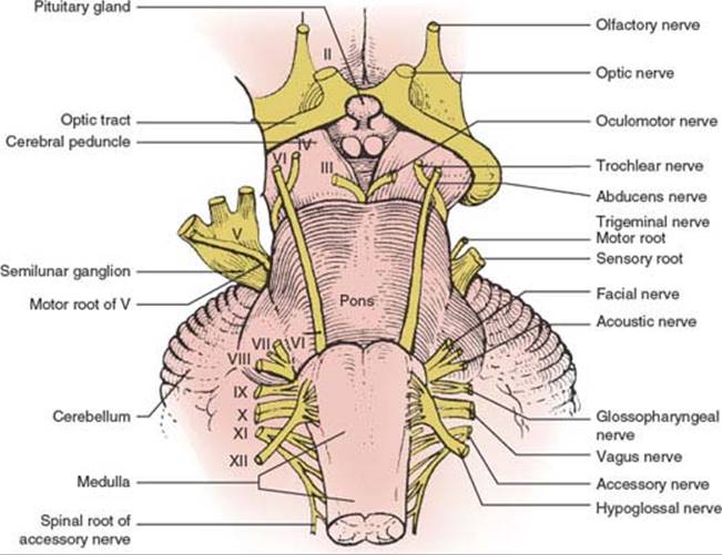
FIG. 15.1. The brainstem (ventral view).
PARAMEDIAN BULBAR BRANCHES
The paramedian portion of the medulla (the hypoglossal nucleus and emergent nerve fibers, the medial longitudinal fasciculus, the medial lemniscus, the pyramids, and the medial part of the inferior olivary nucleus) are supplied by the vertebral artery. At lower medullary levels, the anterior spinal artery also contributes to the paramedian zone.
LATERAL BULBAR BRANCHES
The lateral portion of the medulla is supplied by the intracranial vertebral artery (fourth segment) or the posterior inferior cerebellar artery. Occasionally, the basilar artery or the anterior inferior cerebellar artery also contributes.
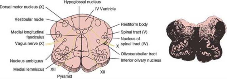
FIG. 15.2. Midportion of the medulla at the origin of the hypoglossal and vagus nerves. Myelin-stained section is shown at right. (From Daube JR, Reagan TJ, Sandok BA, et al. Medical neurosciences: an approach to anatomy, pathology, and physiology by system and levels, 2nd ed. Boston, MA: Little, Brown and Company, 1986. By permission of Mayo Foundation.)
Medullary Syndromes
MEDIAL MEDULLARY SYNDROME (DEJERINE’S ANTERIOR BULBAR SYNDROME)
This syndrome often results from atherosclerotic occlusion of the vertebral artery, anterior spinal artery, or the lower segment of the basilar artery. Vertebrobasilar dissection, dolichoectasia of the vertebrobasilar system, embolism, and meningovascular syphilis are less common causes of the medial medullary infarction [155]. The anterior spinal artery supplies the paramedian region of the medulla oblongata, which includes the ipsilateral pyramid, medial lemniscus, and hypoglossal nerve and nucleus (Fig. 15.3). Its occlusion therefore results in the following signs:
1. Ipsilateral paresis, atrophy, and fibrillation of the tongue (due to cranial nerve XII affection). The protruded tongue deviates toward the lesion (away from the hemiplegia). Cranial nerve XII function may be spared [127].
2. Contralateral hemiplegia (due to involvement of the pyramid) with sparing of the face.
3. Contralateral loss of position and vibratory sensation (due to involvement of the medial lemniscus). The more the dorsolateral spinothalamic tract is unaffected, the more the pain and temperature sensation are spared.
4. Occasionally, upbeat nystagmus may occur because of dorsal extension of the infarct toward the medial longitudinal fasciculus [70]. Unilateral lesions of the nucleus intercalatus can account for primary position upbeat nystagmus due to a unilateral medial medullary infarction [69]. It has also been proposed that damage to the uncrossed climbing fibers from the inferior olivary nucleus to the contralateral cerebellar Purkinje cells results in ocular contrapulsion from rostral medial medullary infarctions [81].
The medial medullary syndrome may occur bilaterally [57,99] resulting in flaccid quadriplegia (with facial sparing), bilateral lower motor neuron lesions of the tongue, complete loss of position and vibratory sensation affecting all four extremities and respiratory failure, or acute onset of triparesis (with involvement of both lower limbs and contralateral upper extremity), suggestive of a possible fiber segregation of the descending tracts of different extremities [58]. Located in the caudal medullary tegmentum, both hypoglossal nuclei have been involved in isolation in a small medullary infarction [12].

FIG. 15.3. Cross section of medulla oblongata showing area involved in medial medullary infarction and lateral medullary infarction (Wallenberg syndrome). CN = cranial nerve.
Because the hypoglossal fibers run somewhat laterally to the medial lemniscus and pyramid, they are occasionally spared in cases of anterior spinal artery occlusion. Occasionally, only the pyramid is damaged, resulting in a pure motor hemiplegia that spares the face [30,128,135]. Central facial paresis may also result from a unilateral contralateral medullary infarction, suggesting that some of the facial corticobulbar fibers descend ipsilaterally before making a loop as low as the medulla oblongata before decussating and ascending to the contralateral facial nucleus that innervates the perioral musculature [26,148]. A crossed motor hemiparesis (hemiplegia cruciata), with paralysis of the ipsilateral arm and the contralateral leg (resulting from a lower medullary lesion compromising the crossed fibers to the arm as well as the uncrossed fibers to the leg), is an extremely rare occurrence [14].
Apart from incomplete syndromes (e.g., medial medullary syndrome presenting as pure motor hemiparesis, or medial medullary syndrome without tongue paralysis), other unusual neurologic findings may be observed including contralateral paralysis of the pharyngeal constrictor muscle [111] and contralateral tongue paralysis [27].
LATERAL MEDULLARY (WALLENBERG) SYNDROME
This syndrome [33,34,54,80,107,114,132] is most often secondary to intracranial vertebral artery or posterior inferior cerebellar artery occlusion [78]. The presumed pathogenesis among 130 consecutive patients with pure lateral medullary infarctions included large vessel disease in 50%, arterial dissection in 15%, small vessel disease in 13%, and cardioembolism in 5% [78]. Spontaneous dissections of the vertebral arteries are a common cause [75,105]. Dissections were observed more often with caudal lesions [78]. The syndrome has also been described with cocaine abuse [104], medullary neoplasms (usually metastases), abscess, demyelinating disease [141], radionecrosis, hematoma (secondary to rupture of a vascular malformation), neck manipulation [56], trauma, bullet injury to the vertebral artery [102], and posterior spinal fusion surgery with instrumentation in a patient with a previously undiagnosed Chiari 1 malformation [122]. The characteristic clinical picture results from damage to a wedge-shaped area of the lateral medulla (Fig. 15.3) and inferior cerebellum and consists of several signs:
1. Ipsilateral facial hypalgesia and thermoanesthesia (due to trigeminal spinal nucleus and tract involvement). Ipsilateral facial pain is common [34].
2. Contralateral trunk and extremity hypalgesia and thermoanesthesia (due to damage to the spinothalamic tract).
3. Ipsilateral palatal, pharyngeal, and vocal cord paralysis with dysphagia and dysarthria (due to involvement of the nucleus ambiguus).
4. Ipsilateral Horner syndrome (due to affection of the descending sympathetic fibers). Ipsilateral hypohidrosis of the body may occur, probably due to interruption of the mostly uncrossed excitatory sweating pathway, which descends from the hypothalamus through the tegmental area of the mesencephalon and pons and, more caudally, through the posterolateral area of the medulla to synapse with the sympathetic sudomotor neurons of the intermediolateral cell column of the spinal cord [84].
5. Vertigo, nausea, and vomiting (due to involvement of the vestibular nuclei).
6. Ipsilateral cerebellar signs and symptoms (due to involvement of the inferior cerebellar peduncle and cerebellum).
7. Occasionally, hiccups (singultus) attributed to lesions of the dorsolateral region of the middle medulla [117] and diplopia (perhaps secondary to involvement of the lower pons).
Lateral lesions located in the rostral medulla are associated with more severe dysphagia, hoarseness, and the presence of facial paresis, whereas more caudal lesions situated in the lateral surface of the medulla, correlate with more marked vertigo, nystagmus, and gait ataxia [80]. Nausea, vomiting, and Horner syndrome are common regardless of the location of the lesion in the lateral medulla; lesions that extend more ventromedially cause facial sensory changes on the contralateral side of the lesion [80]. The motor system (pyramids), tongue movements, and vibration and position sense are typically spared with lateral medullary lesions because the corresponding anatomic structures are located in the medial medulla. The triad of Horner syndrome, ipsilateral ataxia, and contralateral hypalgesia clinically identifies patients with lateral medullary infarction [132]. Cerebellar infarcts only infrequently accompany the lateral medullary syndrome, suggesting that most of the posterior inferior cerebellar artery territory is spared, despite the high frequency of vertebral artery occlusion as a cause of this syndrome [132].
Headache, especially unilateral headache localized to the upper posterior cervical region, is relatively common with the lateral medullary syndrome, particularly when the syndrome is due to cervical vertebral artery dissection [64,105]. This type of headache should be distinguished from the rare paroxysmal retro-orbital hemicranial-like attacks reported after strokes involving the dorsal medulla and high cervical spinal cord at the C1 level [36].
The sensory defect in the lateral medullary syndrome usually affects the ipsilateral face and the contralateral leg, arm, and trunk. However, several patients with lateral brainstem lesions developed a sensory defect involving the ipsilateral face and the contralateral foot, with the latter defect extending upward to end in a sensory level [96]. These patients with a crossed pattern of sensory defect had far lateral lesions of the lateral medulla and pons, with the leg and lower torso involvement due to selective partial disruption of the somatotopically organized sacral and lumbar afferent fibers of the lateral spinothalamic tract (located far laterally in the brainstem), with sparing of the more medial thoracic and cervical fibers [96]. Several patients have also been described with a continuous hemisensory defect of the face, arm, and trunk (unilateral pattern), with the lower border demarcated at a sensory level [96]. These patients were thought to have mediolateral medullary and pontine lesions contralateral to the side of the sensory defect, which affected the medial cervical and thoracic afferents of the lateral spinothalamic tract (i.e., spared the lateral sacral and lumbar afferents) and the ventral trigeminothalamic tract (accounting for contralateral facial sensory loss), but spared the spinal nucleus and tract of the trigeminal nerve. In rare instances of infarcts involving the pontomedullary sulcus, sensory symptoms electively involve the contralateral upper limb and base of the neck resulting in loss of pain and temperature, and reinforcing the notion that a somatotopic arrangement of the spinothalamic tract in its medullary course [160].
Rare manifestations of the Wallenberg syndrome include the following:
1. Wild arm ataxia probably related to involvement of the lateral cuneate nucleus [32,33].
2. Clumsiness of the ipsilateral upper limb resulting from extension of the injury into the subolivary area [22].
3. Central pain associated with allodynia [121].
4. Contralateral hyperhidrosis with ipsilateral anhidrosis due to interruption of the sympathetic pathways (noted a few months after infarction) [130].
5. An inability to sneeze due to compromise of the sneezing center located at the ventromedial margin of the descending tract and nucleus (spinal nucleus) of the trigeminal nerve [68].
6. Paroxysmal sneezing due to presumed involvement of the hypothetical human “sneezing center” in the rostral dorsolateral medulla [45,113,137].
7. Loss of taste that results from involvement of the rostral and the lateral zone of the nucleus tractus solitarius [59].
8. Autonomic dysfunction including tachycardia, blood pressure lability, and respiratory failure from the involvement of the caudal and medial zone of the nucleus tractus solitarius [25].
9. Failure of automatic breathing (Ondine’s curse) due to discrete lesions of the nucleus ambiguus and the adjacent reticular formation.
10. Transient urinary retention from interruption of descending fibers from facilitatory pontine micturition centers [89].
11. Body lateropulsion without limb ataxia from the involvement of the descending lateral vestibulospinal tract, or body lateropulsion with limb ataxia due to interruption of the ascending dorsal spinocerebellar tract [151].
12. Axial lateral pulsion that results from the involvement of the vestibulospinal and spinocerebellar tracts as well as central vestibular pathways [7].
13. Isolated ipsiversive lateropulsion [3].
14. Pure sensory stroke with loss of pain and temperature involving the face, arm, trunk, and leg as the only manifestations of the lateral medullary infarction [8,15].
15. Ipsilateral sensory symptoms predominantly involving the upper extremities, especially the fingers, with occasional impairment of vibration and position sense from caudal lesions involving the dorsal columns or decussating lemniscal fibers [79].
16. Ipsilateral hemiparesis from the involvement of the lower most caudal end of the medulla just below the pyramidal decussation [38]. An ipsilateral spastic hemiplegia associated with a lateral medullary syndrome is also known as the submedullary syndrome of Opalski (see subsequent text) [106].
17. Central hypoventilation is seen along with vasomotor instability [87].
18. Poststroke facial pain that results from the involvement of the primary afferent fibers in the descending spinal trigeminal tract [53].
Various abnormalities of eye movements and vision have been described with the lateral medullary syndrome (Table 15.1) [18,21,29,39,100]. These include the following:
1. Dysfunction of ocular alignment. Lateral medullary lesions damage the otolithic vestibular nuclei and, therefore, patients with Wallenberg syndrome often demonstrate skew deviation with hypotropia on the side of the lesion [77]. Brandt and Dieterich have called this type 2 skew deviation and stated that this skew results from elevation of the contralateral eye, without vertical displacement of the ipsilateral eye [19,20]. Some patients also show an ipsilateral head tilt and a disconjugate ocular torsion (the ocular tilt reaction, see Chapter 8) with excyclodeviation of the ipsilateral lower eye but with little or no incyclodeviation of the contralateral higher eye [20,39,107]. Therefore, patients may complain of diplopia with images displaced vertically and tilted with respect to each other. Some patients with Wallenberg syndrome may also exhibit ocular ipsipulsion due to damage to the climbing fibers from the contralateral inferior olivary nucleus to the dorsal vermis [82] or complain of the unusual (and almost unbelievable) sensation of environmental tilt, in which the whole room is tilted on its side or even upside down (“floor-on-ceiling” phenomenon) [39,127]. This syndrome is also probably caused by a disturbance of vestibular-otolith central connections [127]. Environmental tilt or “upside down” reversal of vision may also occur with vertebrobasilar transient ischemic attacks [143], vertebrobasilar ischemia [144], encephalitis, head injury [100], demyelinating disease [138], or after third ventriculostomy for hydrocephalus [116].
TABLE 15.1 Ocular Motor Abnormalities in Wallenberg Lateral Medullary Syndrome

Damage to otolithic central projections mediating ocular counter-roll may also contribute to the genesis of torsional nystagmus (see subsequent text) in the lateral medullary syndrome [107]. Central otolithic involvement may also be responsible for the see-saw nystagmus observed in occasional patients [63,103]. See-saw nystagmus is a disjunctive, vertical-torsional nystagmus half cycle, which consists of elevation and intorsion of one eye with synchronous depression and extorsion of the other eye; the next half cycle consists of the reversal of these vertical and torsional movements. This type of nystagmus is usually pendular and noted especially with large, extensive suprasellar lesions that compress or infiltrate the mesodiencephalon bilaterally. With lateral medullary lesions, however, a jerk see-saw nystagmus may occur [63,107]. The torsional component of this nystagmus is conjugate with the fast component contraversive to the side of the lesion [63]. This contrasts with the jerk see-saw nystagmus described with unilateral, focal mesodiencephalic lesions, in which the quick phase of the torsional component is toward the side of the lesion [63].
2. Nystagmus. Nystagmus in the lateral medullary syndrome may be due to direct damage to the vestibular nuclei or their cerebellar, semicircular canal, or otolithic connections. Nystagmus in the lateral medullary syndrome is usually positional and can be horizontal [42], torsional [107], or mixed, with torsion, vertical, and horizontal components [10]. Typically, horizontal nystagmus beats away from the side of the lesion, with the horizontal drift velocity directed toward the side of the lesion being influenced by eye position and by fixation. Occasionally, the nystagmus may beat with the fast component ipsilaterally during gaze toward the side of the lesion or during eye closure [10]. A vertical nystagmus is usually upbeating [10]. The nystagmus is often evident only in the initial days after dorsolateral medullary infarction, and rapidly declines over the following days [125]. Torsional nystagmus is common with Wallenberg syndrome, with the upper pole of the iris beating away from the side of infarction [107]. Torsional nystagmus has been attributed to an imbalance of central projections from the anterior and posterior semicircular canals and the otolithic receptors that mediate ocular counter-roll [107].
As mentioned in the preceding text, see-saw nystagmus may also occur with lateral medullary lesions [103]. Gaze-evoked eyelid nystagmus associated with ocular nystagmus has been described, in which a clinically obvious upward jerking of the lids occurred synchronously with the fast phase of a gaze-evoked horizontal nystagmus [35]. This eyelid nystagmus was inhibited or totally arrested by the near reflex.
3. Smooth pursuit and gaze-holding abnormalities. Structures and pathways located in the lateral medulla are also concerned with smooth pursuit eye movements and gaze holding [162]. The cerebellar flocculus, paraflocculus, and vermis climbing fibers pass through the inferior cerebellar peduncle and are concerned with these functions.
Patients with the lateral medullary syndrome may complain of a sensation of their bodies being pulled to one side and attempt to counteract this lateropulsion of the body by leaning toward the opposite side. Because of gaze-holding impairment, ocular movements may be similarly affected, with a tendency for the eyes to be “pulled” toward the involved medulla (lateropulsion or ipsipulsion of eye movements) [10,42,55,98,157,161]. If a patient is asked to fixate straight ahead and close the eyelids, the eyes will deviate toward the side of the medullary lesion (reflected by a series of small corrective hypometric saccadic [fast] eye movements in the opposite direction, which are directed to fixation when the eyes are again opened). Even blinking may induce this lateropulsion. These abnormalities of gaze holding may also be reflected in saccadic eye movement abnormalities. Smooth pursuit eye movements tracking targets moving away from the side of the lesion are also impaired with lateral medullary lesions, whereas pursuit toward the side of the lesion is normal, or nearly so [10,98,162].
4. Abnormalities of saccades. The cerebellum may be involved in modulating the amplitude but not the speed of saccadic (fast) eye movements. Interruption of cerebellar central connections that traverse the lateral medulla probably accounts for some of the observed ocular motor deficits [132]. Damage to the juxtarestiform body, which carries signals from the fastigial nucleus to the brainstem reticular formation, may account for a saccadic abnormality referred to as lateropulsion of saccadic eye movements [90].
As noted in the preceding text, gaze-holding abnormalities in patients with Wallenberg syndrome may result in ipsipulsion of eye movements. This disorder of gaze holding may also induce saccadic abnormalities. Horizontal saccades away from the side of the lesion are hypometric (undershoot the target), whereas saccades directed toward the side of the lesion are hypermetric (overshoot the target) [161]. Quick phases of nystagmus are similarly affected. Ipsipulsion with lateral medullary lesions is therefore opposite to the contrapulsion of saccades that occurs with lesions of the superior cerebellar peduncle [126,157].
Patients with Wallenberg syndrome may have permanent saccadic dysmetria (hypermetria to the side of the lesion and hypometria to targets contralateral to the lesion) and a reduced capability to readjust saccadic amplitude [161]. This horizontal saccade bias with lateral medullary lesions is also reflected in vertical eye movements. On attempting to make a purely vertical saccade, an oblique or elliptical saccade directed toward the lesion (in the direction of lateropulsion) is made, requiring corrective saccades away from the side of the lesion to bring the eyes back toward the intended target. Later, attempted vertical saccades may take on S-shaped trajectories as an adaptive strategy to correct the saccadic dysmetria [90]. Even a torsional component of this bias may occur (torsipulsion), with inappropriate torsional fast eye movements induced during saccades toward or away from the side of the medullary lesion [107].
The medial branch of the posterior inferior cerebellar artery supplies the dorsolateral medulla; infarcts of this branch may be clinically silent, cause isolated vertigo often misdiagnosed as labyrinthitis, cause vertigo associated with ipsilateral lateropulsion of the trunk and gaze and dysmetria or unsteadiness, or cause a full Wallenberg syndrome [5,6,62,73]. Bilateral cerebellar infarction in the territory of the medial branches of the posterior inferior cerebellar arteries may cause vertigo, dysarthria, dysequilibrium with retropulsion, bilateral gaze-evoked nystagmus, and marked gait ataxia without brainstem signs [145]. Vertigo and upside-down vision have been described because of an infarct in the cerebellar flocculus and nodulus due to affection of the medial branch of the posterior inferior cerebellar artery [28].
Atherosclerotic occlusion or dissection of the intracranial vertebral artery can lead to a total unilateral hemimedullary (Babinski-Nageotte) syndrome, a combination of the medial and lateral medullary syndromes [109]. This rare syndrome is characterized by contralateral hemiplegia and sensory loss of the limbs and trunk, ipsilateral hemiataxia, and facial sensory loss, along with dysphagia, dysphonia, and dysarthria. Ipsilateral hemiparesis is extremely rare [93]. Some authorities have suggested Reinhold’s syndrome as the proper eponym for the hemimedullary syndrome [85]. Because of the separate arterial topography supplying the medulla, the simultaneous occurrence of ischemic lesions involving the lateral and medial parts of the medulla is extremely rare [109]. Combinations of the two major syndromes may also occur as bilateral medial and bilateral lateral medullary syndromes [59].
Tegmental medullary lesions (e.g., glioma) may cause lack of appetite and early satiety (medullary satiety), implying that the medulla may play a role in the regulation of feeding behaviors [94]. Lesions affecting the obex of the medulla may result in neurogenic pulmonary edema [140]. This supports the hypothesis that lesions of caudal brainstem structures, especially the nucleus tractus solitarius, the dorsal motor nucleus of the vagus, and the medial reticular formation are responsible for the generation of neurogenic pulmonary edema. Lesions of the area postrema, an emetic center located in the caudal part of the fourth ventricle and lacking a blood—brain barrier, lesions of the dorsolateral pontine tegmentum, as well as other lesions of the lower brainstem, may account for vomiting, often out of proportion to dizziness [50].
OPALSKI (SUBMEDULLARY) SYNDROME
When ipsilateral hemiplegia is associated with symptoms of a lateral medullary syndrome, it corresponds to the submedullary syndrome of Opalski. Opalski syndrome results from an occlusion of the vertebral artery. The ipsilateral hemiplegia is due to a lesion of the lower medulla involving the corticospinal tract after the pyramidal decussation [71,115].
LATERAL PONTOMEDULLARY SYNDROME
This syndrome [48] may result from occlusion of an aberrant arterial branch arising from the upper vertebral artery and running superiorly and laterally to the region of exit of cranial nerves VII and VIII from the pons. It may also occur with pontine hemorrhage [4]. The clinical findings are those seen in the lateral medullary syndrome plus several pontine findings, which includes the following:
1. Ipsilateral facial weakness (due to involvement of cranial nerve VII)
2. Ipsilateral tinnitus and, occasionally, hearing disturbance (due to involvement of cranial nerve VIII)

FIG. 15.4. Cross section of the lower pons at the level of cranial nerves VI and VII. Myelin-stained section is shown on the right. (From Daube JR, Reagan TJ, Sandok BA, et al. Medical neurosciences: an approach to anatomy, pathology, and physiology by system and levels, 2nd ed. Boston, MA: Little, Brown and Company, 1986. By permission of Mayo Foundation.)
The Pons
Anatomy of the Pons
The pons (Fig. 15.1) [23] is part of the metencephalon (pons and cerebellum), and extends from a caudal plane, which passes from the striae medullaris posteriorly through the pontomedullary sulcus anteriorly, to a cephalad plane, which passes immediately caudal to the inferior colliculi (dorsally) and to the cerebral peduncles (ventrally). The dorsal part of the pons is referred to as the tegmentum, and the ventral portion is referred to as the basis pontis, basilar pons, or pontocerebellar portion (Fig. 15.4). The pontine tegmentum is composed largely of the pontine reticular formation, which is a rostral continuation of the medullary reticular formation. This central core is generally divided into a medial region of primarily large neurons (magnocellular region) and a lateral region of mainly small neurons (parvocellular region). The basis pontis contains the pontine nuclei and multidirectional nerve fiber bundles.
Cranial nerve nuclei in the pons include the nucleus of the abducens nerve (cranial nerve VI), which is located in the dorsomedial pons just beneath the floor of the fourth ventricle. Fibers from this nucleus pass ventrally between bundles of corticospinal tract fibers to exit at the pontomedullary junction. Ventromedial to the abducens nucleus is the paramedian pontine reticular formation (PPRF), which plays an important role in the control of saccadic eye movements (see Chapter 8). The motor nucleus of the facial nerve (cranial nerve VII) is situated ventrolaterally. Fibers from this nucleus run dorsomedially toward the floor of the fourth ventricle, make an acute bend around the abducens nucleus, and then turn laterally through the pons to exit lateral to the abducens nerve fibers. The main motor and main sensory nuclei of the trigeminal nerve (cranial nerve V) are located dorsolaterally, as are the cochlear nuclei and the lateral and superior vestibular nuclei (cranial nerve VIII). The superior and inferior salivatory nuclei and the lacrimal nucleus (cranial nerves VII and IX) are also located in the pons.
Fiber tracts within the pons include the medial longitudinal fasciculus, which is situated dorsomedially, and the medial lemniscus, which lies dorsal to the corticospinal, corticobulbar, and corticopontine fiber bundles. Other tracts within the pons include the ventral spinocerebellar, spinothalamic, lateral tectospinal, rubrospinal, and corticopontocerebellar tracts. The pons also contains auditory connections, including the lateral lemniscus, the nucleus of the lateral lemniscus, the trapezoid body, and the superior olivary nuclear complex. The brachium pontis or middle cerebellar peduncle connects the ventral pons with the cerebellum.
Vascular Supply of the Pons
At the lower border of the pons, the paired vertebral arteries fuse in the midline to form the basilar artery. The first branch of the basilar artery is the anterior inferior cerebellar artery. Then comes a series of paramedian and short circumferential pontine branches, and penultimately, the superior cerebellar artery. Finally, the basilar artery divides into the two posterior cerebral arteries. The blood supply to the pons may be divided into three groups.
PARAMEDIAN VESSELS
The paramedian vessels (four to six in number) arise from the basilar artery and penetrate perpendicularly into the pontine parenchyma. They supply the medial basal pons, including the pontine nuclei, the corticospinal fibers, and the medial lemniscus.
SHORT CIRCUMFERENTIAL ARTERIES
The short circumferential arteries also arise from the basilar artery and enter the brachium pontis. These vessels supply the ventrolateral basis pontis.
LONG CIRCUMFERENTIAL ARTERIES
The long circumferential arteries supply most of the pontine tegmentum and part of the middle cerebellar peduncles and include the following:
1. The superior cerebellar artery, which arises from the basilar artery near its bifurcation, supplies the dorsolateral pons and brachium pontis, the dorsal reticular formation, and the periaqueductal region (occasionally, the ventrolateral pontine tegmentum is also supplied by this vessel).
2. The anterior inferior cerebellar artery, which most often arises from the basilar artery and supplies the lateral tegmentum of the lower two-thirds of the pons and the ventrolateral cerebellum.
3. The internal auditory artery, which arises from the anterior inferior cerebellar artery (occasionally from the basilar artery) and supplies the auditory, vestibular, and facial cranial nerves.
Pontine Syndromes
Numerous classical brainstem (eponymous) syndromes featuring cranial nerve palsies, cerebellar signs, long tract signs, and sensory disturbances were instrumental in establishing the seminal guidelines for brainstem localization [139].
VENTRAL PONTINE SYNDROMES
Millard-Gubler Syndrome. A unilateral lesion of the ventrocaudal pons may involve the basis pontis and the fascicles of cranial nerves VI and VII. This involvement results in the following:
1. Contralateral hemiplegia (sparing the face) is due to pyramidal tract involvement.
2. Ipsilateral lateral rectus paresis (cranial nerve VI) with diplopia that is accentuated when the patient “looks toward” the lesion.
3. Ipsilateral peripheral facial paresis (cranial nerve VII).
Raymond Syndrome. A unilateral lesion of the ventral medial pons, which affects the ipsilateral abducens nerve fascicles and the corticospinal tract but spares cranial nerve VII, may cause this rare syndrome (also called alternating abducens hemiplegia) [134], which consists of the following:
1. Ipsilateral lateral rectus paresis (cranial nerve VI)
2. Contralateral hemiplegia, sparing the face, due to pyramidal tract involvement
Pure Motor Hemiparesis. Lesions (especially lacunar infarction) involving the corticospinal tracts in the basis pontis may produce a pure motor hemiplegia with or without facial involvement [48,52,101,112]. Patients often have severe dysarthria and dysphagia. Bouts of uncontrollable laughter may also occur [136]). Other locations of lesions causing pure motor hemiplegia include the posterior limb of the internal capsule, the cerebral peduncle, and the medullary pyramid [30]. A combination of dysarthria and a history of previous transient gait abnormality or vertigo favor a pontine lesion as the cause of pure motor hemiparesis rather than a more common capsular lesion [112].
Dysarthria—Clumsy Hand Syndrome. Vascular lesions in the basis pontis (especially lacunar infarction) [46,48,55,60,101] at the junction of the upper one-third and lower two-thirds of the pons may result in dysarthria—clumsy hand syndrome. In this syndrome facial weakness and severe dysarthria and dysphagia occur along with clumsiness, impaired finger dexterity, and paresis of the hand. Hyperreflexia and a Babinski’s sign may occur on the same side as the arm paresis, but sensation is spared. A similar clinical presentation may occur with lesions in the genu of the internal capsule or with small, deep cerebellar hemorrhages [131].
Ataxic Hemiparesis. A lesion (usually a lacunar infarction) [47,48,51,101] in the basis pontis at the junction of the upper one-third and the lower two-thirds of the pons may result in the ataxic hemiparesis (homolateral ataxia and crural paresis) syndrome. In this syndrome hemiparesis that is more severe in the lower extremity, is associated with ipsilateral hemiataxia and occasionally dysarthria, nystagmus, and paresthesias. The hemiparesis is also associated with hyperreflexia and a Babinski’s sign. The lesion is located in the contralateral pons. The ataxia is unilateral, probably because transverse fibers originating from the contralateral pontine nuclei (and projecting to the contralateral cerebellum) are spared [110]. This syndrome has also been described with contralateral thalamocapsular lesions, lesions of the contralateral posterior limb of the internal capsule, lesions of the contralateral red nucleus, and with superficial anterior cerebral artery territory infarcts in the paracentral area [17,67].
As a rare occurrence, focal infarcts in the basilar pons have been associated with dysarthria-dysmetria, dysarthria-facial paresis, or ipsilateral gaze paresis and internuclear ophthalmoplegia [136].
Locked-in Syndrome. Bilateral ventral pontine lesions (infarction, tumor, hemorrhage, trauma, cervical manipulation, tumor, pontine abscess, encephalitis, arteritis, neuro-Behcet’s, multiple sclerosis, air embolism, heroin abuse, diazepam toxicity, or central pontine myelinolysis) may result in the locked-in syndrome (de-efferented state) [66,118,123]. This syndrome consists of the following signs:
1. Quadriplegia due to bilateral corticospinal tract involvement in the basis pontis
2. Aphonia due to involvement of the corticobulbar fibers innervating the lower cranial nerve nuclei
3. Occasional impairment of horizontal eye movements due to bilateral involvement of the fascicles of cranial nerve VI
Because the reticular formation is not injured, the patient is fully awake. The supranuclear ocular motor pathways lie dorsally and are therefore spared; therefore, vertical eye movements and blinking are intact (the patient may actually convey his wishes in Morse code). In thrombosis of the basilar artery, not infrequently a hemiparesis is present at an early stage (“herald hemiparesis” of basilar artery occlusion), when brainstem signs may be absent or few [49]. Therefore, a cerebral hemisphere localization is suggested, but in a few hours bilateral hemiplegia appears, associated with a locked-in syndrome or coma [49]. De-efferentation may also occur with purely peripheral lesions (e.g., polio, polyneuritis, myasthenia gravis).
DORSAL PONTINE SYNDROMES
Foville Syndrome. This syndrome is due to lesions involving the dorsal pontine tegmentum in the caudal third of the pons. It consists of the following:
1. Contralateral hemiplegia (with facial sparing) which is due to interruption of the corticospinal tract.
2. Ipsilateral peripheral-type facial palsy which is due to involvement of the nucleus and fascicle (or both) of cranial nerve VII.
3. Inability to move the eyes conjugately to the ipsilateral side (gaze is “away from” the lesion) due to involvement of the PPRF or abducens nucleus, or both.
Raymond-Cestan Syndrome. The Raymond-Cestan syndrome is seen with rostral lesions of the dorsal pons. It includes the following:
1. Cerebellar signs (ataxia) with a coarse “rubral” tremor which is due to the involvement of the cerebellum.
2. Contralateral hypesthesia with reduction of all sensory modalities (face and extremities) which is due to the involvement of the medial lemniscus and the spinothalamic tract.
3. With ventral extension, there may be contralateral hemiparesis (due to corticospinal tract involvement) or paralysis of conjugate gaze toward the side of the lesion (due to involvement of the PPRF).
PARAMEDIAN PONTINE SYNDROMES
Several clinical syndromes of paramedian pontine infarction have been described [11].
1. Unilateral mediobasal infarcts. These patients present with severe facio-brachio-crural hemiparesis, dysarthria, and homolateral or bilateral ataxia.
2. Unilateral mediolateral basal infarcts. Most patients show slight hemiparesis with ataxia and dysarthria, ataxic hemiparesis, or dysarthria—clumsy hand syndrome.
3. Unilateral mediocentral or mediotegmental infarcts. Presentations include dysarthria—clumsy hand syndrome, ataxic hemiparesis with prominent sensory or eye movement disorders, and hemiparesis with contralateral facial or abducens palsy.
4. Bilateral centrobasal infarcts. These patients have pseudobulbar palsy and bilateral sensorimotor disturbances.
The most common etiology for paramedian pontine infarcts is small vessel disease; vertebrobasilar large vessel disease and cardiac embolism are less common causes [11].
An unusual finding observed in patients with unilateral paramedian pontine infarction consists of bilateral Wallerian degeneration of the middle cerebellar peduncles [156].
LATERAL PONTINE SYNDROMES
Marie-Foix Syndrome. This syndrome is seen with lateral pontine lesions, especially those affecting the brachium pontis. It consists of the following:
1. Ipsilateral cerebellar ataxia due to involvement of cerebellar connections
2. Contralateral hemiparesis due to involvement of the corticospinal tract
3. Variable contralateral hemihypesthesia for pain and temperature due to involvement of the spinothalamic tract
Rostral lateral pontine infarcts can present with contralateral crural predominant hemiparesis or crural monoparesis. Lesions associated with crural hemiparesis primarily involve the lateral and dorsal pontine base, while lesions responsible for crural monoparesis primarily involve the dorsolateral pontine base [76].
As a rare occurrence, pontine lesions have been associated with anosognosia for the hemiplegia [43], blepharospasm [9], brief clonic jerking and other convulsive-like movements [133], jaw-opening dystonia, [40] hemidystonia [146], a focally enhanced startled response [163], symptomatic orthostatic tremor [13], dysarthria-dysmetria or dysarthria-facial paresis [136], body lateropulsion from paramedian tegmental involvement ventral to the fourth ventricle [164], truncal ataxia without limb ataxia [102], isolated bilateral ataxia due to selective involvement of part of the decussation of the superior cerebellar peduncle [88], bilateral deafness [154], cheiro-pedal syndrome with numbness of hand and foot associated with hypesthesia and hypalgesia [74], painful Horner syndrome [31], contralateral hemihyperhidrosis [120], intraoral sensory loss [44], trigeminal neuralgia [119], ipsilateral transient eye and nose pain [41], isolated cranial nerve palsies [149], transient hemiageusia [86], disturbances of cognition and affect, pathologic crying, prodrome of inappropriate or pathological laughter (fou rire prodromique) resulting from rostral and medial pontine involvement [61,136,147], rapid eye movement sleep behavior disorder occurring either in isolation or in association with narcolepsy [83,95]. In other circumstances, they have mimicked an acute peripheral vestibulopathy [150]. While lesions in the dorsolateral pontine tegmentum may cause vomiting, medial tegmental upper pontine lesions, probably affecting the PPRF bilaterally, may cause central reflex hyperpnea, formerly called central neurogenic hyperventilation. Volitional central facial paresis results from lesions involving the contralateral corticobulbar fibers. Emotional innervation of the muscles of facial expression is involuntary and of uncertain origin. Volitional type of facial paresis with unimpaired emotional movements to emotional stimuli has also been described indicating that the pathways subserving volitional and emotional input to the facial nucleus are still anatomically separated in the upper pons [153,158]. Conversely, emotional (mimetic) facial paresis has been noted with dorsolateral pontine lesions involving structures distinct from the corticobulbar fibers that mediate volitional facial innervation [72].
The Syndrome of Universal Dissociative Anesthesia
Universal dissociative anesthesia is a rare syndrome that has been described in a patient affected by combined right superior cerebellar artery occlusion, resulting in lateral superior pontine infarction, and left posterior inferior cerebellar artery occlusion, resulting in a left Wallenberg lateral medullary syndrome [159]. The patient had loss of pain and temperature sensation over the face, neck, trunk, and all extremities, whereas light touch, vibration, position, and deep pain sensation were preserved (dissociated sensory loss). This interesting lesson in localization was due to bilateral discrete interruption of spinothalamic fibers and the spinal nucleus and tract of the trigeminal nerve.
The clinical findings with pontine hemorrhage are discussed in Chapter 21.
The Mesencephalon
Anatomy of the Mesencephalon
The rostral boundary of the mesencephalon is the superior colliculi—mammillary bodies’ plane; the caudal boundary is the plane just caudal to the inferior colliculi (Fig. 15.1). The midbrain (Fig. 15.5) may be divided into the dorsal tectum or quadrigeminal plate (containing the colliculi), the central tegmentum, and the ventrally located cerebral peduncles [1].
The dorsal tectum contains the corpora quadrigemina, made up of four rounded eminences arranged in pairs: the superior and inferior colliculi. The tegmentum contains ascending and descending tracts, reticular nuclei, and well-delineated nuclear masses. The cerebral peduncles are ventral and contain corticopontine fibers (frontopontine projection) in their medial fifth, corticospinal tract fibers in their middle three-fifths, and temporopontine fibers in their lateral fifth. Fibers in the corticospinal tract are somatotopically arranged with the fibers destined to the arm medially placed and those to the leg laterally located, with the trunk fibers in between. The substantia nigra is a pigmented layer possessing melanin granules, dorsal to the peduncles and ventral to the red nucleus, composed of a dorsal zona compacta and a ventral zona reticulata.

FIG. 15.5. Cross-section of the mesencephalon. A: Lower mesencephalon at the level of inferior colliculus. B: Upper midbrain at the level of superior colliculus. Myelin-stained sections are shown below. (From Daube JR, Reagan TJ, Sandok BA, et al. Medical neurosciences: an approach to anatomy, pathology, and physiology by system and levels, 2nd ed. Boston, MA: Little, Brown and Company, 1986. By permission of Mayo Foundation.)
The nucleus of the trochlear nerve (cranial nerve IV) is located in the ventral part of the central gray matter at the level of the inferior colliculus; the nucleus of the oculomotor nerve (cranial nerve III) lies rostral to the trochlear nucleus beneath the superior colliculus, just posterior to the medial longitudinal fasciculus. Mesencephalic tracts include the crus cerebri, the dentatorubrothalamic tract, the medial tegmental tract, the medial longitudinal fasciculus, the posterior commissure, the spinothalamic tract, and the medial lemniscus.
Vascular Supply of the Mesencephalon
The mesencephalon receives its blood supply from branches of the basilar, posterior cerebral, superior cerebellar, posterior communicating, anterior and posterior choroidal arteries.
The mesencephalon’s vascular supply includes the paramedian and the circumferential vessels.
PARAMEDIAN VESSELS
The paramedian vessels (the retromamillary trunk) arise from the origins of the posterior cerebral arteries and include the thalamoperforating arteries (supplying the thalamus) and the peduncular arteries (supplying the medial peduncles and the midbrain tegmentum, including the oculomotor nucleus, the red nucleus, and the substantia nigra).
CIRCUMFERENTIAL ARTERIES
The circumferential (peripeduncular) arteries include the following:
1. The quadrigeminal arteries (arising from the posterior cerebral arteries), which supply the superior and inferior colliculi.
2. The superior cerebellar arteries, which send branches to the cerebral peduncles and brachium conjunctivum before supplying the superior cerebellum.
3. The posterior choroidal arteries, which supply the cerebral peduncles, the lateral superior colliculi, the thalamus, and the choroid plexus of the third ventricle.
4. The anterior choroidal arteries (from the internal carotids or middle cerebral arteries), which in some cases help supply the cerebral peduncles as well as supramesencephalic structures.
5. The posterior cerebral arteries, which also give rise to some mesencephalic branches.
Mesencephalic Syndromes
VENTRAL CRANIAL NERVE III FASCICULAR SYNDROME (WEBER’S SYNDROME)
A lesion affecting the cerebral peduncle, especially the medial peduncle, may damage pyramidal fibers and the fascicle of cranial nerve III [16] (Fig. 15.6). This results in the Weber’s syndrome, which consists of the following:
1. Contralateral hemiplegia (including the lower face) due to corticospinal and corticobulbar tract involvement
2. Ipsilateral oculomotor paresis, including parasympathetic cranial nerve III paresis (i.e., dilated pupil)
This syndrome may be seen with intrinsic or extrinsic brainstem lesions and may even be the presenting sign of multiple sclerosis [92]. When supranuclear fibers for horizontal gaze are interrupted in the medial peduncle, a supranuclear-type conjugate gaze palsy to the opposite side may occur (the midbrain syndrome of Foville).
DORSAL CRANIAL NERVE III FASCICULAR SYNDROMES (BENEDIKT’S SYNDROME)
A lesion affecting the mesencephalic tegmentum may affect the red nucleus, the brachium conjunctivum, and the fascicle of cranial nerve III (Fig. 15.6). More ventral tegmental lesions result in Benedikt’s syndrome, [91] which consists of the following:
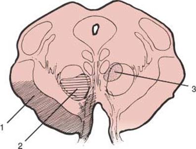
FIG. 15.6. Diagram of a section through the mesencephalon showing regions in which the oculomotor nerve fascicle may be injured, causing specific neurologic syndromes. (1) Weber’s syndrome; (2) Benedikt’s syndrome; (3) Claude’s syndrome.
1. Ipsilateral oculomotor paresis, usually with a dilated pupil
2. Contralateral involuntary movements, including intention tremor, hemichorea, or hemiathetosis, due to destruction of the red nucleus
Similar clinical manifestations are noted with more dorsal midbrain tegmental lesions (Fig. 15.6) that injure the dorsal red nucleus and brachium conjunctivum (Claude’s syndrome) but with prominent cerebellar signs (e.g., asynergia, ataxia, dysmetria, dysdiadochokinesia) and no hemiballismus [16]. The Nothnagel’s syndrome is a variant of the dorsal midbrain syndrome (see subsequent text), and may not include a fascicular third nerve palsy.
DORSAL MESENCEPHALIC SYNDROMES
Dorsal rostral mesencephalic lesions produce mainly neuro-ophthalmologic abnormalities. The dorsal mesencephalic syndrome (also known as the Sylvian aqueduct syndrome, the Koeber-Salus-Elschnig syndrome, or Parinaud’s syndrome) [65] is most often seen with hydrocephalus or tumors of the pineal region. This syndrome includes all or some of the following signs:
1. Paralysis of conjugate upward gaze (occasionally down-gaze)
2. Pupillary abnormalities (pupils are usually large with light-near dissociation)
3. Convergence-retraction nystagmus on upward gaze (especially elicited by inducing upward saccades by a down-moving optokinetic target)
4. Pathologic lid retraction (Collier’s sign)
5. Lid lag
6. During horizontal refixations, the abducting eye may move more slowly than the adducting eye (“pseudoabducens palsy”), perhaps reflecting excess convergence tone
TOP OF THE BASILAR SYNDROME
Occlusive vascular disease of the rostral basilar artery, usually embolic, frequently results in the “top of the basilar” syndrome [24,97] due to infarction of the midbrain, thalamus, and portions of the temporal and occipital lobes. An uncommon variant of this syndrome may also result in bilateral paramedian midbrain ischemia [142]. This syndrome may also occur in patients with giant basilar artery tip aneurysms, in patients with vasculitis, and after cerebral angiography [97]. This syndrome variably includes the following:
1. Disorders of eye movements. Unilateral or bilateral paralysis of upward or downward gaze, disordered convergence, pseudoabducens palsy, convergence-retraction nystagmus, ocular abduction abnormalities, elevation and retraction of the upper eyelids (Collier’s sign), skew deviation, and lightning-like eye oscillations.
2. Pupillary abnormalities. Small and reactive, large or midposition and fixed, corectopia, occasionally oval pupil.
3. Behavioral abnormalities. Somnolence, sleep-wake cycle abnormalities, peduncular hallucinosis, memory difficulties, agitated delirium.
4. Visual defects. Hemianopia, cortical blindness, Balint’s syndrome.
5. Motor and sensory deficits. Lesions causing pseudoabducens palsy with convergence-retraction nystagmus have been further mapped to the midbrain-diencephalic junction [124]. In addition, isolated unilateral superior oblique palsies have been described in patients with contralateral tegmental lesions of the trochlear nucleus and adjacent intraaxial trochlear nerve [152]. Likewise, isolated cranial nerve palsies and cheiro-oral syndrome have been reported as the sole manifestation of small mesencephalic infarcts [2,149]. Furthermore, strategically placed unilateral caudal paramedian midbrain lesions may produce bilateral cerebellar dysfunction [108].
References
1. Afifi AK, Bergman RA. Functional neuroanatomy. Text and atlas. New York, NY: McGraw-Hill, 2000: 105–146.
2. Aizawa H, Najuguchi N, Katayama T, et al. Cheiro-oral syndrome due to midbrain lesion. Neurology 2002;58:1414.
3. Akdal G, Thurtell MJ, Halmagyi GM. Isolated lateropulsion in acute lateral medullary infarction. Arch Neurol 2007;64:1542–1543.
4. Aleksic S, Budzilovich G. Lateral inferior pontine syndrome. Clinico-pathologic study and review of literature. J Neurol Sci 1973;18:317–322.
5. Amarenco P. The spectrum of cerebellar infarctions. Neurology 1991;41:973–979.
6. Amarenco P, Roullet E, Hommel M, et al. Infarction in the territory of the medial branch of the posterior inferior cerebellar artery. J Neurol Neurosurg Psychiatry 1990;53:731–735.
7. Arai M. Ipsilateral axial lateropulsion as an initial symptom of vertebral artery occlusion. J Neurol Neurosurg Psychiatry 2004;75(11):1648.
8. Arai M. Isolated thermoanesthesia with a midlateral medullary infarction. Neurology 2002;58(11):1695.
9. Aramideh M, Ongerboer de Visser BW, Holstege G, et al. Blepharospasm in association with a lower pontine lesion. Neurology 1996;46:476–478.
10. Baloh RW, Yee RD, Honrubia V. Eye movements in patients with Wallenberg’s syndrome. Ann NY Acad Sci 1981;374:600–613.
11. Bassetti C, Bogousslavsky J, Barth A, et al. Paramedian pontine infarct: clinical-topographic correlation. Neurology (Suppl) 1993;43:159.
12. Benito-León J, Alvarez-Cermeño JC. Isolated tongue paralysis as a manifestation of bilateral medullary infarction. J Neurol Neurosurg Psychiatry 2003;74: 1698–1699.
13. Benito-León J, Modriguez J, Orti-Pareja M, et al. Symptomatic orthostatic tremor in pontine lesions. Neurology 1997;49:1439–1441.
14. Bing R. Textbook of nervous diseases. New York, NY: Rebmen, 1915.
15. Blitshteyn S, Rubino FA. Pure sensory stroke as an isolated manifestation of the lateral medullary infarction. J Neuroimaging 2005;15(1):82–84.
16. Bogousslavsky J, Maeder R, Regli F, et al. Pure midbrain infarction: clinical syndromes, MRI, and etiologic patterns. Neurology 1994;44:2032–2040.
17. Bogousslavsky J, Martin R, Moulin T. Homolateral ataxia and crural paresis: a syndrome of anterior cerebral artery territory infarction. J Neurol Neurosurg Psychiatry 1992;55:1146–1149.
18. Bogousslavsky J, Meienberg O. Eye movement disorders in brainstem and cerebellar stroke. Arch Neurol 1987;44:141–148.
19. Brandt T, Dieterich M. Different types of skew deviation. J Neurol Neurosurg Psychiatry 1991;54:549– 550.
20. Brandt T, Dieterich M. Pathological eye-head coordination in roll: tonic ocular tilt reaction in mesencephalic and medullary lesions. Brain 1987;110: 649–666.
21. Brazis PW. Subject review. Ocular motor abnormalities in Wallenberg’s lateral medullary syndrome. Mayo Clin Proc 1992;67:365–368.
22. Brochier T, Ceccaldi M, Milandre L, et al. Dorsolateral infarction of the lower medulla: clinical MRI study. Neurology 1999;52:190–193.
23. Brodal A. Neurological anatomy in relation to clinical medicine, 3rd ed. New York, NY: Oxford University Press, 1981.
24. Caplan LR. “Top of the basilar” syndrome. Neurology 1980;30:72–79.
25. Caplan LR. Vertebrobasilar system syndrome. In: Vinken PJ, Bruyn WG, Klawans HL, eds. Handbook of clinical neurology, Vol. 53. New York, NY: Elsevier Science, 1988:371–408.
26. Cavazos JE, Bulsara K, Caress J, et al. Pure motor sensory hemiplegia including the face induced by an infarct of the medullary pyramid. Clin Neurol Neurosurg 1996;98:21–23.
27. Chang D, Cho SH. Medial medullary infarction with contralateral glossoplegia. J Neurol Neurosurg Psychiatry 2005;76(6):888.
28. Charles N, Froment C, Rode G, et al. Vertigo and upside down vision due to an infarct in the territory of the medial branch of the posterior inferior cerebellar artery caused by dissection of a vertebral artery. J Neurol Neurosurg Psychiatry 1992;55:188–192.
29. Choi K-D, Oh S-Y, Park S-H. Head shaking nystagmus in lateral medullary infarction. Patterns and possible mechanisms. Neurology 2007;68:1337–1344.
30. Chokroverty S, Rubino FA, Haller C. Pure motor hemiplegia due to pyramidal infarction. Arch Neurol 1965;13:30.
31. Crevits L, D’Herde K, Deblacre K. Painful isolated Horner’s syndrome caused by pontine ischemia. Graef Arch Clin Exp Ophthalmol 2004;242(2):181–183.
32. Currier RD, Bebin J. A medullary syndrome characterized by wild arm ataxia. Neurology 1999;53:1068–1069.
33. Currier RD, Dejong RN. The lateral medullary (Wallenberg’s) syndrome. Univ Mich Med Bull 1962;28:106.
34. Currier RD, Giles GL, Dejong RN. Some comments on Wallenberg’s lateral medullary syndrome. Neurology (Minneap.) 1961;11:778.
35. Daroff RB, Hoyt WF, Sanders MD, et al. Gaze-evoked eyelid and ocular nystagmus inhibited by the near reflex: unusual ocular motor phenomena in a lateral medullary syndrome. J Neurol Neurosurg Psychiatry 1968;31:362–368.
36. de la Sayette V, Leproux F, Letellier P. Cervical cord and dorsal medullary infarction presenting with retro-orbital pain. Neurology 1999;53:632–634.
37. DeMyer W. Neuroanatomy, 2nd ed. Baltimore, MD: Williams and Wilkins, 1998.
38. Dhamon SK, Ikbal J, Collins GH. Ipsilateral hemiplegia and the Wallenberg syndrome. Arch Neurol 1984;41:179–180.
39. Dieterich M, Brandt T. Wallenberg’s syndrome: lateral-pulsion, cyclorotation, and subjective visual vertical in thirty-six patients. Ann Neurol 1992;31:399–408.
40. Dietrichs E, Heier MS, Nakstad PH. Jaw-opening dystonia presumably caused by a pontine lesion. Mov Disord 2000;15(5):1026–1028.
41. Doi H, Nakamura M, Suenaga T, et al. Transient eye and nose pain as an initial manifestation of pontine infarction. Neurology 2003;60:521–523.
42. Estanol B, Lopez-Rios G. Neuro-otology of the lateral medullary syndrome. Arch Neurol 1982;39:176–179.
43. Evyapan D, Kumral E. Pontine anosognosia for hemiplegia. Neurology 1999;53:647–649.
44. Ferro JM. Intraoral sensory loss as the presenting feature in pontine hematoma. Cerebrovasc Dis 2000; 10(6):486–487.
45. Fink JN. Localization of the sneeze center. Neuroimages. Neurology 2001;56:138.
46. Fisher CM. A lacunar stroke: the dysarthria-clumsy hand syndrome. Neurology 1967;17:614–617.
47. Fisher CM. Ataxic hemiparesis: a pathologic study. Arch Neurol 1978;35:126–128.
48. Fisher CM. Lacunar strokes and infarcts: a review. Neurology 1982;32:871–876.
49. Fisher CM. The ‘herald hemiparesis’ of basilar artery occlusion. Arch Neurol 1988;45:1301–1303.
50. Fisher CM. Vomiting out of proportion to dizziness. Neurology 1996;46:267.
51. Fisher CM, Cole M. Homolateral ataxia and crural paresis: a vascular syndrome. J Neurol Neurosurg Psychiatry 1965;28:48.
52. Fisher CM, Curry HB. Pure motor hemiplegia of vascular origin. Arch Neurol 1965;13:30.
53. Fitzek S, Baumgartner U, Fitzek C, et al. Mechanisms and predictors of chronic facial pain in lateral medullary infarction. Ann Neurol 2001;49(4):493–500.
54. Freddo L, Sacco RL, Bello JA, et al. Lateral medullary syndrome: clinicoanatomical features studied by magnetic resonance and vascular imaging. Ann Neurol 1989;26:157.
55. Frisen L. Lateropulsion of the eyes–a localizing brainstem sign. J Neurol 1978;218:171–177.
56. Frumkin LR, Baloh RW. Wallenberg’s syndrome following neck manipulation. Neurology 1990;40: 611–615.
57. Fukuda M, Aiba T, Takahashi S. Bilateral medial medullary infarction due to bilateral vertebral artery dissection. Clin Neurol Neurosurg 2004;106(2): 132–135.
58. Fung HC, Chen ST, Tang LM, et al. Triparesis: MRI documentation of bipyramidal medullary infarction. Neurology 2002;58(7):1130–1131.
59. Gan R, Noronha A. The medullary vascular syndromes revisited. J Neurol 1995;242:195–202.
60. Glass JD, Levey AL, Rothstein JD. The dysarthria-clumsy hand syndrome: a distinct clinical entity related to pontine infarction. Ann Neurol 1990;27:487–494.
61. Gondini F de AA, Parks BJ, Cruz-Flores S. “Fou rire prodromique” as the presentation of pontine ischaemia secondary to vertebrobasilar stenosis. J Neurol Neurosurg Psychiatry 2001;71:802–804.
62. Guiang RL, Ellington OB. Acute pure vertiginous dysequilibrium in cerebellar infarction. Eur Neurol 1977;16:11–15.
63. Halmagyi GM, Hoyt WF. See-saw nystagmus due to unilateral mesodiencephalic lesion. J Clin Neuroophthalmol 1991;11:79–84.
64. Hart RG, Easton JD. Dissections. Stroke 1985;16: 925–927.
65. Hatcher MA, Klintworth GK. The Sylvian aqueduct syndrome. Arch Neurol 1966;15:215–222.
66. Hawkes CH. “Locked-in” syndrome: report of seven cases. Br Med J 1974;4:379–382.
67. Helweg-Larsen S, Larsson H, Henriksen O, et al. Ataxic hemiparesis: three different locations studied by MRI. Neurology 1988;38:1322–1324.
68. Hersch M. Loss of ability to sneeze in lateral medullary syndrome. Neurology 2000;54:520–521.
69. Hirose G, Ogasawara T, Shirakawa T, et al. Primary position upbeat nystagmus due to unilateral medial medullary infarction. Ann Neurol 1998;43:404–406.
70. Ho KL, Meyer KR. The medial medullary syndrome. Arch Neurol 1981;38:385–387.
71. Hommel M, Pollak P, Gaio JM, et al. Imagerie par resonance magnetique et infarctus laterobulbaire. Rev Neurol (Paris) 1988;144:272–278.
72. Hopf HC, Fitzek C, Marx J, et al. Emotional facial paresis of pontine origin. Neurology 2000;54:1217.
73. Huang CY, Yu IL. Small cerebellar strokes may mimic labyrinthine lesions. J Neurol Neurosurg Psychiatry 1985;48:263–265.
74. Igarashi O, Aoyagi J, Kawase Y, et al. Cheiro-pedal syndrome following pontine infarction. Source Neurol Res 2005;27(1):103–104.
75. Kameda W, Kawanami T, Kurita K, et al. Lateral and medial medullary infarction: a comparative analysis of 214 patients. Stroke 2004;35(3):694–699.
76. Kataoka S, Miaki M, Saiki M, et al. Rostral lateral pontine infarction. Neurological/topographical correlations. Neurology 2003;61:114–117.
77. Keane J. Ocular skew deviation. Analysis of 100 cases. Arch Neurol 1975;32:185–190.
78. Kim JS. Pure lateral medullary infarction: clinical-radiological correlation of 130 acute, consecutive patients. Brain 2003;126:1864–1872.
79. Kim JS. Sensory symptoms in ipsilateral limbs/body due to lateral medullary infarction. Neurology 2001; 57:1230–1234.
80. Kim JS, Lee JH, Suh DC, et al. Spectrum of lateral medullary syndrome. Correlation between clinical findings and magnetic resonance imaging in 33 subjects. Stroke 1994;25:1405–1410.
81. Kim JS, Moon SY, Kim K-Y, et al. Ocular contrapulsion in rostral medial medullary infarction. Neurology 2004;63:1325–1327.
82. Kim JS, Moon SY, Park SH, et al. Ocular lateropulsion in Wallenberg syndrome. Neurology 2004;62(12): 2287.
83. Kimura K, Tachibana N, Kohyama J, et al. Shizuoka City Hospital, Shizuoka, Japan. A discrete pontine ischemic lesion could cause REM sleep behavior disorder. Neurology 2000;55(6):894–895.
84. Korpelainen JT, Sotaniemi KA, Myllyla VV. Ipsilateral hypohidrosis in brain stem infarction. Stroke 1993;24:100–104.
85. Krasminski M, Neudecker S, Schluter A, et al. Babinski-Nageotte syndrome and hemimedullary (Reinhold’s) syndrome are clinically and morphologically distinct conditions. J Neurol 2003;250:938– 942.
86. Landis BN, Leuchter I, Ruiz D, et al. Transient hemiageusia in cerebrovascular lateral pontine lesions. J Neurol Neurosurg Psychiatry 2006;77:680–683.
87. Lassman AB, Mayer SA. Paroxysmal apnea and vasomotor instability following medullary infarction. Arch Neurol 2005;62(8):1286–1288.
88. Lee H, Cho Y-N. Bilateral cerebellar ataxia as the sole manifestation of a unilateral rostral pontine tegmental infarct. J Neurol Neursurg Psych 2003;74:1445.
89. Lee K, Jang IM, Roh H, et al. Transient urinary retention in acute right lateral medullary infarction. Neurologist 2008;14:312–315.
90. Leigh R, Zee D. Neurology of eye movements, 2nd ed. Philadelphia, PA: Davis, 1991:423–424.
91. Liu GT, Crenner CW, Logigian EL, et al. Midbrain syndromes of Benedikt, Claude, and Nothnagel: setting the record straight. Neurology 1992;42:1820–1822.
92. Malik NN, Day AC, Clifton A, et al. Weber’s syndrome as the presenting sign of multiple sclerosis. Neuroophthalmology 2007;31:15–17.
93. Masayoshi T, Mari T, Hideakitt T, et al. Babinski-Nageotte syndrome with ipsilateral hemiparesis. Arch Neurol 2005;62(4):676–677.
94. Masdeu JC, Ross ER. Medullary satiety. Neurology 1988;38:1643–1645.
95. Mathis J, Hess CW, Bassetti C. Isolated mediotegmental lesion causing narcolepsy and rapid eye movement sleep behaviour disorder: a case evidencing a common pathway in narcolepsy and rapid eye movement sleep behaviour disorder. J Neurol Neurosurg Psychiatry 2007; 78:427–429.
96. Matsumoto S, Okuda B, Imai T, et al. A sensory level on the trunk in lower lateral brainstem lesions. Neurology 1988;38:1515.
97. Mehler MF. The rostral basilar artery syndrome: diagnosis, etiology, prognosis. Neurology 1989;39:9–16.
98. Meyer KT, Baloh RW, Krohel GB, et al. Ocular lateropulsion. A sign of lateral medullary disease. Arch Ophthalmol 1980;98:1614–1616.
99. Meyer JS, Herndon RM. Bilateral infarction of the pyramidal tracts in man. Neurology 1962;12:637.
100. Miller NR. Walsh and Hoyt’s clinical neuroophthalmology, 4th ed. Baltimore, MD: Williams & Wilkins, 1985:742–744.
101. Miller VT. Lacunar stroke–-a reassessment. Arch Neurol 1983;40:129–134.
102. Mitoma H, Hayashi R, Yanagisawa N, et al. Gait disturbances in patients with pontine medial tegmental lesions: clinical characteristics and gait analysis. Arch Neurol 2000;57:1048–1057.
103. Moberg A, Preber L, Silfverskiold BP, et al. Imbalance, nystagmus, and diplopia in Wallenberg’s syndrome. Acta Otolaryngol (Stockh) 1962;55:269.
104. Mody CK, Miller BL, McIntyre HB, et al. Neurologic complications of cocaine abuse. Neurology 1988; 38:1189–1193.
105. Mokri B, Houser OW, Sandok BA, et al. Spontaneous dissections of the vertebral arteries. Neurology 1988;38:880–885.
106. Montaner J, Alvarez-Sabin J. Opalski syndrome. J Neurol Neurosurg Psychiatry 1999;67:688–689.
107. Morrow MJ, Sharpe JA. Torsion nystagmus in the lateral medullary syndrome. Ann Neurol 1988;24:390.
108. Mossuto-Agatiello L. Caudal paramedian midbrain syndrome. Neurology 2006;66:1668–1671.
109. Mossuto-Agatiello L, Kniahynicki C. The hemimedullary syndrome: case report and review of the literature. J Neurol 1990;237:208–212.
110. Nabatame H, Fukuyama H, Akiguchi I, et al. Pontine ataxic hemiparesis studied by a high-resolution magnetic imaging system. Ann Neurol 1987;21:204–207.
111. Nakajima M, Inoue M, Sakai Y. Contralateral pharyngeal paralysis caused by medial medullary infarction. J Neurol Neurosurg Psychiatry 2005;76(9): 1292–1293.
112. Nighoghossian N, Ryvlin P, Trouillas P, et al. Pontine versus capsular pure motor hemiparesis. Neurology 1993;43:2197–2201.
113. Nonaka S, Unno T, Ohta Y, Mori S. Sneeze-evoking region within the brainstem. Brain Res 1990;551: 265–270.
114. Norrving B, Cronqvist S. Lateral medullary infarction: prognosis in an unselected series. Neurology 1991;41:244–248.
115. Osetowska E, Krasnicka Z. Sur le syndrome sousbulbaire d’Opalski avec une etude anatomique (contribution aux aspects paradoxaus du syndrome de Wallenberg). Rev Neurol (Paris) 1961;104:314–320.
116. Pamir MN, Ozer AF, Siva A, et al. “Upside down” reversal of vision after third ventriculostomy. J Clin Neuroophthalmol 1990;10:271–272.
117. Park MH, Kim BJ, Koh SB, et al. Lesional location of lateral medullary infarction presenting with hiccups (singultus). J Neurol Neurosurg Psychiatry 2005;76(1): 95–98.
118. Patterson JR, Grabois M. Locked-in syndrome: a review of 139 cases. Stroke 1986;17(4):758–764.
119. Peker S, Kansel G, Sun I, et al. Trigeminal neuralgia due to pontine infarction. Headache 2004;144(10): 1043–1044.
120. Pellecchia MT, Criscuolo C, DeJoanna G, et al. Pure unilateral hyperhidrosis after pontine infarct. Neurology 2003;61:1305.
121. Peyron R, Garcia-Larrea L, Gregoire MC, et al. Allodynia after lateral medullary (Wallenberg) infarct. A PET study. Brain 1998;121:345–356.
122. Pikielny R, Parera IC, Micheli F. Wallenberg’s syndrome secondary to bullet injury of the vertebral artery. Stroke 1993;24:141–142.
123. Plum F, Posner JB. The diagnosis of stupor and coma, 3rd ed. Philadelphia, PA: FA Davis Co, 1980:9.
124. Pullicino P, Lincoff N, Truax BT. Abnormal vergence with upper brainstem infarcts: pseudoabducens palsy. Neurology 2000;55:352–358.
125. Rambold H, Helmchen C. Spontaneous nystagmus in dorsolateral medullary infarction indicates vestibular semicircular canal imbalance. J Neurol Neurosurg Psychiatry 2005;76(1):88–94.
126. Ranalli PJ, Sharpe JA. Contrapulsion of saccades and ipsilateral ataxia: a unilateral disorder of the rostral cerebellum. Ann Neurol 1986;20:311–316.
127. Ropper AH. Illusion of tilting of the visual environment. Report of 5 cases. J Clin Neuroophthalmol 1983;3:147–151.
128. Ropper AH, Fisher CM, Kleinman GM. Pyramidal infarction in the medulla: a cause of pure motor hemiplegia sparing the face. Neurology 1979;29:91–95.
129. Rose JB, Markowitz SD, Gundlapalli SP, et al. Posterior inferior cerebellar artery infarction: an unusual complication of posterior spinal fusion surgery in an adolescent with idiopathic scoliosis. Anesthesiology2004;100(5):1308–1311.
130. Rousseaux M, Hurtevent JF, Benaim C, et al. Late contralateral hyperhidrosis in lateral medullary infarcts. Stroke 1996;27:991–995.
131. Roy EP III, Keefover RW, Riggs JE, et al. Dysarthria-clumsy hand syndrome and cerebellar hemorrhage. Ann Neurol 1987;21:415–416.
132. Sacco RL, Freddo L, Bello JA, et al. Wallenberg’s lateral medullary syndrome. Clinical-magnetic resonance imaging correlations. Arch Neurol 1993;50: 609–614.
133. Saposnik G, Caplan LR. Convulsive-like movements in brainstem stroke. Arch Neurol 2001;58(4):654–657.
134. Satake M, Kira J, Yamada T, et al. Raymond syndrome (alternating abducent hemiplegia) caused by a small haematoma at the medial pontomedullary junction. J Neurol Neurosurg Psychiatry1995;58:261.
135. Sawada H, Seriu N, Udaka F, et al. Magnetic resonance imaging of medial medullary infarction. Stroke 1990;21:963–966.
136. Schmahmann JD, Ko R, Mac More J. The human basis pontis: motor syndromes and topographic organization. Brain 2004;127(6):1269–1291.
137. Seijo-Martinez M, Varela-Freijanes A, Grandes J, et al. Sneeze related area in the medulla: localization of the human sneezing centre? J Neurol Neurosurg Psychiatry 2006;77:559–561.
138. Siegel AM. Inverted vision in multiple sclerosis. Neurology 1988;38:1335.
139. Silverman IE, Liu GT, Volpe NJ, et al. The crossed paralyses. The original brainstem syndromes of Millard-Gubler, Foville, Weber, and Raymond-Cestan. Arch Neurol 1995;52:635–638.
140. Simon RP, Gean-Marton AD, Sander JE. Medullary lesion inducing pulmonary edema: a magnetic resonance imaging study. Ann Neurol 1991;30:727.
141. Smith DB, Demasters BKK. Demyelinative disease presenting as Wallenberg’s syndrome. Report of a patient. Stroke 1981;12:877–878.
142. Spengos K, Wohrle JC, Tsivgoulis G, et al. Bilateral paramedian midbrain infarct: an uncommon variant of the “top of the basilar” syndrome. J Neurol Neurosurg Psychiatry 2005;76(5):742–743.
143. Steiner I, Shahin R, Melamed E. Acute “upside down” reversal of vision in transient vertebrobasilar ischemia. Neurology 1987;37:1685–1686.
144. Stracciari A, Guarino M, Ciucci G, et al. Acute upside down reversal of vision in vertebrobasilar ischaemia. J Neurol Neurosurg Psychiatry 1993;56:423.
145. Tada Y, Mizutani T, Nishimura T, et al. Acute bilateral cerebellar infarction in the territory of the medial branches of posterior inferior cerebellar arteries. Stroke 1994;25:686.
146. Tan EK, Chan LL, Auchus AP. Hemidystonia precipitated by acute pontine infarct. J Neurol Sci 2005;234 (1–2):109–111.
147. Tatemichi TK, Nichols FT, Mohr JP. Pathologic crying: a pontine pseudobulbar syndrome. Ann Neurol 1987;22:133.
148. Terao S, Takatsu S, Izumi M, et al. Central facial weakness due to medial medullary infarction: the course of facial corticobulbar fibers. J Neurol Neurosurg Psychiatry 1997;63:391–393.
149. Thömke F, Guttman L, Stoeter P, et al. Cerebrovascular brainstem diseases with isolated cranial nerve palsies. Cerebrovasc Dis 2002;13(3):147–155.
150. Thömke F, Hopf HC. Pontine lesions mimicking acute peripheral vestibulopathy. J Neurol Neurosurg Psychiatry 1999;66:340–349.
151. Thömke F, Marx JJ, Iannetti GD, et al. A topodiagnostic investigation on body lateropulsion in medullary infarcts. Neurology 2005;64(4):716–718.
152. Thömke F, Ringel K. Isolated superior oblique palsies with brainstem lesions. Neurology 1999;53:1126–1127.
153. Töpper R, Kosinski C, Mull M. Volitional type of facial palsy associated with pontine ischemia. J Neurol Neurosurg Psychiatry 1995;58:732–734.
154. Toyoda K, Hirano T, kumai Y, et al. Bilateral deafness as a prodromal symptom of basilar artery occlusion. J Neurol Sci 2002;193(2):147–150.
155. Tyler KL, Sandberg E, Baum KF. Medial medullary syndrome and meningovascular syphilis: a case report in an HIV-infected man and a review of the literature. Neurology 1994;44:2231–2235.
156. Uchino A, Sawada A, Takase Y, et al. Wallerian degeneration of the middle cerebellar peduncle after pontine infarction: MR imaging. Radiat Med 2004; 22(1):37–41.
157. Uno A, Mukuno K, Sekiya H, et al. Lateral pulsion in Wallenberg’s syndrome and contrapulsion in the proximal type of superior cerebellar artery syndrome. Neuroophthalmology 1989;9:75.
158. Urban PP, Wicht S, Marx J, et al. Isolated voluntary facial paresis due to pontine ischemia. Neurology 1998; 50:1859–1862.
159. Venna N, Sabin TD. Universal dissociated anesthesia due to bilateral brainstem infarcts. Arch Neurol 1985; 42:918–922.
160. Vuillier F, Tatu L, Dietsch E, et al. Pontomedullary sulcus infarct: a variant of lateral medullary syndrome. J Neurol Neurosurg Psychiatry 2006;77:1276–1278.
161. Waespe W, Baumgartner R. Enduring dysmetria and impaired gain adaptivity of saccadic eye movements in Wallenberg’s lateral medullary syndrome. Brain 1992; 115:1123–1146.
162. Waespe W, Wichmann W. Oculomotor disturbances during visual-vestibular interaction in Wallenberg’s lateral medullary syndrome. Brain 1990;113:821–846.
163. Watson SR, Colbach JG. Focal pathological startle following pontine infarction. Mov Disord 2002;17(1): 212–218.
164. Yi H-A, Kim H-A, Lee H, et al. Body lateropulsion as an isolated or predominant symptom of a pontine infarction. J Neurol Neurosurg Psychiatry 2007;78: 372–374.