
Organization of the Autonomic Nervous System
This section considers only the general anatomic principles of the autonomic nervous system (ANS) that should assist the examiner in determining whether significant autonomic dysfunction is present. For more detailed information, readers are referred to corresponding chapters on this textbook, and standard neuroanatomy textbooks [33,52,84,100,101,117].
The ANS is commonly divided into two major components: (a) sympathetic and (b) parasympathetic, each usually made of preganglionic and postganglionic neurons (Fig. 21.1). These two major components provide a dual autonomic innervation to every organ in the body, which have in general opposite effects. Most authors now consider the enteric nervous system (ENS), also referred to as the gut brain, to be a third component of the ANS.
The sympathetic and parasympathetic components have two neuronal populations interposed between the central nervous system and the effector organs. The ANS is bidirectionally connected with a central autonomic network (CAN), which includes the medial prefrontal cortex, insular cortex, central nucleus of the amygdala, hypothalamus, periaqueductal gray region, parabrachial/Kölliker-Fuse nuclear complex, nucleus ambiguous, and nucleus tractus solitarius. This extensively interconnected network controls sympathetic and parasympathetic, neuroendocrine, and respiratory motor neurons [14,77,104].
The ANS consists of both afferent and efferent fibers. Basic morphologic and physiologic differences between the sympathetic and parasympathetic systems are shown on Table 21.1. Sensory afferent signals that drive autonomic activity originate from viscera (visceral sensory), body surface (somatic sensory), and external environment (special sensory). The perikarya of visceral afferent axons are located in dorsal root ganglia.
A two-neuron pathway conveys motor impulses to the autonomic effectors. The perikaryon of the first visceral efferent neuron resides inside the central nervous system (brainstem or spinal cord); its axon (preganglionic) synapses on a ganglion or plexus of neurons. The perikaryon of the second visceral efferent neuron resides outside the central nervous system in a ganglion or plexus of neurons; its axon (postganglionic) synapses on an autonomic effector.
Sympathetic Nervous System
The majority of the sympathetic preganglionic neurons (or primary efferent neurons) are located in the intermediolateral gray cell column (lamina VII or intermediate zone) of spinal cord levels T1 to L2 (or L3). These neurons are organized somatotopically; rostral neurons supply the head and neck, while those in more caudal segments supply the heart, lungs, abdominal viscera, and pelvic viscera in that order. Their axons project onto a postganglionic neuron (or secondary efferent neuron) with its cell body in one of the 22 to 23 pairs of paravertebral sympathetic ganglia (3 cervical, 10–12 thoracic, 4 lumbar, 4–5 sacral) located on either side of the vertebral column or in related prevertebral sympathetic ganglia (celiac, superior mesenteric, and inferior mesenteric). Preganglionic nerve fibers reach the ganglionic neurons via the ventral spinal roots and the myelinated white rami communicantes (14 pairs). Once preganglionic sympathetic fibers reach the chain ganglion, they may synapse with the first paravertebralganglion they encounter at the level of entry, they may ascend or descend to synapse on neighboring ganglia forming a sympathetic chain, or they may bypass the paravertebral ganglia, joining with other preganglionic fibers to terminate in one of the prevertebral ganglia.
The paravertebral ganglia in the neck fuse to form three ganglia: superior cervical ganglion, middle cervical ganglion, and inferior cervical ganglion. Sometimes, the inferior cervical ganglion (or lower two cervical ganglia) fuses with the first thoracic (sometimes the second thoracic ganglia) sympathetic chain ganglion to form the cervicothoracic or stellate ganglion. At the coccygeal region, the caudal-most ganglia of the right and left paravertebral chains fuse to form the unpaired ganglion impar.

FIG. 21.1. Diagram of the autonomic nervous system (ANS). The parasympathetic division (left) arises from CN III, CN VII, CN IX, and CN X and from spinal cord segments S2 to S4. The sympathetic division (right) arises from spinal cored segments T1 to L2.
From a paravertebral ganglion, postganglionic fibers are distributed as branches to the spinal nerves through the gray rami communicantes (31 pairs), or cranial nerves (IX, X, XII), or they travel along the wall of a blood vessel, or proceed directly to their destination in the various organs including the heart, blood vessels, broncho-pulmonary system, sweat glands, smooth muscles within the gut and bladder, and sexual organs.
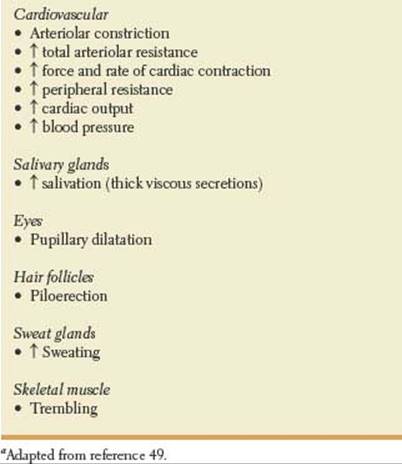
Some fibers pass through the splanchnic ganglia without synapsing, providing innervation to the adrenal medulla. Thus, the adrenal medulla is the only gland innervated by preganglionic (cholinergic) sympathetic nerve fibers, ending directly on modified neuronal cells that secrete adrenaline and noradrenaline into the blood stream. Table 21.2 illustrates the main effects of sympathetic stimulation [49].
TABLE 21.1 Basic Characteristics of Sympathetic and Parasympathetic Systes

Parasympathetic Nervous System
The parasympathetic autonomic nervous system has a cranial (brainstem) division and a sacral division. Parasympathetic preganglionic neurons of the cranial division are located within the nuclei of CN III (Eddinger-Westphal nucleus situated in the rostral midbrain at the level of the superior colliculus), CN VII (superior salivatory and lacrimal nuclei in the pontine tegmentum a), CN IX (inferior salivatory nucleus in the periventricular gray matter of the rostral medulla), and CN X (nucleus ambiguous in the reticular formation of the medulla, and dorsal motor nucleus of the vagus situated on the floor of the fourth ventricle where it forms the vagal trigone). The largest group of parasympathetic nerve fibers in the body is in the vagus nerves (CN X). Myelinated fibers of the cranial outflow synapse in peripheral ganglia located near or within the innervated organs: ciliary ganglion [CN III], submandibular and pterygopalatine ganglia [CN VII], otic ganglion [CN IX], and cardiac ganglion and plexuses [CN X]. Parasympathetic nerve fibers of CN III flow to the pupillary sphincters and ciliary muscles of the eyes. Parasympathetic fibers of CN VII flow to the lacrimal, nasal, and submandibular glands. Fibers from CN IX pass to the parotid gland. The vagus (CN X) supplies parasympathetic innervation to the heart, lungs, esophagus, stomach, small intestine, proximal half of the colon up to the splenic flexure, liver, gallbladder, pancreas and upper ureters [52,84,101].
TABLE 21.2 Main Effects of Diffuse Sympathetic Stimulationaa
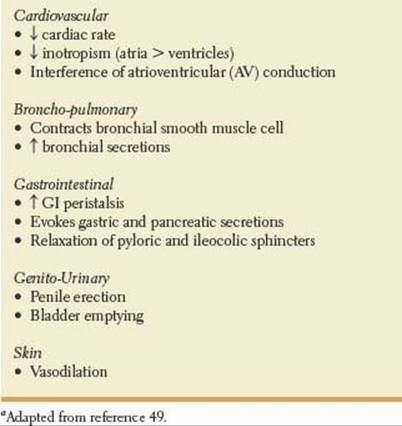
The sacral parasympathetic division resides in the intermediolateral cell columns of the second to fourth segments of the sacral (S2–S4) spinal cord. Myelinated axons exit through the ventral roots forming the sacral plexus, and then enter the pelvic splanchnic nerves also called nervi erigentes. Myelinated efferent fibers of the sacral outflow synapse in the intramural ganglion and hypogastric (pelvic) plexuses. The sacral parasympathetic outflow distributes its fibers to the descending colon, rectum, lower portion of the ureters, bladder, and external genitalia [52,84,101]. The genital organs are innervated by three sets of peripheral nerves, namely the sacral parasympathetic (pelvic nerves or nervi erigentes), the thoracolumbar sympathetic (hypogastric and lumbar sympathetic chain), and the sacral somatic nerves (pudendal nerves). The pelvic nerves contain the axons of sacral parasympathetic preganglionic neurons located in the intermediolateral cell column of S2–S4. The hypogastric nerves contain the axons of the sympathetic preganglionic neurons located in the intermediolateral cell column of T11–L2 spinal segments. The pudendal nerves contain the axons of somatic motor neurons located in the ventral horn of S2–S4. Parasympathetic fibers play a major role in penile erection and micturition, while parasympathetic efferents in the pelvic nerves promote bladder emptying (see Chapter 5). The main effects of vagal (cholinergic) stimulation are show in Table 21.3.
Enteric Nervous System
The ENS, derived primarily from the vagal neural crest cells, consists of an extrinsic and an intrinsic component. The intrinsic component (500 million neurons) consists of two interconnected plexuses, the Meissner’s submucosal plexus, and the Auerbach’s myenteric plexus, within the gastrointestinal wall. In humans, the submucosal plexus of Meissner consists of three interconnected layers. The outer layer is also known as the outer submucosal plexus of Schadabasch’s or Henle’s plexus[15]. The extrinsic innervation of the ENS depends on preganglionic parasympathetic and sympathetic outputs that regulate peristalsis and secretion. The parasympathetic output arises in the dorsal motor nucleus of the vagus and in the sacral parasympathetic nucleus of the spinal cord. The sympathetic output originates in the prevertebral ganglia. The primary targets of the ENS are mucosal secretory cells, gastrointestinal neuroendocrine cells, gastrointestinal microvasculature, and immunomodulatory and inflammatory cells in the gut. The ENS influences these effector cells in the gut directly or indirectly through its actions on intermediate cells including neuroendocrine cells, cells of the immune system, and the interstitial cells of Cajal. The interstitial cells of Cajal are a network of small anastomosing cells located within the gut muscle layers, and an integral part of normal gastrointestinal motility and modulation of enteric neural activity [4,17,21,22,39,42,56,68,70,71,80,88]. Disorders of the enteric nervous system may produce motor, secretor, inflammatory, or immune dysfunction.
TABLE 21.3 Main Effects of Cholinergic Stimulationa a
CENTRAL AUTONOMIC NETWORK
Tightly interconnected neuroanatomical structures contribute to regulation of the autonomic circuits.
Medial Prefrontal Cortex. The medial prefrontal cortex is highly activated by stress and plays a key role in autonomic and affective responses. The medial prefrontal cortex is implicated in complex cognitive and emotional states and modulation of neuroendocrine and autonomic function [7,54,102,103]. In addition, the anterior cingulate cortex is implicated in modulating sympathetic nervous system tone [30,75].
Insular Cortex. The insular cortex plays an important role in the CAN, and is the most important cortical area involved in cardiovascular regulation. Insular cortex neurons have connections with the limbic system and lateral frontal cortical system, and project to the central nucleus of the amygdala and autonomic nuclei of the medulla oblongata [95]. The insula also has wide connections with temporal and parietal cortices, basal ganglia, thalamus, and olfactory cortex. Moreover, the anterior insula has connections to primary and supplementary motor cortices, ventroposterior medial nucleus of the thalamus, and nucleus tractus solitarius, all of them relevant to oropharyngeal swallowing [31]. A possible lateralization in autonomic activity in the insular cortex has been proposed. Some evidence exists for cortical lateralization in the regulation of cardiovascular functions. Patients with right insular cortex lesions seem to be more susceptible to develop cardio-autonomic dysfunction, while the left insular cortex is predominantly responsible for parasympathetic cardiovascular effects [18,29,59,77,83,104–105]. Strokes restricted to the insular cortex have been associated with arterial hypertension, cardiac arrhythmias, increased risk of myocardial injury, raised catecholamines levels, and an increased susceptibility to sudden death [6,11,20,27,44,106].
Central Nucleus of the Amygdala. The amygdala has a key role in regulating arousal and vigilance and responds to the acquisition and expression of conditional fear. It is particularly activated by stimuli such as a fearful face [24,51,115,116]. Moreover, the central nucleus of the amygdala is involved in cardiovascular regulation [94].
Hypothalamus. Anatomically, the hypothalamus is the highest level of integration of autonomic function. The hypothalamus controls the autonomic nervous system by means of the pituitary gland and by direct descending pathways. Connections to the pituitary enable the hypothalamus to influence activities of the endocrine glands. Descending sympathetic fibers from the hypothalamus are uncrossed, and by way of the lateral tegmentum of the brainstem and lateral medullary formation influence the craniosacral parasympathetic and thoracolumbar sympathetic outflows. In particular, the paraventricular nucleus of the hypothalamus has emerged as one of the most important control centers in the brain involved in cardiovascular regulation. This nucleus receives afferents from the insula, prefrontal cortex, amygdala, and other hypothalamic nuclei [100]. The lateral hypothalamus, in addition to the insula and the amygdala also play a key role in the autonomic control of the heart.
Periaqueductal Gray Region. The periaqueductal gray is divided into four regions, namely the dorsomedial, dorsolateral, lateral, and ventrolateral subdivisions. Chemical stimulation of the dorsolateral gray produces hypertension and tachycardia, while activation of the ventrolateral gray accounts for the opposite effect. Different sets of neurons located in different regions of the periaqueductal gray have been implicated in coordinated patterns of somatic and autonomic changes characteristics of defensive responses [12,19,40,41,110]. A major function of the periaqueductal gray is pain processing and modulation, particularly in descending pain inhibition [12,19,40,41,110].
Parabrachial Nuclear Complex. The parabrachial/Kölliker-Fuse nuclear complex located in the dorsolateral pontine tegmentum is divided into three well-defined regions: the medial parabrachial nucleus (corresponding to the classic “pneumotaxic center”), the lateral parabrachial nucleus, and the Kölliker-Fuse nucleus. This nuclear complex plays a key role in the sensory processing of gustatory and visceral information, pain modulation, and automatic control of respiration [13,46,48,67,118]. Moreover, vestibular input projecting to the nucleus tractus solitarius, dorsal motor nucleus of the vagus, and the parabrachial/Kölliker-Fuse nuclear complex, may account for the autonomic effects observed in cases of acute vestibular dysfunction [9,10].
Nucleus Ambiguous. The nucleus ambiguous, located in the ventrolateral part of the medullary tegmentum, extends rostrally to the dorsal aspect of the facial nucleus, and caudally to the level of the pyramidal decussation, and with the nucleus tractus solitarii, and the dorsal motor nucleus of the vagus participate in the control of automatic respiration [46]. Involvement of the nucleus ambiguous in the rostral part of the medulla oblongata is more likely to cause dysphagia than lesions involving the nucleus ambiguous in the middle-lower part of the medulla oblongata [58,61].
Nucleus Tractus Solitarius. The nucleus tractus solitarius in the dorsal medulla contains the medullary respiratory and lower brainstem hypnogenic neurons. It receives afferents from the cardiovascular and respiratory systems for autonomic control of cardiac rhythm, circulation, and respiration, and is believed to be of major importance in cardiovascular and respiratory regulation. Bilateral lesions in the nucleus tractus solitarius cause acute neurogenic hypertension [34].
Localization Principles. Autonomic dysfunction may be localized or generalized, primary or secondary, and is usually associated with underactivity (Table 21.4). However, severe paroxysmal sympathetic overactivity, including neurogenic pulmonary edema, may result from acute brain or spinal cord injuries [8,98].
A few examples of localized dysautonomias include Adie’s tonic pupil and Horner’s syndrome described in Chapter 8. Crocodile tears or Bogorad syndrome (gustatolacrimal syndrome) is a rare complication of facial paralysis with incomplete recovery and characterized by excessive lacrimation during food consumption. Gustatory sweating (Frey’s syndrome) is a potential complication of parotid surgery [36,86]. Primary hyperhidrosis, a common and troublesome condition, primarily involves the axillae as well as the palms and soles, and is characterized by an excessive and uncontrolled production of sweat by the sweat glands [92]. Reduced tear secretion is observed in many patients with acute pandysautonomias, multisystem atrophy (Shy Drager syndrome) and Riley-Day syndrome (Chapter 8). Finally, vasovagal syncope in the young, and carotid sinus hypersensitivity in the elderly represent examples of intermittent localized dysautonomias.
TABLE 21.4 Cardinal Signs of Autonomic Dysfunctionaa
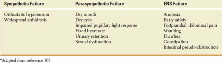
As various visceral functions are controlled by extrinsic pathways which involve neuronal circuits in the brain, hypothalamus, brainstem, spinal cord, peripheral ganglia, or peripheral nerves, the following considerations appear logical steps when attempting localization. First, is there a significant ANS dysfunction present (Table 21.5)? Second, does the dysautonomia involve the sympathetic, parasympathetic, or ENS? Third, is the involvement central, preganglionic, ganglionic, postganglionic, or localized to the neuroeffector [26]?
Symptoms of dysautonomia are variable, and include cardiovascular symptoms, respiratory abnormalities, gastrointestinal, urogenital, sudomotor, and thermoregulatory manifestations, pupillary abnormalities, and sleep disorders.
Autonomic nervous system dysfunction may manifest with symptoms of orthostatic intolerance, including orthostatic hypotension. Orthostatic hypotension is polyetiological. Several potential causes are non-neurogenic, including medications, hypovolemia, impaired venous return, and cardiac insufficiency. Neurologic disorders likely to cause orthostatic hypotension include multisystem atrophy (MSA), pure autonomic failure (PAF), diabetic neuropathy, autonomic neuropathies, baroreflex failure, and dopamine beta hydroxylase (DBH) deficiency [63,90]. Patients with autonomic failure secondary to DBH, a condition characterized by severe orthostatic hypotension, have normal parasympathetic and sympathetic cholinergic function but lack sympathetic noradrenergic function [25,99]. Rarely, orthostatic hypertension, thought to involve activation of the sympathetic nervous system, has been associated with diabetes mellitus and dysautonomias [43].
TABLE 21.5 Clinical Presentation of Autonomic Disordersaa

Autonomic dysfunction complicates a multiplicity of central nervous system and peripheral nervous system conditions. In many instances, the target organ is obvious and the clinical task is to uncover the pathway involved (e.g., pupillary abnormalities). Autonomic dysfunction is a prominent feature in acute spinal cord injury; acutely, its effects on cardiovascular regulation, respiratory system, and thermoregulation, may be life threatening [2,3,60,82]. Low blood pressure, bradycardia, inability to lose excess heat by sweating, atonic bladder, and paralytic ileus are prominent manifestations of acute spinal cord injury. Autonomic dysfunction is also a major feature of other neurological diseases such as Parkinson’s disease, pure autonomic failure, MSA, dementia with Lewy bodies, and Wernicke’s encephalopathy [13,61,79]. Autoimmune autonomic ganglionopathies are examples of severe but potentially treatable antibody-mediated forms of autonomic failure [62,76,107]. Autonomic dysfunction commonly occurs with multiple sclerosis [53,76]. Disturbances of autonomic function are more common among cluster headache sufferers; parasympathetic overactivity in the greater superficial petrosal nerve causes facial flushing and lacrimation in these patients [35]. In addition, hypothalamic activation also occurs in the trigeminal autonomic cephalgias [69]. Epileptic seizures may manifest autonomic dysfunction including convulsive apnea, abnormal sexual function, and life threatening cardiovascular effects, including sudden unexpected death in epilepsy (SUDEP) [89,97,113].
Most autonomic neuropathies occur in association with a somatic neuropathy, but they can occur in isolation. Autonomic neuropathies may be immune-mediated, infectious, nutritional, toxic, drug-induced, paraneoplastic, hereditary, or idiopathic, and may involve the parasympathetic, sympathetic, or enteric nervous system [38]. Sensory abnormalities predominate in hereditary sensory and autonomic neuropathies (HSAN) [109]. Widespread autonomic dysfunction is prominent in Guillain-Barré syndrome, acute intermittent porphyria, amyloidosis, and diabetes mellitus [82,111,112]. Acute porphyric neuropathy is predominantly motor; autonomic dysfunction is mainly due to vagal insufficiency [87]. Typical symptoms of acute porphyric neuropathy include abdominal pain, vomiting, constipation, tachycardia, hypertension, and bladder dysfunction [78].
Autonomic dysfunction is also the most prominent manifestation of small diameter lightly myelinated or unmyelinated fiber neuropathies, and sudomotor dysfunction is one of the earliest neurophysiologic abnormalities in these neuropathies [37,45,58,66]. Known causes of small fiber neuropathy include diabetes mellitus, amyloidosis, toxins, and inherited sensory and autonomic neuropathies. Neuropathic pain, acroparesthesias, hypohidrosis, or anhydrosis often associated with decreased saliva and tear production, are frequently observed signs in Fabry disease, a rare X-linked disorder caused by a deficiency of the lysosomal enzyme a-gal A (a-galactosidase) [28]. Involvement of the peripheral nervous system in Fabry disease affects mainly small A delta and C fibers [96].
Diabetic neuropathies involve the somatic and autonomic nerves. Symptoms of diabetic neuropathy are often vague. Gastrointestinal dysmotility is common in diabetes, and the role of the ENS has gained considerable importance in accounting for many of these manifestations [23]. Cardiovascular autonomic neuropathy is also one of the most clinically relevant complications of diabetes mellitus. Besides increasing morbidity and mortality, cardiovascular autonomic neuropathy may account for exercise intolerance, cardiac arrhythmias, heart rate variability, and an increased rate of silent myocardial ischemia [32,57]. Ventricular arrhythmias due to relative sympathetic overactivity with early autonomic neuropathy may account for sudden nocturnal death (“dead in bed”) in young people with Type I diabetes mellitus [114]. Rarely, an acute painful autonomic neuropathy may appear shortly after initiation of intensive glycemic control [47].
Autonomic symptoms have also been described in uremic neuropathy, alcoholic neuropathy, familial dysautonomia, HIV infection, leprosy, botulism, tetanus, and venoms of jellyfish and other marine animals [1,16,55].
Finally, many neurogenic and muscle conditions are associated with disorders of gut motility. Gastrointestinal manifestations are prominent manifestations of Parkinson’s disease, Down’s syndrome, neurofibromatosis type 1, Riley-Day syndrome, enteric neuropathies (e.g., Hirschsprung’s disease, celiac disease, intestinal neuronal dysplasia, mitochondriopathies), enteric myopathies (e.g., muscular dystrophies, myotonic dystrophy, familial visceral myopathies), and collagenopathies (e.g., desmosis, connective tissue disorders) [4,17,21,42,68,81,85,88,108]. A rare form of familial visceral neuropathy with neuronal intranuclear inclusions within the neurons of the ENS may also present with a variable phenotype including achalasia, gastroesophageal reflux, intestinal dysmotility and pseudo-obstruction, peripheral neuropathy, and pupillary abnormalities [91].
References
1. Agrawal A, Pandit L, Dalal M, et al. Neurological manifestations of Hansen’s disease and their management. Clin Neurol Neurosurg 2005;107(6):445–454.
2. Alexander MS. Autonomic function and spinal cord injury: are we at a crossroads? Spinal Cord 2008;46: 402–405.
3. Alexander MS, Biering-Sorensen F, Bodner D, et al. International standards to document remaining autonomic function after spinal cord injury. Spinal Cord 2009;47:36–43.
4. Altaf MA, Sood MR. The nervous system and gastrointestinal function. Dev Disabil Res Rev 2008;14: 87–95.
5. Apostolidis AN, Fowler CJ. Evaluation and treatment of autonomic disorders of the urogenital system. Semin Neurol 2003;23(4):443–452.
6. Ay H, Koroshetz WJ, Benner T, et al. Neuroanatomic correlates of stroke-related myocardial injury. Neurology 2006;66:1325–1329.
7. Bacon SJ, Smith AD. A monosynaptic pathway from an identified vasomotor centre in the medial prefrontal cortex to an autonomic area in the thoracic spinal cord. Neuroscience 1993:54(3):719–728.
8. Baguley IJ. Autonomic complications following central nervous system injury. Semin Neurol 2008;28(5): 716–725.
9. Balaban CD. Projections from the parabrachial nucleus to the vestibular nuclei: potential substrates for autonomic and limbic influences on vestibular responses. Brain Res 2004;996(1):126–137.
10. Balaban CD. Vestibular nucleus projections to the parabrachial nucleus in rabbits: implications for vestibular influences on the autonomic nervous system. Exp Brain Res 1996;108(3):367–381.
11. Baranchuk A, Nault MA, Morillo CA. The central nervous system and sudden cardiac death: what should we know? Cardiol J 2009;16(2):105–112.
12. Behbehani, MM. Functional characteristics of the midbrain periaqueductal gray. Prog Neurobiol 1995; 46:575–605.
13. Benarroch EE. Brainstem respiratory control: substrates of respiratory failure of multiple system atrophy. Mov Disord 2007;22(2):155–161.
14. Benarroch EE. The central autonomic network: functional organization, dysfunction, and perspective. Mayo Clin Proc 1993;68(10):988–1001.
15. Brehmer A, Rupprecht H, Neuhuber W. Two submucosal nerve plexus in human intestines. Histochem Cell Biol 2010;133(2):149–161.
16. Burnett JW, Weinrich D, Williamson JA, et al. Autonomic neurotoxicity of jellyfish and marine animal venoms. Clin Auton Res 1998;8(2):125–130.
17. Burns AJ. Disorders of interstitial cells of Cajal. J Pediatr Gastroenterol Nutr 2007;45(2):S103-S106.
18. Butcher KS, Cechetto DF. Autonomic responses of the insular cortex in hypertension and normotensive rats. Am J Physiol 1995;268(1 pt 2):R214–222.
19. Carrive P, Bandler R, Dampney RA. Somatic and autonomic integration in the midbrain of the unanesthetized decerebrate cat: a distinctive pattern evoked by excitation of neurons in the subtentorial portion of the midbrain periaqueductal grey. Brain Res 1989; 483(2):251–258.
20. Cereda C, Ghika J, Maeder P, et al. Strokes restricted to the insular cortex. Neurology 2002;59:1950–1955.
21. Cerosimo MG, Benarroch EE. Neural control of the gastrointestinal tract: implications for Parkinson’s disease. Mov Disord 2008;23(8):1065–1075.
22. Champaneria MC, Modlin IM, Kidd M, et al. Friedrich Feyrter: a precise intellect in a diffuse system. Neuroendocrinology 2006;83(5–6):394–404.
23. Chandrasekharan B, Srinivasan S. Diabetes and the enteric nervous system. Neurogastroenterol Motil 2007;19(12):951–960.
24. Cheng DT, Richards J, Helmstetter FJ. Activity in the human amygdala corresponds to early, rather than late period autonomic responses to a signal for shock. Learn Mem 2007;14(7):485–490.
25. Cheshire WP Jr, Dickson DW, Nahm KF, et al. Dopamine beta-hydroxylase deficiency involves the central autonomic network. Acta Neuropathol 2006; 112(2):227–229.
26. Cheshire WP Jr, Kuntz NL. Clinical evaluation of the patient with an autonomic disorder. In: Phillip AL, Eduardo EB, eds. Clinical autonomic disorders. 3rd ed. Wolter Kluwer/Lippincott Williams & Wilkins, 2008;Ch 10:112–129.
27. Cheshire WP Jr, Saper CB. The insular cortex and cardiac response to stroke. Neurology 2006;66:1296– 1297.
28. Clavelou P, Besson G. Neurological aspects of Fabry disease. Presse Med 2007;36 (Spec No 1):1s65– 1s68.
29. Colivicchi F, Bassi A, Santini M, et al. Cardiac autonomic derangement and arrhythmias in right-sided stroke with insular involvement. Stroke 2004;35:2094– 2098.
30. Critchley HD, Mathias CJ, Josephs O, et al. Human cingulated cortex and autonomic control: converging neuroimaging and clinical evidence. Brain 2003;126: 2139–2152.
31. Daniels SK, Foundas AL. The role of the insular cortex in dysphagia. Dysphagia 1997;12(3):146–156.
32. Debono M, Cachia E. The impact of cardiovascular autonomic neuropathy in diabetes: is it associated with left ventricular dysfunction? Auton Neurosci 2007; 132(1–2):1–7.
33. De Myer W. Neuroanatomy. Williams and Wilkins. Baltimore. Maryland, 1998.
34. Doba N, Reis DJ. Acute fulminant neurogenic hypertension produced by brainstem lesions in the rat. Circ Res 1973;32:584–593.
35. Drummond PD. Autonomic disturbances in cluster headache. Brain 1988;111:1199–1203.
36. Eckardt A, Kuettner C. Treatment of gustatory sweating (Frey’s syndrome) with botulinum toxin A. Head Neck 2003;25(8):624–628.
37. England JD, Gronseth GS, Franklin G, et al. Practice parameter: evaluation of distal symmetric polyneuropathy: role of autonomic testing, nerve biopsy, and skin biopsy (an evidence–based review). Report of the American Academy of Neurology, American Association of Neuromuscular and Electrodiagnostic Medicine, and American Academy of Physical Medicine and Rehabilitation. Neurology2009;72(2): 177–184.
38. Etienne M, Weimer LH. Immune-mediated autonomic neuropathies. Curr Neurol Neurosci Rep 2006; 6(1):57–64.
39. Fang S, Wu R, Christensen J. Intramucosal nerve cells in human small intestine. J Auton Nerv Syst 1993;44(2–3):129–136.
40. Farkas E, Jansen ASP, Loewy AD. Periaqueductal gray matter input to cardiac-related sympathetic premotor neurons. Brain Res 1998;792:179–192.
41. Farkas E, Jansen ASP, Loewy AD. Periaqueductal gray matter projection to vagal preganglionic neurons and the nucleus tractus solitarius. Brain Res 1997; 764(1–2):257–261.
42. Feichter S, Meier-Ruge WA, Bruder E. The histopathology of gastrointestinal motility disorders in children. Semin Pediatr Surg 2009;18:206–211.
43. Fessel J, Robertson D. Orthostatic hypertension: when pressor reflexes overcompensate. Nat Clin Pract Nephrol 2006;2(8):424–431.
44. Fink JN, Selim MH, Kumar S, et al. Insular cortex infarction in acute middle cerebral artery territory stroke. Arch Neurol 2005;62:1081–1085.
45. Freeman R. Autonomic peripheral neuropathy. Lancet 2005;365(9466):1259–1270.
46. Giangaspero F, Schiavina M, Sturani C, et al. Failure of automatic control of ventilation (Ondine’s curse) associated with viral encephalitis of the brainstem: a clinicopathologic study of one case. Clin Neuropathol1988;7(5):234–237.
47. Gibbons CH, Freeman R. Treatment-induced diabetic neuropathy: a reversible painful autonomic neuropathy. Ann Neurol 2010;67:534–541.
48. Gioia M, Rodella L, Petruccioli MG, et al. The cytoarchitecture of the adult human parabrachial nucleus: a Nissl and Golgi study. Arch Histol Cytol 2000;63(5):411–424.
49. Goldstein DS. The Autonomic Nervous System in Health and Disease. New York: Marcel Dekker, Inc., 2001.
50. Grigg-Damberger M, Wells A. Central congenital hypoventilation syndrome: changing face of a less mysterious but more complex genetic disorder. Semin Respir Crit Care Med. 2009;30(3):262–274.
51. Grossberg S, Bullock D, Dranias MR. Neural dynamics underlying impaired autonomic and conditioned responses following amygdala and orbitofrontal lesions. Behav Neurosci 2008:122(5):1100–1125.
52. Guyton AC. Basic neuroscience. Anatomy and physiology. 2nd ed. W.B. Philadelphia: Saunders Company, 1991.
53. Haensch C-A, Jörg J. Autonomic dysfunction in multiple sclerosis. J Neurol 2006;253(Suppl 1):1/3–1/9.
54. Hänsel A, von Känel R. The ventro-medial prefrontal cortex: a major link between the autonomic nervous system, regulation of emotion, and stress reactivity? Biopsychosoc Med 2008;2:21.
55. Hilz MJ, Dütsch M, Neundörfer B. Autonomic disorders in polyneuropathies. Med Klin (Munich) 1998;93(9):533–540.
56. Hoyle CHV, Burnstock G. Neuronal populations in the submucous plexus of the human colon. J Anat. 1989;166:7–22.
57. Ieda M, Kimura K, Kanazawa H, et al. Regulation of cardiac nerves: a new paradigm in the management of sudden cardiac death? Curr Med Chem 2008;15(17): 1731–1736.
58. Illigens BM, Gibbons, CH. Sweat testing to evaluate autonomic function. Clin Auton Res 2009;19(2): 79–87.
59. Kapp BS, Schwaber JS, Driscoll PA. The organization of insular cortex projections to the amygdaloid central nucleus and autonomic regulatory nuclei of the dorsal medulla. Brain Res 1985;360(1–2):355–360.
60. Karlsson A-K. Autonomic dysfunction in spinal cord injury: clinical presentation of symptoms and signs. Prog Brain Res 2006;152:1–8.
61. Kaufmann H, Biaggioni I. Autonomic failure in neurodegenerative disorders. Semin Neurol 2003;23(4) 351–363.
62. Kimpinski K, Iodice V, Sandroni P, et al. Sudomotor dysfunction in autoimmune autonomic ganglionopathy. Neurology 2009;73:1501–1506.
63. Klein CM. Evaluation and management of autonomic nervous system disorders. Semin Neurol 2008; 28(2):195–204.
64. Komisaruk BR, Mosier KM, Liu W-C, et al. Functional localization of brainstem and cervical spinal cord nuclei in humans with fMRI. AJNR 2002;23: 609–617.
65. Kurono H, Uesaka Y, Kunimoto M, et al. The correlation between dysphagia and involvement of the ambiguous nucleus on MRI in acute-phase lateral medullary syndrome [in Japanese]. Rinsho Shinkeigaku 2006;46(7):461–466.
66. Lacomis D. Small-fiber neuropathy. Muscle Nerve 2002;26(2):173–188.
67. Lavezzi AM, Ottaviani G, Ballabio G, et al. Preliminary study on the cytoarchitecture of the human parabrachial/Kölliker-fuse complex, with reference to sudden infant death syndrome and sudden intrauterine unexplained death. Pediatr Dev Pathol 2004; 7(2):171–179.
68. Lechin F, van der Dijs B. Central nervous system plus autonomic nervous system disorders responsible for gastrointestinal and pancreatobiliary diseases. Dig Dis Sci 2009;54:458–470.
69. Leone M, Bussone G. Pathophysiology of trigeminal autonomic cephalalgias. Lancet Neurol 2009;8:755– 764.
70. Linden DR, Farrugia G. Autonomic control of gastrointestinal function. In: Phillip AL, Eduardo EB, eds. Clinical autonomic disorders. 3rd ed. Philadelphia: Wolter Kluwer/Lippincott Williams & Wilkins, Ch 8, 2008:88–105.
71. Mannl A, Pospischil A, Dahme E. The plexus submucosus (Meissner and Schabadasch) in the pig intestine. I. Light and electron microscopic studies of the normal structure. Zentralbl Veterinarmed A1986; 33(9):647–659.
72. Mathias CJ. Autonomic neuropathy-aspects of diagnosis and management. In: Asbury AD, Thomas PK, eds. Peripheral nerve disorders. II. Oxford: Butterworth-Heinemann, 1995:95–117.
73. Mathias CJ. Autonomic nervous system disorders and erectile dysfunction. Int J STD AIDS 1996;7(3): 5–8.
74. Mathias CJ. Disorders of the autonomic nervous system. In: Bradley WG, Daroff RB, Fenichel GM, Marsden CD, eds. Neurology in clinical practice. 2nd ed. Boston, MA: Butterworth-Heinemann, 1996:82: 1953–1981.
75. Matthews SC, Paulus MP, Simmons AN, et al. Functional subdivisions within anterior cingulate cortex and their relationship to autonomic nervous system function. NeuroImage 2004;22:1151–1156.
76. Merkelbach S, Haensch C-A, Hemmer B, et al. Multiple sclerosis and the autonomic nervous system. J Neurol 2006;253(Suppl 1):1/21–1/25.
77. Meyer S, Strittmatter M, Fischer C, et al. Lateralization in autonomic dysfunction in ischemic stroke involving the insular cortex. Neuroreport 2004;15(2): 357–361.
78. Meyer UA, Schuurmans MM, Lindberg RL. Acute porphyrias: pathogenesis of neurological manifestations. Semin Liver Dis 1998;18(1):43–52.
79. Miceli G, Tosi P, Marcheselli S, et al. Autonomic dysfunction in Parkinson’s disease. Neurol Sci 2003; 24:S32–S34.
80. Modlin IM, Champaneria MC, Bornschein J, et al. Evolution of the diffuse neuroendocrine system—clear cells and cloudy origins. Neuroendocrinology 2006;84(2):69–82.
81. Moore SW. Down syndrome and the enteric nervous system. Pediatr Surg Int 2008;24:873–883.
82. Niakan E, Yadollah H, Comstock JP. Diabetic autonomic neuropathy. Metabolism 1986;35(3):224–234.
83. Oppenheimer SM, Kedem G, Martin WM. Left-insular cortex lesions perturb cardiac autonomic tone in humans. Clin Auton Res 1996;6(3):131–140.
84. Patestas MA, Gartner LP. Autonomic nervous system. In: A textbook of neuroanatomy. Malden, MA: Blackwell Publishing, 2006;Ch 9:118–133.
85. Perkin GD, Murray-Lyon I. Neurology and the gastrointestinal system. J Neurol Neurosurg Psychiatry 1998;65(3):291–300.
86. Pino Rivero V, González Palomino A, Trinidad Ramos G, et al. Bogorad syndrome or crocodile tears alter Ramsay-Hunt. An Otorrinolaringol Ibero Am 2006;33(3):225–229.
87. Pischik E, Kauppinen R. Neurological manifestations of acute intermittent porphyria. Cell Mol Biol 2009;55(1):72–83.
88. Quigley EMM. What we have learned about colonic motility: normal and disturbed. Curr Opin Gastroenterol 2010;26:53–60.
89. Rees PM, Fowler CJ, Maas CP. Sexual function in men and women with neurological disorders. Lancet 2007;369:512–525.
90. Robertson D. The pathophysiology and diagnosis of orthostatic hypotension. Clin Auton Res 2008;18(1): 2–7.
91. Roper EC, Gibson A, McAlindon ME, et al. Familial visceral neuropathy: a defined entity? Am J Med Genet A 2005;137(3):249–254.
92. Rusciani L, Severino E, Rusciani A. Type A botulinum toxin: a new treatment for axillary and palmar hyperhidrosis. J Drugs Dermatol 2002;1(2):147–151.
93. Sakakibara R, Yamaguchi C, Tomoyuki U, et al. Pelvic autonomic dysfunction without paraplegia: a sequel of spinal cord stroke. Eur Neurol 2008;60: 97–100.
94. Salomé N, Viltart O, Leman S, et al. Activation of ventrolateral medullary neurons projecting to spinal autonomic areas after chemical stimulation of the central nucleus of amygdala: a neuroanatomical study in the rat. Brain Res 2001;890(2):287–295.
95. Saper CB. Convergence of autonomic and limbic connections in the insular cortex of the rat. J Comp Neurol 1982;210(2):163–173.
96. Schiffman CB, Murray GJ, Treco D, et al. Infusion of alpha-galactosidase a reduces tissue, globotriaosylceramide storage in patients with Fabry disease. Proc Natl Acad Sci USA. 2000;97:365–370.
97. Scorza FA, Arida RM, Cysneiros RM, et al. The brain-heart connection: implications for understanding sudden unexpected death in epilepsy. Cardiol J 2009;16(5):394–399.
98. ˇSed´y J, Zicha J, Kunesˇ J, et al. Mechanisms of neurogenic pulmonary edema development. Physiol Res 2008;57:499–506.
99. Senard JM, Rouet P. Dopamine beta-hydroxylase deficiency. Orphanet J Rare Dis 2006;30(1):7.
100. Snell RS. The hypothalamus and its connections. In: Clinical neuroanatomy. 7th ed. Wolters Kluwer/Lippincott Williams & Wilkins, 2010;Ch 13:382–395.
101. Snell RS. The autonomic nervous system. In: Clinical neuroanatomy. 7th ed. Wolters Kluwer/Lippincott Williams & Wilkins, 2010;Ch 14:396–426.
102. Sullivan RM, Gratton A. Lateralized effects of medial prefrontal cortex lesions on neuroendocrine and autonomic stress responses in rats. J Neurosci 1999;19(7): 2834–2840.
103. Teves D, Videen TO, Cryer PE, et al. Activation of human medial prefrontal cortex during autonomic responses to hypoglycemia. Proc Natl Acad Sci USA 2004;101(16):6217–6221.
104. Thayer JF. Vagal tone and the inflammatory reflex. Clev Clin J of Med 2009;76(2):523–526.
105. Tokgözoglu SL, Batur, MK, Topçuoglu MA, et al. Effects of stroke localization on cardiac autonomic balance and sudden death. Stroke 1999;30:1307–1311.
106. Varnavas G, Corand W. The insular cortex: morphological and vascular anatomic characteristics. Neurosurgery 1999;44(1):127–136.
107. Vernino S. Antibody testing as a diagnostic tool in autonomic disorders. Clin Auton Res 2009;19(1):13–19.
108. Vernino S, Sandroni P, Singer W, et al. Autonomic ganglia. Target and novel therapeutic tool. Neurology 2008;70:1926–0932.
109. Verpoorten N, De Jonghe P, Timmerman V. Disease mechanisms in hereditary sensory and autonomic neuropathies. Neurobiol Dis 2006;21(2):247–255.
110. Vianna DML, Brandão ML. Anatomical connections of the periaqueductal gray: specific neural substrates for different kinds of fear. Braz J Med Biol Res 2003; 36:557–566.
111. Vinik AI, Maser RE, Mitchell BD, et al. Diabetic autonomic neuropathy. Diabetes Care 2003;26(5): 1553–1579.
112. Wang AK, Fealey RD, Gehrking TL, et al. Patterns of neuropathy and autonomic failure in patients with amyloidosis. Mayo Clin Proc 2008;83(11):1226– 1230.
113. Wannamaker BB. Autonomic nervous system and epilepsy. Epilepsia 1985;Suppl 1:s31–s39.
114. Weston PJ, Gill GV. Is undetected autonomic dysfunction responsible for sudden death in Type 1 diabetes mellitus? The ‘dead in bed’ syndrome revisited. Diabet Med 1999;16(8):626–631.
115. Williams LM, Barton MJ, Kem AH, et al. Distinct amygdala-autonomic arousal profiles in response to fear signals in healthy males and females. Neuroimage 2005;28(3):618–626.
116. Williams LM, Brown KJ, Das P, et al. The dynamics of cortico-amygdala and autonomic activity over the experimental time course of fear perception. Brain Res Cogn Brain Res 2004;21(1):114–123.
117. Wilson-Pauwels L, Stewart PA, Akesson EJ. Autonomic nerves. Basic science. Clinical aspects. Case studies. BC Decker Inc. Hamilton. London, 1997.
118. Young RF, Tronnier V, Rinaldi PC. Chronic stimulation of the Kölliker-Fuse nucleus region for relief of intractable pain in humans. J Neurosurg 1992;76: 979–985.