
Most causes of coma speedily threaten life or recovery of neurological function. Thus, they must be promptly identified and treated. Unfortunately, patients with a depressed level of alertness cannot give an account of the events leading to their situation, and often no one who has observed the patient before admission is available to provide such information. Thus, the physician has to rely on examination of the patient, not only to localize the damaged anatomic structures but also to identify the offending agent. The examination should be thoughtful and well informed but not necessarily long. A delay in protecting the airway of a poorly responsive patient may cause irreparable neurological damage [123].
The diagnosis of impaired alertness and coma is well reviewed in some excellent monographs [118,189]. The following pages draw heavily from these sources. Unlike in previous editions of this book, the diagnosis of death based on neurological criteria (brain death) is discussed at the end of this chapter. Despite the availability of neurophysiological and neuroimaging tools to help make this diagnosis, the neurological examination, object of this chapter, remains critical [178].
The Unresponsive Patient
Terms such as coma, stupor, lethargy, and the like indicate a depressed level of alertness. These terms, however, fail to convey vital information needed for neurologic localization and management. Rather than using one of these terms, a description of the patient’s level of responsiveness (incorporating some detail of the patient’s responses to diverse reproducible stimuli) facilitates communication among members of the health care team, enhances consistency in successive evaluations of the patient, and sets the basis for a rational diagnostic assessment. For instance, stating that “the patient was stuporous” provides little information. Instead, the real situation can be much better conveyed by explaining in everyday English that “Mr. Z lay motionless in bed unless called loudly by name, when he opened his eyes briefly and looked to the left. He failed to answer any questions or to follow instructions.”
Two terms have gained acceptance among neurologists and are widely used: akinetic mutism and locked-in syndrome. Akinetic mutism [141] refers to a state in which the patient, although seemingly awake remains silent and motionless. Only the eyes dart in the direction of moving objects, such as the examiner approaching the patient’s bed. The examiner, attempting to converse with a patient in this state, gets the distinct impression of failing to draw the patient’s attention and interest. Despite the lack of movement, there are few signs indicative of damage to the descending motor pathways. Instead, “frontal release signs,” such as grasp or sucking, may be present. Patients who remain completely motionless are not seen as often as those who move one side or one arm in a stereotyped fashion but in every other respect fit into the syndrome of akinetic mutism. In such cases, the paralyzed side may display signs of corticospinal tract involvement, such as hyperreflexia and a Babinski sign.
If a history is available, akinetic mutism can usually be distinguished from psychogenic (often catatonic) unresponsiveness. Otherwise, the diagnosis may be difficult. Particularly when exposed to painful stimuli (such as those caused by soiled linen or a decubitus ulcer) or to infection, patients with akinetic mutism appear excited and tachycardic and perspire heavily, thus superficially resembling a catatonic patient. Signs of frontal release or corticospinal tract damage favor the diagnosis of akinetic mutism. In the catatonic patient, the electroencephalogram (EEG) is normal (often desynchronized, with low-voltage fast activity), but in the patient with akinetic mutism, the EEG may show slow-wave abnormalities [102].
Lesions that cause akinetic mutism affect bilaterally the frontal region (anterior cingulate gyri), the diencephalo-mesencephalic reticular formation, the globus pallidus, or the hypothalamus [31,92,101]. Common causes are anoxia, head trauma, cerebral infarction, severe acute hydrocephalus, and direct compression by tumors [95,172]. Other extensive lesions, such as air embolism or end-stage degenerative or infectious brain disorders, such as Creutzfeldt–Jakob disease, can also cause this syndrome [3,51,109,150,175]. Metabolic or ictal disorders disrupting the same areas may give rise to a transient disorder of alertness similar to akinetic mutism [7,143]. A syndrome of transient mutism and relative akinesia may occur a few days after midline cerebellar or fourth ventricular surgery [18,21,36]. One such patient improved after dopaminergic stimulation, suggesting involvement of the dopaminergic pathways, at least in some cases [18]. Hyperkinetic mutism, with continuous bilateral ballism and dystonia, has been described in a diabetic with multiple subcortical and cortical infarcts [53].
When the cerebral hemispheres have sustained severe and widespread damage (such as that due to severe trauma, anoxia, or encephalitis), the patient may, after some weeks of complete unresponsiveness, evolve into a situation similar to akinetic mutism, with the return of sleep–wake cycles. These patients, however, demonstrate obvious signs of pronounced bilateral corticospinal tract damage. This situation, in which the patient’s functions are restricted to the autonomic sphere, has been termed the vegetative state [4]. The vegetative state has been defined as a chronic neurologic condition characterized by lack of awareness of self and external stimuli, accompanied by sleep–wake cycles, with preservation of vital vegetative functions, such as cardiac function, respiration, and maintenance of blood pressure. Patients in a vegetative state show no evidence of sustained, reproducible, purposeful, or voluntary behavioral responses to visual, auditory, tactile, or noxious stimuli; show no evidence of language comprehension or expression; have bowel and bladder incontinence; and have variably preserved cranial nerve and spinal reflexes. Persistent vegetative state is defined as a vegetative state present one month after acute traumatic or nontraumatic brain injury or lasting for at least one month in patients with degenerative or metabolic disorders or developmental malformations [4].
In addition to its pejorative connotation, the term persistent vegetative state assumes that it is possible from the physical examination combined with available clinical information to define accurately the patient’s state of awareness. Given that motor responses on the part of the patient are needed to evaluate the presence of awareness of self and external stimuli, in many of these patients with severe damage to motor mechanisms it is very difficult to assess the degree of awareness. The presence of extensive damage on MRI or CT is helpful but does not define this issue either [181]. In some patients meeting diagnostic criteria for the chronic vegetative state, functional neuroimaging has documented awareness and the ability to follow commands [97]. Given the shortcomings of the term persistent vegetative state, it is better to refer to this state as persistent unresponsiveness. Responses are all that the examiner can evaluate. Persistent unresponsiveness is a descriptive term reflecting accurately the information on the patient, and avoids the pitfall of making inaccurate assumptions as to the degree of awareness or the level of brain function in a given patient.
Recovery of consciousness from a posttraumatic persistent unresponsive state is unlikely after 12 months in adults and children [91]. Recovery from a nontraumatic persistent unresponsive state after three months is rare in both adults and children, but it can happen, usually with residual severe disability [9,37].
Even more vague and inaccurate than the term “vegetative state” is the more recent “minimally conscious state.” If a short description is to be used, “minimally responsive” seems much more appropriate [10]. Recovery from the minimally responsive state has been documented even after 19 years from onset [174].
The locked-in syndrome refers to a condition in which the patient is mute and motionless (deefferented) but remains awake, alert, aware of self, and capable of perceiving sensory stimuli. Although horizontal eye movements are often impaired due to involvement of the paramedian pontine reticular formation, the patient’s level of alertness can be gleaned from her response to commands involving vertical eye movements or eyelid movements. The EEG reflects the patient’s state of wakefulness. The locked-in syndrome is usually due to basilar artery thrombosis with ventral pontine infarction, pontine hemorrhage or tumor, or central pontine myelinolysis (osmotic demyelination syndrome) [50,96]. These lesions involve the descending motor pathways bilaterally in the basis pontis but spare the more dorsal reticular formation). Bilateral ventral midbrain lesions [23,41,94], tentorial herniation [68,183], Guillain–Barré syndrome [8,124], or myasthenia gravis may rarely cause this syndrome. In locked-in syndrome due to mesencephalic lesions, bilateral ptosis and vertical (as well as horizontal) ophthalmoplegia are present. Fou rire prodromique (pathologic laughter at the onset of a stroke) may rarely herald the onset of a bilateral ventral pontine stroke leading to a locked-in syndrome [176].
Anatomic Substrate of Alertness
In general, the maintenance of consciousness depends on interaction between the ascending reticular activating system (ARAS) and the cerebral hemispheres. Damage to the ARAS, described in animals by Moruzzi and Magoun in 1949 [99], induces a state of coma in which the animal becomes unresponsive and its EEG shows sleep patterns despite vigorous sensory stimulation. In humans, the ARAS lies in the paramedian tegmental region of the posterior portion of pons and midbrain [110]. It is a complex polysynaptic fiber system that extends from the superior half of the pons through the midbrain to the posterior portion of the hypothalamus and to the thalamic reticular formation (Fig. 23.1). The thalamus is the source of diffuse thalamocortical projections that regulate and coordinate cortical activity [58,84]. Sedative drugs act, at least in part, by interfering with the synaptic network of the ARAS, which is played on by sensory stimuli.

FIG. 23.1. Ascending reticular activating system (ARAS). The dotted area in this midsagittal section of the brain corresponds to the approximate location of the ARAS in the upper brainstem and the diencephalon.
The medial longitudinal fasciculi, which connect the abducens and oculomotor nuclei, and the oculomotor and trochlear nuclei themselves are situated amid the neurons of the pontine and midbrain portions of the ARAS. Thus, when unresponsiveness is caused by brainstem damage, the lesion affects the mechanisms of ocular motility as well, and its location can often be determined by abnormal patterns of ocular motility.
Bilateral cerebral hemispheric lesions may cause transient coma, particularly when they involve the mesial frontal region. Large unilateral lesions of the dominant hemisphere may occasionally cause transient unresponsiveness, even in the absence of a mass effect [1].
In the diencephalon, posterior hypothalamic lesions induce prolonged hypersomnia. Acute bilateral damage of the paraventricular thalamic nuclei is attended by transient unresponsiveness, followed, when the lesions are large, by severe amnestic dementia (see Chap. 18).
Signs with Localizing Value in Coma
In a comatose patient, the respiratory pattern, pupillary response, eye movements, and position or movements of the limbs provide important clues to the anatomic site and nature of the injury.
Respiratory Patterns
Although the respiratory pattern of a patient in coma may be helpful in localizing the level of structural dysfunction in the neuraxis [156], metabolic abnormalities may affect the respiratory centers of the pons (pneumotaxic and apneustic) and medulla (expiratory and inspiratory) and result in patterns resembling those due to neurologic disease (Fig. 23.2). Thus, caution and a thorough evaluation of the metabolic status of the patient must guide the interpretation of respiratory changes.
POSTHYPERVENTILATION APNEA
This condition reflects mild bilateral hemispheric dysfunction. Because demonstration of this respiratory abnormality requires the patient’s active cooperation, this sign is mentioned here mainly to clarify the genesis of other respiratory patterns. To elicit this phenomenon, the patient is simply asked to take five deep breaths. This maneuver normally decreases arterial pCO2 by about 10 mm Hg and, in the healthy patient, is followed by a very brief period of apnea (<10 seconds). The stimulus for rhythmic breathing when the pCO2 is lowered probably originates in forebrain structures, because sleep, obtundation, or bilateral hemispheric dysfunction abolishes it. Thus, when bilateral hemispheric lesions are present, the posthyperventilation apnea lasts for as long as 20 or 30 seconds.

FIG. 23.2. Respiratory patterns characteristic of lesions at different levels of the brain.
CHEYNE–STOKES RESPIRATION
This type of respiration consists of brief periods of hyperpnea alternating regularly with even shorter periods of apnea. After the apneic phase, the amplitude of respiratory movements increases gradually to a peak and then slowly wanes to apnea. During the hyperpneic stage, the patient becomes more alert, the pupils may dilate toward normal from the miosis characteristic of diencephalic dysfunction, and the motor behavior reflects control by higher centers (e.g., decorticate posturing yields to semipurposeful movements). The eyelids may open during the rapid breathing phase and close during the slow breathing phase.
Cheyne–Stokes respiration represents a more severe degree of posthyperventilation apnea in which the respiratory drive becomes more closely dependent on the pCO2. Because the “smoothing effect” provided by forebrain structures has been removed, pCO2 accumulation causes hyperpnea, which in turn induces a drop in pCO2. With this drop, the respiratory stimulus ceases, and a period of apnea ensues.
This respiratory pattern may follow bilateral widespread cortical lesions but is more likely to be associated with bilateral thalamic dysfunction and has also been described with lesions of the descending pathways anywhere from the cerebral hemispheres to the level of the upper pons [106]. Metabolic disturbances, such as uremia, diffuse anoxia, and heart failure, often underlie this breathing disorder. This pattern of respiration may also be seen in some elderly individuals during sleep and in some normal individuals at high altitudes. Cheyne–Stokes respiration in patients with supratentorial mass lesions may indicate incipient transtentorial herniation [118].
HYPERVENTILATION WITH BRAINSTEM INJURY
Patients with lesions of the midbrain and pons often have prolonged and rapid hyperpnea. Because most of these patients are relatively hypoxic despite the excessive ventilatory effort, this type of breathing cannot truly be called neurogenic hyperventilation. In a few cases where pulmonary or metabolic causes of hyperventilation were absent, brainstem tumors were found at autopsy. In these cases, tumoral metabolism may have lowered the pH of the local cerebrospinal fluid, thereby providing a stimulus to the respiratory center of the medulla [117]. Central neurogenic hyperventilation, responsive to morphine and methadone, occurred with an astrocytoma centered in the medial tegmental parapontine reticular formation [56].
APNEUSTIC BREATHING
Apneustic breathing is characterized by a long inspiratory pause, after which the air is retained for several seconds and then released. This abnormality appears with lesions of the lateral tegmentum of the lower half of the pons.
CLUSTER BREATHING
Breathing with a cluster of breaths following each other in an irregular sequence may result from low pontine or high medullary lesions.
ATAXIC BREATHING
This type of breathing has a completely irregular pattern (also called the atrial fibrillation of respiration) in which inspiratory gasps of diverse amplitude and length are intermingled with periods of apnea. This respiratory abnormality, often present in agonal patients, heralds complete respiratory failure and follows damage of the dorsomedial medulla. The most common etiologies for this pattern include cerebellar or pontine hemorrhages, trauma, and posterior fossa tumors. Less often, a paramedian medullary infarct (usually due to severe atherosclerosis of a vertebral artery) may cause this syndrome. The classic breathing pattern described by Biot was ataxic breathing in patients with severe meningitis [118].
“ONDINE CURSE”
Pathways from the cerebral cortex subserving voluntary respiration are separate from those descending from the medulla subserving automatic respiration; thus, selective impairment of automatic or voluntary breathing is possible [156]. Descending pathways that are under voluntary control travel within the dorsal cord in the region of the corticospinal tract, whereas pathways from primary medullary respiratory centers travel in the ventrolateral cord, with anatomic separation of inspiratory and expiratory pathways [156].
Ondine curse refers to the loss of automatic breathing during sleep. This respiratory pattern, obviously absent in comatose patients, is mentioned here because it occurs with lower brainstem dysfunction. Responsible lesions have a similar or somewhat lower location than those that cause ataxic breathing but are smaller or develop more slowly. Both unilateral [5,82] and bilateral [30] medullary tegmental infarcts have produced this syndrome. This disorder has also been recorded with high cervical cord lesions after surgical section of the ventrolateral spinal cord for pain relief [75,170], probably because of reticulospinal tract interruption. Of 12 patients who died with presumed Ondine curse after high cervical percutaneous cordotomy for pain, all had lesions involving the region of the anterolateral funiculus in the C2 segment containing pain fibers activated from the second to fifth thoracic dermatomes [75].
Central hypoventilation may be caused by unilateral caudal brainstem infarction [12]. One patient with nearly complete loss of ventilation involving both automatic and voluntary components had an infarct involving the reticular formation, nucleus tractus solitarius, nucleus ambiguus, and nucleus retroambiguus on the right, which spared the dorsal motor nucleus of the vagus nerve and sensory and corticospinal tracts. A second patient with hypoventilation more selectively involving automatic responses (Ondine curse) had an infarct involving the medullary reticular formation and nucleus ambiguus that spared the nucleus tractus solitarius. These cases suggest that unilateral involvement of the pontomedullary reticular formation and nucleus ambiguus is sufficient for generating loss of automatic respiration, whereas an associated lesion of the nucleus tractus solitarius may lead to more severe respiratory failure involving automatic and voluntary responses [12]. Central hypoventilation or apnea has also been reported with bilateral damage to the high cervical spinal cord or with dorsolateral lesions of the tegmentum of the medulla. A selective paresis of voluntary but not automatic respiration has been described with a discrete infarction of the ventral basis pontis, further suggesting that automatic and voluntary respirations are controlled by anatomically independent pathways [100].
In patients who have lost all respiratory reflexes and are intubated, the self-cycling of the ventilator may erroneously suggest that the patient is triggering it [182].
Temperature Changes
Hyperthermia is not uncommon in coma caused by severe traumatic brain injury [165]. Patients in coma are predisposed to infection, but in a proportion of patients, hyperthermia may be neurogenic, that is, related to an altered temperature regulation system. In many of these instances, they correspond to hypothalamic dysfunction (see Chapter 17). Neurogenic hyperthermia has also been described with pontine tegmental lesions [110].
The Pupils
Pupillary shape, size, symmetry, and response to light provide valuable clues to brainstem and third cranial nerve function. The pupillary light reflex is very resistant to metabolic dysfunction. Abnormalities of this reflex, particularly when unilateral, indicate structural lesions of the midbrain or oculomotor nerve. A few exceptions are noteworthy. Atropinic agents, instilled into the eyes, applied on the skin (e.g., transdermal scopolamine) [24], ingested, or given during cardiopulmonary resuscitation, may cause pharmacologic iridoplegia. In these cases, a solution of 1% pilocarpine applied to the eye will fail to constrict the pupils, whereas in the case of anoxic pupillary dilation, this cholinergic agent, acting directly on the constrictor of the iris, produces miosis. Because many patients in coma have small pupils, anticholinergic agents are sometimes used to facilitate visualization of the optic fundi, thus eliminating a potentially useful diagnostic indicator. In many cases, a better way to obtain pupillary dilation is by pinching the skin of the neck (ciliospinal reflex). Glutethimide (Doriden) induces unequal pupils that are midsized or slightly dilated and poorly responsive to light. Other agents that may cause unreactive pupils include barbiturates (the pupillary light reflex is more often retained), succinylcholine, and, rarely, other anticonvulsants, lidocaine, phenothiazines, methanol, and aminoglycoside antibiotics [25,42]. Agents other than glutethimide or anticholinergic drugs cause pupillary dilation only when taken in massive amounts, enough to eliminate respiratory reflexes or, in the cases of succinylcholine and aminoglycoside antibiotics, generalized neuromuscular junction blockade. Usually, the amount of sedative drug is insufficient to abolish the pupillary light reflex. Hypothermia and acute anoxia may also cause unreactive pupils, which, if persistent beyond several minutes after an anoxic insult, carry a poor prognosis [38].
The areas of the brain and anatomic pathways that mediate the pupillary light reflex are reviewed in Chapter 8, in which the origin and course of sympathetic and parasympathetic influences on the iris muscle are described.
Various structural lesions causing coma may be associated with pupillary abnormalities (Fig. 23.3):
1. Sleep or bilateral diencephalic dysfunction (metabolic coma) is accompanied by small pupils that react well to light (“diencephalic” pupils).
2. Unilateral hypothalamic damage induces miosis and anhidrosis on the side of the body ipsilateral to the lesion.
3. Midbrain lesions causing coma usually produce distinct pupillary abnormalities. Tectal or pretectal lesions affecting the posterior commissure abolish the light reflex, but the pupils, which are midsized or slightly large, may show spontaneous oscillations in size (hippus) and become larger when the neck is pinched (ciliospinal reflex). Tegmental lesions, which involve the third nerve nucleus, may cause irregular constriction of the sphincter of the iris, with a resultant pear-shaped pupil or displacement of the pupil to one side (midbrain corectopia) [151]. The pupils, often unequal, tend to be midsized and lack light or ciliospinal responses. Unilateral or bilateral oval pupils (which may be fixed to light) may occur with severe cerebrovascular lesions that injure the oculomotor or pupillomotor fibers. The oval shape is due to nonuniform paresis or paralysis of the pupil sphincter, with resultant eccentric antagonistic effects of pupil dilators.
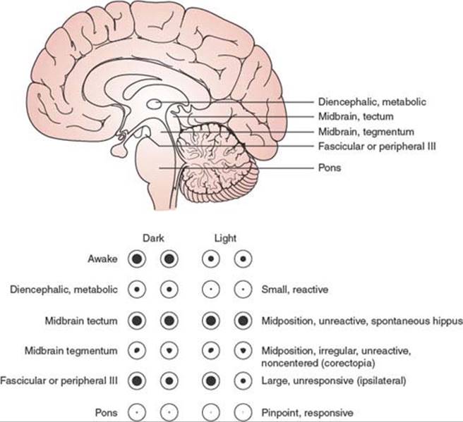
FIG. 23.3. Pupillary responses characteristic of lesions at different levels of the brain.
4. Pontine tegmental lesions cause small pupils due to interruption of descending sympathetic pathways. Pinpoint pupils, when observed with a magnifying glass, may be seen to constrict to light, may occur with pontine hemorrhage, and are due to a combination of sympathetic damage and parasympathetic irritation.
5. Lateral pontine, lateral medullary, and ventrolateral cervical cord lesions produce an ipsilateral Horner syndrome.
6. Oculomotor nerve compression and elongation by herniation of the uncus of the temporal lobe (through the tentorial incisura) affect pupillary function earlier and more noticeably than the extrinsic eye movements subserved by this cranial nerve. Possible explanations for pupillary dilation on the side of a mass lesion include compression of the third cranial nerve by uncal herniation beneath the tentorial edge; compression of the nerve by the posterior cerebral artery or by the hippocampal gyrus; stretching or buckling of the nerve by traction at the superior orbital fissure, posterior clinoid, or clivus; or compression of the midbrain oculomotor complex [137]. The light reflex is sluggish or absent, and, unlike the situation with midbrain involvement, the pupil becomes widely dilated owing to sparing of the sympathetic pathways (Hutchinson pupil).
Ropper studied the pupil opposite the one already enlarged from transtentorial herniation in 13 patients [135]. In most patients, the pupil was initially 2.5 to 4 mm in diameter with a diminished or absent light reaction; this initial phase was followed by a slight reduction in pupil size, then reenlargement to greater than the original pupil size, all with preserved roundness. Subsequent deterioration varied, but a transitional oval pupil shape was infrequent, and oculomotor function was otherwise preserved until both pupils were enlarged and fixed to light. Thus, subsequent neurologic deterioration in a patient with transtentorial herniation can often be appreciated by change in the reactivity and size of the opposite pupil [135]. Other reports have demonstrated a paradoxical initial enlargement of the pupil opposite the side of a mass lesion, especially with acute subdural hematoma [115] or intraparenchymal [22] or subarachnoid [88] hemorrhage.
7. Other oculomotor nerve lesions causing pupillary abnormalities are less likely to impair consciousness, except when associated with a subarachnoid hemorrhage. Posterior communicating artery aneurysms can compress the third nerve and a massive subarachnoid hemorrhage may result in coma. Rarely, with the Guillain–Barré syndrome patients may become completely paralyzed and lose even their pupillary response [8,124]. This complete locked-in state may be mistaken for severe anoxic brain damage in these patients who are obviously prone to anoxic events [80]. However, in a locked-in patient, the electroencephalogram will show normal or slightly slow brain activity.
With acute neurosurgical lesions, fixed pupils are not necessarily a sign of irreversible coma. In a series of 40 patients with fixed pupils, 25% of them made a functional recovery [146]. None of these patients recovered after more than 6 hours with fixed pupils.
Eye Movements
The anatomic pathways subserving eye movements were reviewed in Chapter 8. In the comatose patient, the assessment of eye movements helps to determine the level of structural brainstem damage (Fig. 23.4) or the depth of coma induced by metabolic agents.
In the absence of voluntary eye movements, the assessment of ocular motility in comatose patients relies heavily on reflex eye movements, including the oculocephalic reflex, elicited by the doll’s eye maneuver, and the oculovestibular reflex, elicited by instillation of cold or warm water into the external auditory canal [16,118]. Caloric testing with 50 mL of ice water instilled over 30 seconds into the external auditory canal, after the head is raised 30 degrees and an intact tympanic membrane is documented, provides a stronger stimulus than the oculocephalic reflex. If only the latter reflex is present, either caloric stimulation has been performed inadequately (e.g., hindered by the presence of wax in the external auditory canal) or there is damage to the labyrinth (e.g., by ototoxic antibodies) or the vestibular nuclei in the laterosuperior medulla.
Because of the absence of cortical control of eye movements, the comatose patient lacks voluntary saccades, including the quick phase of nystagmus and tracking eye movements. Instead, if the brainstem is intact, the eyelids are closed, and the eyes, slightly divergent, drift slowly from side to side (roving eye movements). Spontaneous blinking requires an intact pontine reticular formation. Blinking induced by a bright light is probably mediated by the superior colliculus and remains intact despite occipital damage. Absence of blinking only on one side indicates unilateral nuclear, fascicular, or peripheral facial nerve dysfunction. The eyelids may remain tonically retracted due to failure of levator inhibition in some cases of pontine infarction (eyes-open coma) [66].
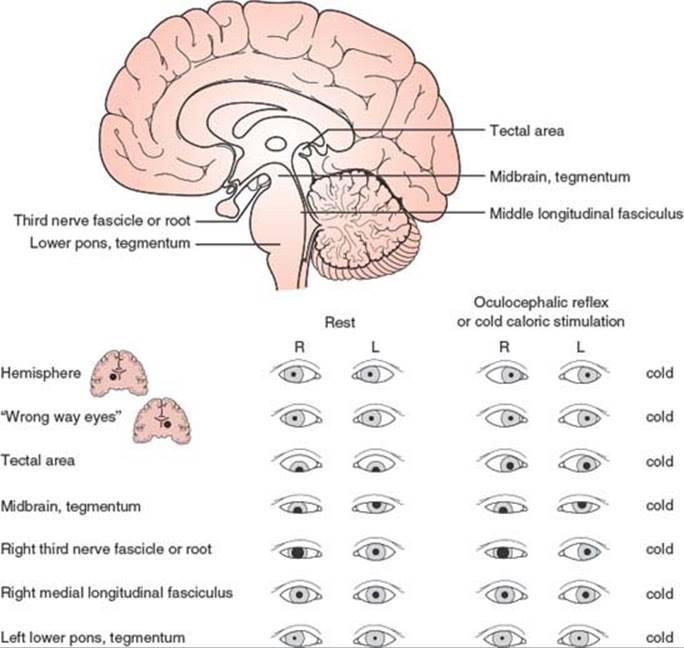
FIG. 23.4. Eye movement abnormalities characteristic of lesions at different levels of the brain. The responses to cold caloric stimulation of the left ear are indicated in the right-hand column. The first two responses at the top show normal extrinsic ocular motility with cold caloric stimulation of the left ear.
The roving eye movements of light coma cannot be voluntarily executed and are therefore incompatible with the diagnosis of feigned unresponsiveness. As coma deepens, roving eye movements disappear first, followed by the oculocephalic reflex; finally, even cold water instilled in the ear fails to induce eye movements. In metabolic coma, the pupils may still react when eye movements cannot be elicited.
Other spontaneous eye movements seen in comatose patients include the following (Table 23.1):
1.Short-cycle periodic alternating gaze (ping-pong gaze), which consists of roving of the eyes from one extreme of horizontal gaze to the other and back, with each oscillating cycle taking 2.5 to 8 seconds [32,54,90,161]. This finding usually indicates bilateral cerebral damage (e.g., bilateral cerebral infarcts) with an intact brainstem, but it has also been described with posterior fossa hemorrhage, basal ganglia infarcts, hydrocephalus, and overdose of the monoamine oxidase inhibitor tranylcypromine [54,77,78,128,153,177]. The disorder may occasionally occur in coma with no structural hemispheric lesion [76,190]. A man in his twenties, with chronic hydrocephalus from infancy and absent vertical eye movements, had ping-pong gaze since childhood only when awake [78]. One case was attributed to bilateral lesions of the cerebral peduncles [76].
TABLE 23.1 Spontaneous Eye Movements in Comatose Patients

Crevits and Decruyenaere described three patients with ping-pong gaze (due to hepatic encephalopathy, carbon monoxide intoxication, and hypoxia, respectively) and proposed that this term be reserved for those forms of periodic alternating gaze without a silent period [26]. They noted that the only constant clinical implication of ping-pong gaze was integrity, at least in part, of the lower brainstem, with lack of cortical inhibition of the horizontal gaze centers in the brainstem. This disorder of ocular motility had no prognostic value [26].
Ping-pong gaze must be differentiated from periodic alternating gaze deviation, which is an alternating horizontal conjugate gaze deviation lasting 1 to 2 minutes in each direction. Periodic alternating gaze deviation usually occurs in alert patients with structural lesions involving the cerebellum and brainstem, such as the Arnold-Chiari malformation or medulloblastoma, but it has been described in obtunded or comatose patients with hepatic encephalopathy [6].
2.Repetitive divergence is rarely seen in patients with coma from metabolic encephalopathy (e.g., hepatic encephalopathy) [104]. With this disorder, the eyes are midposition or slightly divergent at rest. They then slowly deviate out, become fully deviated for a brief period, and then rapidly return to primary position before repeating the cycle. These motions are synchronous in both eyes.
3.Nystagmoid jerking of a single eye, in a vertical, horizontal, or rotatory fashion, may occur with mid-to-lower pontine damage [118]. Pontine lesions occasionally give rise to disconjugate rotatory and vertical movements of the eyes, in which one eye may rise and intort as the other falls and extorts [118]. This type of movement should not be confused with see-saw nystagmus, which is very seldom seen in comatose patients [64].
4.Electrographic status epilepticus without appendicular motor manifestations, due to anoxia, may result in brisk, small amplitude, mainly vertical (occasionally horizontal) eye movements detectable by passive lid elevation [157].
5.Ocular bobbing refers to intermittent, often conjugate, brisk, bilateral downward movement of the eyes with slow return to midposition [39]. Both mesencephalic and medullary burst neuron centers may play a part in its genesis [138]. Cold calorics may increase the amplitude and frequency of the bobbing or have no effect [27]. Ocular bobbing has been associated with intrinsic pontine lesions (e.g., hemorrhage, tumor, infarction, central pontine myelinolysis) [60,79,93,138,167,190], extra-axial posterior fossa masses (e.g., aneurysm rupture or cerebellar hemorrhage or infarction) [13,48,108], diffuse encephalitis [144], Jakob–Creutzfeldt disease [138], and toxic-metabolic encephalopathies (e.g., acute organophosphate poisoning) [34,49,162]. “Typical” ocular bobbing, which is associated with preserved horizontal eye movements, is thought to be specific but not pathognomonic of acute pontine injury, whereas “atypical” ocular bobbing, which is associated with absent horizontal eye movements, is thought to be less helpful in predicting the site of abnormality [162]. Monocular bobbing (paretic bobbing), which consists of a quick downward movement of one eye and intorsion or no movement in the other eye, may occur if there is a coexistent unilateral fascicular oculomotor nerve palsy [162]. Disconjugate ocular bobbing, with movements involving sometimes one eye and sometimes the other, may also occur without oculomotor nerve palsy [43].
6.Inverse ocular bobbing (ocular dipping or fast-upward ocular bobbing) [44,70,85,93,131,145,159,173] consists of a slow-downward eye movement with fast return to midposition, which may occur in anoxic coma or after prolonged status epilepticus [70,133,159]. It probably reflects diffuse brain dysfunction rather than a single structural lesion because brainstem horizontal gaze reflexes are usually intact. Ocular dipping has also been described associated with deafness in a patient with pinealoblastoma [169]. Inverse/ reverse ocular bobbing consists of inverse ocular bobbing in which the eyes do not stop on rapidly returning to primary position but shoot into upgaze and slowly return to midposition [140,168].
7.Reverse ocular bobbing (fast-upward ocular bobbing) consists of fast-upward eye movement with a slow return to midposition, which may occur in patients with metabolic encephalopathy, viral encephalitis, or pontine hemorrhage [15,44,93]. It has been described with coma, due to combined phenothiazine and benzodiazepine poisoning [81]. Occasionally, ocular bobbing, ocular dipping, and reverse bobbing may occur at different times in the same patient [140].
8.Slow-upward ocular bobbing (converse ocular bobbing or reverse ocular dipping) is characterized by slow-upward eye movements followed by fast return to midposition [44,93]. This eye movement disorder has been described with pontine infarction (the patient had a one-and-a-half syndrome) [44] and with metabolic or viral encephalopathy (i.e., diffuse cerebral dysfunction) [93].
9.Pretectal pseudobobbing has been described with acute hydrocephalus [65] and consists of arrhythmic, repetitive downward and inward (“V-pattern”) eye movements at a rate ranging from 1 per 3 seconds to 2 per second and an amplitude of 1/5 to 1/2 of the full voluntary range. These movements may be mistaken for ocular bobbing, but their V pattern, their faster rate, and their pretectal rather than pontine-associated signs distinguished them from true pontine bobbing. Thus, patients with pretectal pseudobobbing may have abnormal pupillary light reactions, intact horizontal eye movements, open and often retracted eyelids, a blink frequently preceding each eye movement, and a mute or stuporous rather than a comatose state. Pretectal pseudobobbing probably represents a variety of convergence nystagmus, and its presence usually indicates the need for prompt surgical attention (e.g., hydrocephalus decompression) [65]. It is possible that some cases of “ocular bobbing” associated with thalamic hemorrhage or tentorial herniation may actually be cases of pretectal pseudobobbing.
10.Vertical ocular myoclonus consists of pendular, vertical isolated movements of the eyes noted in patients either locked-in or comatose after severe pontine strokes [61]. Their frequency is 2 Hz, and other rhythmic body movements at a similar frequency occur after a 6-week to 9-month delay. These movements are generally associated with palatal myoclonus (palatal tremor), with which they share a common mechanism [61].
ABNORMALITIES OF LATERAL GAZE
Conjugate Gaze. When both eyes remain deviated toward the same side in a comatose patient, the lesion may be in the cerebral hemisphere (most often involving the frontal eye fields) or in the pontine tegmentum. In the case of a hemispheric lesion, unless the patient is having a seizure, the eyes “look toward the lesion” (away from the hemiparetic side) but can be brought to the other side with the oculocephalic maneuver, caloric testing, or both. A seizure originating in the frontal or occipital lobes may cause deviation of the eyes and head away from the lesion, but such deviation is brief and usually accompanied by nystagmoid jerks; as soon as the seizure ceases, the eyes return to “look” toward the lesion. Thalamic and, rarely, basal ganglionic lesions, almost always hemorrhagic, may produce forced deviation of the eyes to the side contralateral to the lesion (wrong-way eyes) [166]. Very rarely, frontal-perisylvian lesions may cause wrong-way eyes [113].
Predominantly unilateral lesions affecting the tegmentum of the lower pons cause a horizontal gaze palsy toward the side of the lesion so that the eyes look toward the hemiparetic side. Neither the oculocephalic maneuver nor caloric testing overcomes a pontine gaze palsy.
Coma due to toxic substances is often accompanied by impaired conjugate eye movements, horizontal as well as vertical [119]. Thiamine deficiency, causing Wernicke encephalopathy, is a treatable cause of ophthalmoparesis and coma. It need not accompany alcoholism [191].
Disconjugate Gaze. Isolated failure of ocular adduction, in the absence of pupillary changes and with normal vertical eye movements (elicited by oculocephalic or oculovestibular reflexes), indicates a lesion of the medial longitudinal fasciculus (MLF) in the upper pons ipsilateral to the eye that fails to adduct. MLF involvement is commonly bilateral in comatose patients. Rarely, metabolic coma (such as that due to barbiturates, amitriptyline [52], or hepatic failure [19]) may induce a transient MLF syndrome that can usually be overcome by vigorous caloric testing.
Latent strabismus may become apparent when the level of alertness is mildly impaired but disappears in deep coma. Because strabismus involves a single muscle, it seldom mimics neurogenic oculoparesis except, perhaps, when abduction is reduced.
ABNORMALITIES OF VERTICAL GAZE
In patients in light coma, upward gaze can be tested by holding the eyelids open and gently touching the cornea with a wisp of cotton or a similar object. With this stimulus, the eyeballs tend to roll upward (Bell’s phenomenon).Unless the patient is intubated or has a neck injury, the doll’s head maneuver can be used to elicit the vertical component of the oculocephalic reflex. Irrigation of both ears with cold water induces downward deviation of the eyes; warm water induces upward deviation.
Disconjugate vertical gaze in the resting position (skew deviation) may be seen with lesions at different areas of the brainstem, with increased intracranial pressure, or with hepatic coma (see Chapter 8). Persistent deviation of the eyes below the horizontal meridian signifies brainstem dysfunction, which is often due to a structural lesion that affects the tectum of the midbrain but is occasionally caused by metabolic encephalopathy (e.g., hepatic coma [69]). It is also present frequently after anoxic brain damage caused by cardiac arrest, typically a few days after the event [57]. Tonic downward deviation of the eyes, often accompanied by convergence, may occur with thalamic hemorrhage, probably due to pressure on the dorsal mesencephalon. Forced downward deviation of the eyes has also been reported in patients feigning coma [139]. Forced downward deviation of the eyes during caloric testing often occurs in coma induced by sedative drugs [155]. Tonic upgaze has been reported shortly after severe anoxic encephalopathy [57,67] and with phenothiazine intoxication. Paresis of upward gaze is usually present with bilateral midbrain tectal damage. Downward gaze is preferentially affected by bilateral lesions of the superomedial perirubral region in the ventral portion of the origin of the Sylvian aqueduct from the third ventricle. In many of these patients, the lesion extends into the medial thalamic nuclei [112]. Large midbrain tegmental lesions abolish vertical gaze. At rest, the eyes remain in midposition or may be disconjugately deviated in the vertical plane.
Bilateral ptosis newly developed in patients with evolving massive hemispheric infarction seems to suggest compression of the midbrain [11].
Corneal Reflex
The corneal reflex has a higher threshold in comatose patients. Nonetheless, it must be elicited by a gentle and aseptic stimulus to avoid the risk of an infected corneal ulceration in patients with decreased corneal sensitivity (as with cranial nerve V lesions, ipsilateral lateral pontomedullary lesions, or contralateral parietal lesions) [107], or impaired eye closure (as with cranial nerve VII lesions and low pontine lesions). In the latter cases, the stimulus may induce deviation of the jaw to the opposite side (corneopterygoid reflex), and, given an intact upper pons and midbrain, the eyes may roll upward (Bell’s phenomenon).
Motor Activity of the Body and Limbs
When examining a patient in coma, observation of the movements and of the tone and reflexes of the limbs supplies information that has a less clear-cut localizing value than similar findings in alert patients. Rarely is a metabolic coma (notably hypoglycemic) accompanied by hemiparesis; however, other motor patterns, widely known as decorticate (flexor posturing) and decerebrate (extensor posturing) rigidity, are often produced by metabolic disorders [46] and do not have the structural implications that their names, coined during experimental work, would suggest. Of course, structural damage to the origin or course of the motor pathways may give rise to such patterns, which, in these cases, are often asymmetric. However, because metabolic coma is more frequent than structural coma, metabolic coma is often responsible for the motor patterns discussed below (Fig. 23.5).
In light coma, the general motor responses may oscillate between lying quietly in bed and wildly thrashing about. The latter situation occurs when a painful stimulus (such as that caused by a subarachnoid hemorrhage or a full bladder) rouses the patient, whose diminished attention prevents any coherent sequence of movements. Such patients, however, try to avoid painful stimuli by appropriately withdrawing a limb or using it to brush off the offending agent. Gently sliding a cotton-tipped stick along the patient’s forehead often proves enough of a stimulus to obtain such a response. Asymmetric responses betray a deficit of the motor or sensory pathways, or both.

FIG. 23.5. Decorticate and decerebrate posturing of the limbs in comatose patients.
The tone of the extremities can be checked by lifting the arms from the bed and flexing the patient’s knees and releasing them. In light coma, the limbs fall slowly to the resting position. A paretic limb falls like a “dead weight.” Thus, a hemiparesis, or even monoparesis, can be easily detected.
Anoxic lesions of the cerebral border zones may result in predominant damage to the cortex in the area of representation of the arms. Such patients may have bilateral arm weakness with relatively spared lower extremity function (man-in-the-barrel syndrome) [35].
When the level of coma deepens or a structural lesion affects a cerebral hemisphere and the diencephalon, decorticate rigidity may appear; this rigidity is contralateral to the hemispheric lesion. Decorticate rigidity is characterized by adduction of the shoulder and arm, flexion at the elbow, and pronation and flexion at the wrist; the leg remains extended at the hip and knee (see Fig. 23.5).
Severe metabolic (e.g., anoxic) disorders or lesions of the upper brainstem give rise to decerebrate rigidity, which is characterized by extension and pronation of the upper extremities and forcible plantar flexion of the foot (see Fig. 23.5). Brought about by painful stimuli, opisthotonos develops periodically with hyperextension of the trunk and hyperpronation of the arms. In experimental animals, decerebrate rigidity results from brainstem transection at the collicular level, below the red nuclei but leaving intact the pontine reticular formation and vestibular nuclei. The action of the vestibular nuclei, unchecked by higher centers, may explain the increased extensor tone characteristic of decerebrate rigidity. Structural lesions that cause this motor pattern usually affect the midbrain and upper pons either directly, as in the case of brainstem infarcts, or indirectly, by ischemia produced by pressure arising in the supratentorial compartment (downward herniation) or in the posterior fossa.
In a given patient, one side may demonstrate decorticate posturing, whereas the other side, innervated by the motor pathways that have undergone greater damage, displays decerebrate rigidity.
Abnormal extension of the arms with weak flexion of the legs usually indicates damage of the pontine tegmentum [118]. With even lower lesions involving the medulla, total flaccidity ensues.
Flaccidity in a critically ill patient can also be produced by a polyneuropathy (“critical illness neuropathy”). More recently, it has been recognized that many cases of flaccidity in comatose patients, particularly those on steroid treatment, are caused by a severe necrosis of the thick fibers of striated muscle cells (“critical illness myopathy”) [2,148].
When the entire brain, including the brainstem, has undergone total or subtotal irreversible damage, compatible with the diagnosis of death caused by brain destruction, spontaneous reflex movements of spinal origin can still be witnessed in the corpse [149]. Plantar withdrawal responses, muscle stretch reflexes, undulating toe movements, abdominal contractions, Lazarus sign, and respiratory-like movements, among others, have been described [149]. Lazarus sign refers to complex movements suggesting purposeful activity. In one instance, the arms, with flexed elbows and hands held together, adopted a praying position, which was followed by hand separation as the arms fell to the sides of the torso; the legs also performed walking-like movements [86]. Other manifestations of the Lazarus sign have included shoulder adduction, bringing both arms to the chest, moving the hands to the neck, sometimes crossing and touching each other, and then finally falling to the bed. Passive flexion of the neck may elicit a jerk that raises the four limbs off the bed [136]. Spinal automatisms may be present in as many as 40% of heart-beating cadavers, typically within the first 24 hours after total brain destruction, although elaborate movements are much less frequent [149]. In the absence of any brain stem–mediated response, such as facial movements or a gag reflex, limb or trunk movements are more likely to be mediated by the spinal cord. In doubt, ancillary procedures can confirm the total destruction of the brain.
Clinical Presentations of Coma-Inducing Lesions Depending on Their Location
Metabolic Encephalopathy (Diffuse Brain Dysfunction)
The phylogenetically newer brain structures tend to be more sensitive to metabolic injury. This holds true even though the target of various metabolic abnormalities or toxic agents may vary slightly. For instance, carbon monoxide poisoning causes pallidal necrosis in addition to the widespread cortical damage expected from any hypoxic insult. Functions subserved by complex polysynaptic pathways are affected earlier by metabolic disturbances than those mediated by a few neurons. Thus, higher cortical functions and attention succumb early to metabolic insults, whereas the pupillary light reflex remains to the brink of brainstem death. Survival of other functions ranges between these two extremes. By the time decerebrate posturing appears, the corneal reflexes may be severely depressed, but some eye movements may be elicited by the doll’s eye maneuver or caloric stimulation.
Asymmetric motor findings speak against the diagnosis of metabolic encephalopathy. However, downward deviation of the eyes may be occasionally associated with hepatic encephalopathy [69]. Some toxic substances, like ethylene glycol, produce focal brain injuries with the corresponding neurological localizing findings [98]. Focal seizures are common in metabolic encephalopathies, particularly those coursing with breakdown of the blood–brain barrier, such as eclampsia, malignant hypertension, and acute intermittent porphyria [87].
Toxic-metabolic disorders often induce abnormal movements (tremor, asterixis, myoclonus, and seizures) that seldom accompany focal structural lesions of the brain. But because these two types of etiologic factors often coexist, the diagnostic specificity of these abnormal movements is far from absolute.
The tremor of metabolic encephalopathy is coarse and irregular and ranges from 8 to 10 cycles per second. Its amplitude is greatest when the patient holds his hand outstretched, but in less responsive patients it may be felt by holding the patient’s fingers extended.
Asterixis is a sudden, brief loss of postural tone that is translated into a flapping movement when the hand is held in dorsiflexion at the wrist and the fingers are extended and abducted. This hand posture requires some cooperation from the patient, but asterixis can also be elicited by passively extending the patient’s fingers and wrist. Asterixis at the hip joints can be elicited by a maneuver in which the hips are passively flexed and abducted at about 60 to 90 degrees between the thighs [105]. This asterixis seems to be provoked by involuntary contraction of the hip adductors against gravity and may be especially prominent in stuporous patients with hepatic encephalopathy. Asterixis is present with slight stupor and wanes as coma deepens. Unilateral asterixis may appear when a toxic encephalopathy coexists with a structural lesion of the motor pathways that project to the limb with asterixis. Unilateral asterixis may be seen contralateral to lesions of the mesencephalon, ventrolateral thalamus, primary motor cortex, or parietal lobe [17,28,33,89,103,127,160] or ipsilateral to lesions of the pons or medulla [114]. Episodes of lapses of postural control by the reticular formation may be responsible for midbrain asterixis. For this reason, midbrain asterixis has been considered a “segmental form of drop attack” [14]. Occasionally, bilateral asterixis may occur with bilateral lesions of the mid-pons [14] or with bilateral mesencephalic lesions [73].
Multifocal myoclonus consists of sudden, nonrhythmic twitching that affects first one muscle, then another, without any specific pattern except a tendency to involve the facial and proximal limb musculature. Causes of multifocal myoclonus include uremic and hyperosmolar-hyperglycemic encephalopathy, carbon dioxide narcosis, and a large dose of intravenous penicillin [47].
Generalized myoclonus, usually postanoxic, involves mainly the axial musculature, which contracts suddenly, making the patient jump with a certain periodicity [59]. It may also appear as irregular brief jerks in both face and limbs. The myoclonus, often stimulus sensitive, is most prominent in the first postresuscitation day and tends to abate spontaneously in subsequent days. The patients often have a burst-suppression pattern on EEGs and cerebral edema or infarcts on CT or MRI. Severe anoxic cortical and brainstem damage are the most common pathologic correlates of generalized myoclonus (also called myoclonic status), which carries a poor prognosis [111,184].
In addition to symmetric motor findings, hyperventilation or hypoventilation and the presence of acid–base imbalance are characteristic of metabolic coma. Respiratory depression often follows intoxication with opioid substances and darkens its prognosis [55].
Supratentorial Structural Lesions
To cause coma, supratentorial lesions must affect both cerebral hemispheres (e.g., massive bihemispheric or bilateral thalamic infarction) [74]. The clinical presentation of these lesions, and of subarachnoid hemorrhage, resembles in many aspects the presentation of metabolic disorders. For instance, in postoperative coma, often chalked to metabolic causes, ischemic brain lesions may play a major role [45]. However, many cerebral infarcts, even when bilateral, are often staggered, appear more abruptly than metabolic encephalopathies, and cause asymmetric motor signs, at least early in their course. Sudden onset of severe headache (“thunderclap headache”) and signs of meningeal irritation separate subarachnoid hemorrhage from metabolic encephalopathies [83]. Pituitary apoplexy can also present with headache and stupor [163].
The following discussion deals mainly with supratentorial lesions that cause mass effect and secondarily impair consciousness by compressing the diencephalic and upper brainstem structures. Prime examples are hemispheric tumors, subdural or intracerebral hemorrhage, and massive infarcts. When the intracranial pressure of the supratentorial compartment reaches a certain level, the brain substance is squeezed through the tentorial opening. Occasionally, the syndrome of transtentorial herniation may occur with minimal downward displacement of the upper midbrain [134]. Depending on the supratentorial location of the mass and the size of the tentorial opening, either of two clinically relevant syndromes may result: lateral (uncal) herniation or central (transtentorial) herniation.
LATERAL HERNIATION
In patients with a wide tentorial opening, lateral extracerebral or temporal lobe masses push the mesial temporal lobe (uncus anteriorly, parahippocampal gyrus posteriorly) between the ipsilateral aspect of the midbrain and the free edge of the tentorium (Fig. 23.6). As the tongue of herniated tissue compresses the third cranial nerve and posterior cerebral artery downward, the ipsilateral pupil becomes progressively dilated and responds sluggishly to light (Fig. 23.7). This stage can be rather brief and, depending on the size and acuteness of the lesion, it may last from a few minutes to several hours. Prompt recognition and removal (often surgical) of the offending agent is mandatory at this stage because the usual progression is deadly. The posterior cerebral artery, pinched against the tentorial edge by the herniated parahippocampal gyrus, becomes occluded, giving rise to a hemorrhagic mesial occipital infarct. The herniated hippocampus also pushes the midbrain against the rigid edge of the dura on the opposite side of the tentorial opening. This rigid structure carves out a notch (Kernohan notch) in the lateral aspect of the midbrain, interrupting the cerebral peduncle (particularly the fibers that project to the leg) on the side opposite the original temporal lobe lesion (see Fig. 23.6). This results in hemiparesis ipsilateral to the original lesion (Kernohan notch phenomenon). If misinterpreted, such hemiparesis may prove to be a false localizing sign. Therefore, when a dilated pupil and hemiparesis appear ipsilaterally, the original lesion is likely to be on the side of the abnormal pupil.
At this point, anteroposterior elongation and downward displacement of the midbrain have already caused tearing of the paramedian perforating vessels that feed the midbrain tegmentum. The consequent infarction and hemorrhages (Duret hemorrhages) that involve this structure render recovery almost impossible. The pupil that was larger may become a little smaller as the sympathetic pathway is damaged in the midbrain, while the other pupil becomes midsize and unresponsive. Oculomotor paresis appears, first in the eye originally involved, and shortly afterward in the other eye. Abduction may remain as the only elicitable eye movement.
Many causes of lateral herniation, such as hematomas, can be surgically treated. However, patients with a combination of absent pupillary, corneal, and oculocephalic reflexes and extensor posturing before craniotomy have a very poor prognosis [121]. Survivors of tentorial herniation may be left in a locked-in or chronic vegetative state [68] and may demonstrate oculomotor nerve dysfunction, internuclear ophthalmoplegia, vertical gaze paresis, pretectal signs [20,62], homonymous hemianopsia or blindness [63], parkinsonism and other extrapyramidal syndromes, or spastic limb weakness [20]. A proportion of these patients may recover, particularly with aggressive therapy [120,183]. Bilateral visual loss after tentorial herniation is likely due to bilateral posterior cerebral artery compression or stretch resulting in bilateral occipital infarction; however, occasional patients develop optic atrophy, indicating that a pregeniculate mechanism may also be operant [63].

FIG. 23.6. Lateral transtentorial herniation: (A) basal view, (B) coronal view. In this example, a subdural hematoma is causing a marked shift of the midline structures and herniation of the parahippocampal gyrus through the tentorial notch. Occlusion of the posterior cerebral artery, which is pinched between the herniated hippocampal tissue and the rigid end of the tentorium, has resulted in medial temporo-occipital infarction. The midbrain is compressed against the contralateral free tentorial edge, causing a laceration of the crus cerebri (Kernohan notch). Stretching of the slender perforating branches of the basilar artery has produced petechial hemorrhages in the tegmentum of the midbrain (Duret hemorrhages).
CENTRAL HERNIATION
Unlike temporal masses, frontal, parietal, or occipital masses first compress the diencephalon, which, as the supratentorial pressure increases, shifts downward and buckles over the midbrain. Subsequent flattening of the midbrain and pons in the rostrocaudal direction causes elongation and rupture of the paramedian perforating arteries feeding these structures, resulting in infarction and hemorrhages (Duret) in the tegmentum of the midbrain (first) and pons (afterward) (Fig. 23.8).

FIG. 23.7. Clinical findings with lateral transtentorial herniation. (Reproduced from McNealy DE, Plum F. Brainstem dysfunction with supratentorial mass lesions. Arch Neurol 1962;7:10. Copyright 1962, American Medical Association.)
Paralleling the pathologic changes of central herniation, the clinical picture reflects an orderly rostrocaudal progression of brainstem damage. The characteristic evolution of the clinical picture has been termed the central syndrome of rostrocaudal deterioration [118]. Description of this syndrome enables one to review the characteristic clinical findings with lesions at the different levels of the brainstem (Fig. 23.9). In acute illness, MRI changes of brain herniation (incisural or foramen magnum) tend to parallel the clinical signs of brain herniation; in chronic cases, clinical and MRI scans correlate less well, with MRI sometimes revealing major degrees of anatomic herniation well in advance of clinical abnormalities [126].
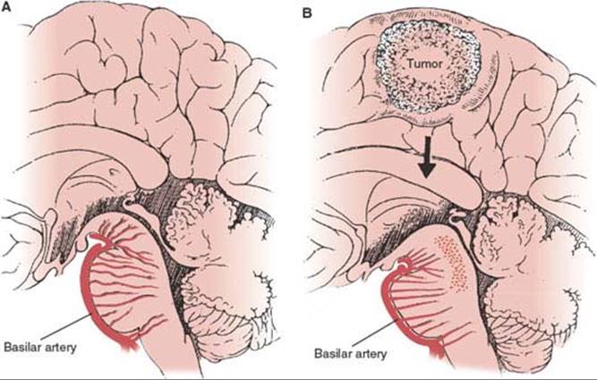
FIG. 23.8. Central transtentorial herniation. A: Normal sagittal section of the brainstem. Note the vascular perforators, branches of the basilar artery. B: Mass effect from a high parietal tumor, resulting in downward displacement and superoinferior flattening of the midbrain and upper pons. The increased cross-sectional diameter of these structures is attended by stretching and rupture of the perforators, with subsequent hemorrhages in the tegmentum of the midbrain and upper pons.
Early Diencephalic Stage. Impaired attention and somnolence appearing in a patient with a supratentorial mass usually herald the beginning of this stage. The respiratory pattern is normal but is punctuated by deep sighs and yawns. In the periods of greater somnolence, the pupils become tiny but react to light, whereas the eyes become slightly divergent, moving slowly from side to side (roving eye movements). Attempts to perform the doll’s eye maneuver may provide enough of a stimulus to awaken the patient, and quick eye movements (saccades) are then elicited rather than the slow adversive movements of the oculocephalic reflex. For the same reason, caloric stimulation may induce nystagmus. The patient resists passive motion of the limbs (paratonia), may have grasp reflexes, and brushes off appropriately any noxious stimulus. Plantar responses are usually bilaterally extensor.
Late Diencephalic Stage. At this stage, the patient cannot be aroused. Cheyne–Stokes respiration replaces normal breathing. The pupils remain small and reactive. Roving eye movements have disappeared, but the doll’s eye maneuver or caloric stimulation easily elicits full and conjugate deviation of the eyes. As the process advances, however, tectal dysfunction may result in restriction of upward gaze. Light painful stimuli fail to elicit any response; heavier ones may induce decorticate posturing, which appears earlier on the side of a previous hemiparesis, opposite the supratentorial lesion. Plantar responses are bilaterally extensor.
Proper diagnosis and treatment at this stage of the syndrome of central herniation may still result in recovery of neurologic function. Once the clinical picture evolves into the next stage (caused by hemorrhages and infarction of the midbrain tegmentum), the prognosis is very poor, except in children.
Midbrain–Upper Pons Stage. The patient now breathes rather quickly and evenly. Temperature oscillations are common, and an occasional patient may develop diabetes insipidus because of stretching of the median eminence of the hypothalamus. The pupils become midsized, unequal, and irregular, often pear-shaped and eccentric. Terminally, generalized anoxia causes a systemic release of epinephrine, and the pupils may be transiently dilated. The doll’s eye maneuver and caloric testing elicit restricted or no vertical eye movements. The eyes often move disconjugately in both the horizontal and the vertical planes. Bilateral impairment of adduction may reflect dysfunction of both third nerve nuclei, of the medial longitudinal fasciculi, or both. Noxious stimuli give rise to decerebrate posturing.
Lower Pontine Stage. Respiration becomes quicker and shallower. Apneustic breathing, common with primary ischemic lesions of this area, is infrequent in patients with transtentorial herniation, perhaps because more medially located structures are preferentially damaged. The pupils remain unchanged from the previous stage, but eye movements are now unobtainable. Decerebrate rigidity decreases. Plantar stimulation may elicit not only bilateral Babinski signs but also withdrawal of the legs, with flexion at the knee and hip.
Medullary Stage. In this agonal stage, ataxic breathing soon gives way to apnea. The blood pressure drops, and the pulse becomes irregular [125].
Large acute supratentorial lesions, particularly massive intraventricular hemorrhage, may cause a quick decompensation of brainstem function, leading to respiratory failure without the step-wise progression described above. Smaller intraventricular hemorrhages may impair reflex eye movements in the horizontal and vertical planes while the patient’s level of consciousness is only mildly depressed. This phenomenon may be secondary to the action of the blood on the floor of the fourth ventricle and may have played a role in an unusual case of transient locked-in syndrome with intraventricular hemorrhage and a transtentorial herniation [183].
Although the clinical deterioration of patients with supratentorial masses often follows the pattern described above, early depression of the level of alertness in patients with an acute hemispheric mass may be more related to distortion of the brain by horizontal rather than vertical displacement of brain tissue [132]. Ropper showed that a horizontal pineal body shift of 0 to 3 mm from the midline was associated with alertness, 3 to 4 mm with drowsiness, 6 to 8.5 mm with stupor, and 8 to 13 mm with coma [132]. With extratemporal masses, the perimesencephalic cistern was often widened, rather than filled by herniated medial temporal lobe. On coronal MRI images, horizontal displacement of the midline of the brain at the level of the incisura correlated better than vertical displacement with the level of alertness [130]. These studies do not disprove that anteroposterior elongation of the midbrain, with subsequent ischemia of the midbrain tegmentum, may play a role in the genesis of coma and subsequent brainstem deterioration, as outlined above.


FIG. 23.9. Clinical findings with central transtentorial herniation, from early (A) to late (D) stages. (Modified from McNealy DE, Plum F. Brainstem dysfunction with supratentorial mass lesions. Arch Neurol1962;7:10. Copyright 1962, American Medical Association.) (continued)
As far as the prognosis of lobar hemorrhages, a Glasgow coma score <14 predicted deterioration [40]. Initial CT characteristics predictive of deterioration include hemorrhage volume >60 mL, shift of the septum pellucidum, effacement of the contralateral ambient cistern, and widening of the contralateral temporal horn [40]. Deeper putaminal hemorrhages fare worse when accompanied by hydrocephalus and the patient has a Glasgow Coma Scale score less than 8 [116]. The Glasgow coma scale is widely used [164], but a new scale (FOUR) predicts better the outcome of the more severely impaired patients [180].
Some patients develop severe brain edema and coma after minor head trauma with a lucid interval. A disturbance of ionic channels has been suspected because this syndrome is more frequent in families with familial hemiplegic migraine, known to be associated with abnormalities of the calcium channel. A S218 L mutation in the CACNA1 A calcium channel has been reported in some of these patients [72].
False localizing signs with supratentorial masses may mislead the observer about the hemisphere involved (e.g., hemiparesis ipsilateral to the lesion due to Kernohan notch) or falsely localize the primary process to the posterior fossa. The latter occurs mainly with lesions located in the midline (e.g., hydrocephalus) or in areas of the frontal and temporal lobes that are clinically “silent.” Extracerebral lesions (e.g., subdural hematoma) in the elderly may behave in a similar manner. These lesions fail to cause focal deficits but raise the pressure of the intracranial contents and produce cranial nerve dysfunction that may be mistaken for evidence of a posterior fossa lesion. Sixth nerve palsy and papilledema are the commonest false localizing signs, but other ophthalmoplegias, trigeminal neuralgia or numbness, unilateral or bilateral deafness, facial palsy, and even weakness in the distribution of the ninth to twelfth cranial nerves may appear as a consequence of raised intracranial pressure with a supratentorial lesion.
Subtentorial Structural Lesions
Destructive lesions (e.g., infarcts, small hemorrhages) of the brainstem can be easily localized clinically. Compressive lesions that cause coma tend to be associated with brisk involvement of the cerebellum or fourth ventricle. Cerebellar hemorrhage is the prime example [187]. Early in the clinical course, occipital headache, vomiting, and ataxia are usually prominent. In the process of rostrocaudal deterioration characteristic of downward transtentorial herniation, all the structures at a particular brainstem level tend to be affected at the same time. This does not happen with compressive lesions of the posterior fossa. Unless massive, these lesions affect one level more than others, often asymmetrically, giving rise to preferentially unilateral brainstem and cerebellar signs.
Lesions that compress the upper brainstem may cause upward transtentorial herniation of the tectum of the midbrain and of the anterior cerebellar lobule, giving rise to signs of midbrain dysfunction with coma, hyperventilation, fixed pupils, and vertical ophthalmoplegia. Lower lesions impinge on the pontine tegmentum, causing somnolence, pinpoint pupils that react briskly to light, oculoparetic nystagmus on lateral gaze, and truncal ataxia. Appendicular ataxia may be so mild as to pass unnoticed. As pontine function becomes worse, horizontal gaze disappears and cannot be elicited with the doll’s eye maneuver or caloric testing, whereas impairment of vertical eye movements clearly lags behind. With lower lesions that impinge mainly on the medulla, respiratory ataxia evolving to apnea and circulatory abnormalities precede changes in the level of alertness. The medulla is particularly resistant to infarction, spontaneous hemorrhages, and even traumatic lesions [179]. However, it is preferentially affected in Listeria monocytogenes rhombencephalitis [171] and in Leigh disease [142,188]
Deterioration with pontine hemorrhages depends on the cause of the lesion. Bleeding from cavernous angiomas has a much better prognosis than hypertensive hemorrhage [122]. More important for surgical decision making are prognostic factors with cerebellar hemorrhages. Anatomic findings that predict deterioration with cerebellar hemorrhage include displacement of the fourth ventricle, brain stem deformity, hydrocephalus and compression of the basal cisterns [71,158]. In some series, a hematoma size of more than 3 cm also worsens prognosis [158]. Clinically, patients fare poorly who have abnormal corneal and oculocephalic responses, a Glasgow coma score less than 8, and motor response less than localization to pain [158].
Mass lesions in the posterior fossa may cause downward herniation of the cerebellar tonsils through the foramen magnum, with subsequent infarction of the tonsils, medulla, and even upper cervical spinal cord. Generalized anoxia and circulatory failure consequent to medullary dysfunction play a role in the genesis of these infarcts.
When patients with preexisting brain stem lesions, most often from strokes or multiple sclerosis, suffer an anoxic or metabolic insult, they may lose all brain stem reflexes transiently [129]. The diagnosis of irreversible damage of the brain stem has to be done more cautiously in these patients.
Psychogenic Unresponsiveness
The patient may hold the eyes forcibly closed and resist eyelid opening or may keep the eyes open in a fixed stare, interrupted by quick blinks. The pupils, which are of normal size and position, react to light unless a cycloplegic drug has been instilled into them. The doll’s eye maneuver elicits random or no eye movements. Caloric testing is more helpful because it gives rise to classic vestibular nystagmus with a quick component that requires activity of the frontal eye fields. This quick component is, conversely, absent in comatose patients. Muscle tone and reflexes are normal. The patient may hyperventilate or breathe normally. Psychogenic unresponsiveness often recurs and as such is frequently misdiagnosed as an epileptic or migrainous disorder [29,147,152].
Diagnosis of Death Caused by Brain Destruction
Although the term “brain death” has been used for years and continues to be widely used, it is not helpful in the clinical setting. Speaking about “brain death” often confuses the families of the so-called brain-dead individual, who end up by asking their physicians whether their loved one—forget about his or her brain—is dead or not [154]. Furthermore, “brain death” implies that in the process of dying there is “brain death,” caused by unavoidable events, and the real death, caused by the physicians turning off ventilation support with the connivance of the relatives. This false impression, source of delays, expenses and guilt, can be avoided by clear explanations and the use of a term similar to the one heading this section.
The American Academy of Neurology criteria for the diagnosis of death caused by brain destruction in adults [186] recommends the following steps:
I. The clinical evaluation (prerequisites).
A. Establish irreversible and proximate cause of coma. The cause of coma can usually be established by history, examination, neuroimaging, and laboratory tests. Exclude the presence of a CNS-depressant drug effect by history, drug screen, calculation of clearance using five times the drug’s half-life (assuming normal hepatic and renal function), or, if available, drug plasma levels below the therapeutic range. Prior use of hypothermia (e.g., after cardiopulmonary resuscitation for cardiac arrest) may delay drug metabolism. The legal alcohol limit for driving (blood alcohol content 0.08%) is a practical threshold below which an examination to determine death could reasonably proceed. There should be no recent administration or continued presence of neuromuscular blocking agents (this can be determined by the presence of a train of four twitches with maximal ulnar nerve stimulation). There should be no severe electrolyte, acid–base, or endocrine disturbance (defined by severe acidosis or laboratory values markedly deviated from the norm).
B. Achieve normal core temperature. In most patients, a warming blanket is needed to raise the body temperature and maintain a normal or near-normal temperature (>36°C). After the initial equilibration of arterial CO2 with mixed central venous CO2, the PaCO2 rises steeply, but then more slowly when the body metabolism raises PaCO2. To avoid delaying an increase in PaCO2, normal or near-normal core temperature is preferred during the apnea test.
C. Achieve normal systolic blood pressure. Hypotension from loss of peripheral vascular tone or hypovolemia (diabetes insipidus) is common; vasopressors or vasopressin are often required. Neurologic examination is usually reliable with a systolic blood pressure of ≥100 mm Hg.
D. Perform one neurologic examination (sufficient to pronounce death in most US states). Only one neurologic examination should be sufficient to pronounce death if a certain period of time (in practice, usually several hours) has passed since the onset of the brain insult, to exclude the possibility of recovery. However, some US state statutes require two examinations separated by a certain amount of time. Legally, all physicians are allowed to determine death in most US states. Neurologists, neurosurgeons, and intensive care specialists may have specialized expertise. It seems reasonable to require that all physicians making a determination of death due to brain destruction be intimately familiar with the diagnostic criteria and have demonstrated competence in this complex examination. “Brain death” statutes in the United States differ by state and institution. Some US state or hospital guidelines require the examiner to have certain expertise.
II.The clinical evaluation (neurologic assessment).
A. Coma. Patients must lack all evidence of responsiveness. Eye opening or eye movement to noxious stimuli is absent. Noxious stimuli should not produce a motor response other than spinally mediated reflexes. The clinical differentiation of spinal responses from retained motor responses associated with brain activity is discussed earlier in this chapter, in the section on “Motor Activity of the Body and Limbs.”
B. Absence of brainstem reflexes.
a. Absence of pupillary response to a bright light is documented in both eyes. Usually the pupils are fixed in a midsize or dilated position (4–9 mm). Constricted pupils suggest the possibility of drug intoxication. When uncertainty exists, a magnifying glass should be used.
b. Absence of ocular movements using oculocephalic testing and oculovestibular reflex testing. Once the integrity of the cervical spine is ensured, the head is briskly rotated horizontally and vertically. There should be no movement of the eyes relative to head movement. The oculovestibular reflex is tested by irrigating each ear with ice water (caloric testing) after the patency of the external auditory canal is confirmed. The head is elevated to 30 degrees. Each external auditory canal is irrigated (one ear at a time) with approximately 50 mL of ice water. Movement of the eyes should be absent during 1 minute of observation. Both sides are tested, with an interval of several minutes.
c. Absence of corneal reflex. Absent corneal reflex is demonstrated by touching the cornea with a piece of tissue paper, a cotton swab, or squirts of water. No eyelid movement should be seen.
d. Absence of facial muscle movement to a noxious stimulus. Deep pressure on the condyles at the level of the temporomandibular joints and deep pressure at the supraorbital ridge should produce no grimacing or facial muscle movement.
e. Absence of the pharyngeal and tracheal reflexes. The pharyngeal or gag reflex is tested by stimulating the posterior pharynx with a tongue blade or suction device. The tracheal reflex is most reliably tested by examining the cough response to tracheal suctioning. The catheter should be inserted into the trachea and advanced to the level of the carina followed by one or two suctioning passes.
C. Apnea.
a. Absence of a breathing drive is tested with a CO2 challenge. Documentation of an increase in PaCO2 above normal levels is typical practice. It requires preparation before the test.
b. Prerequisites: (i) normotension, (ii) normothermia, (iii) euvolemia, (iv) eucapnia (PaCO2 35–45 mm Hg), (v) the absence of hypoxia, and (vi) no prior evidence of CO2 retention (i.e., chronic obstructive pulmonary disease, severe obesity).
c. Procedure:
1. Adjust vasopressors to a systolic blood pressure ≥100 mm Hg.
2. Preoxygenate for at least 10 minutes with 100% oxygen to a PaO2 ≥200 mm Hg.
3. Reduce ventilation frequency to 10 breaths per minute to eucapnia.
4. Reduce positive end-expiratory pressure (PEEP) to 5 cm H2O (oxygen desaturation with decreasing PEEP may suggest difficulty with apnea testing).
5. If pulse oximetry oxygen saturation remains >95%, obtain a baseline blood gas (PaO2, PaCO2, pH, bicarbonate, base excess).
6. Disconnect the patient from the ventilator.
7. Preserve oxygenation (e.g., place an insufflations catheter through the endotracheal tube to a point near the level of the carina and deliver 100% O2 at 6 L/min).
8. Look closely for respiratory movements for 8 to 10 minutes. Respiration is defined as abdominal or chest excursions and may include a brief gasp.
9. Abort if systolic blood pressure decreases to <90 mm Hg.
10. Abort if oxygen saturation measured by pulse oximetry is <85% for >30 seconds. Retry procedure with T-piece, CPAP 10 cm H2O, and 100% O2 12 L/min.
11. If no respiratory drive is observed, repeat blood gas (PaO2, PaCO2, pH, bicarbonate, base excess) after approximately 8 minutes.
12. If respiratory movements are absent and arterial PCO2 is ≥60 mm Hg (or 20 mm Hg increase in arterial PCO2 over a baseline normal arterial PCO2), the apnea test result is positive (i.e., supports the clinical diagnosis of death).
13. If the test is inconclusive but the patient is hemodynamically stable during the procedure, it may be repeated for a longer period of time (10–15 minutes) after the patient is again adequately preoxygenated.
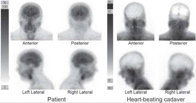
FIG. 23.10. 99mTc HM-PAO brain perfusion studies of a patient (left) and a heart-beating cadaver having undergone death by brain destruction a few hours prior to the scan (right). Scintigraphy was obtained from anterior, posterior and both lateral projections, after injection in the ICU and a brief scanning procedure at the nuclear medicine service. Note that in the corpse, instead of normal brain perfusion, there is an empty area outlined by the skull. In the corpse, the activity in extracerebral tissues appears higher than in the patient because the threshold has been lowered for the cadaver study, to increase sensitivity for the detection of any remaining brain activity. Images courtesy of Dr. Javier Arbizu, from the Nuclear Medicine Service of the Clínica Universidad de Navarra, Pamplona, Spain.
An apnea test can be performed in up to about 90% of patients suspected of death by brain destruction [185]. In some cases, ancillary tests, such as electroencephalography or neuroimaging, are needed. But although ancillary tests can render impressive findings (Fig. 23.10) and may help the patient’s relatives to understand the nature of the event, the diagnosis most often rests on the clinical evaluation [178].
References
1. Albert ML, Silverberg R, Reches A, et al. Cerebral dominance for consciousness. Arch Neurol 1976; 33:453–454.
2. Allen DC, Arunachalam R, Mills KR. Critical illness myopathy: further evidence from muscle-fiber excitability studies of an acquired channelopathy. Muscle Nerve 2008;37:14–22.
3. Allroggen H, Dennis G, Abbott RJ, et al. New variant Creutzfeldt-Jakob disease: three case reports from Leicestershire. J Neurol Neurosurg Psychiatry 2000; 68:375–378.
4. ANA Committee on Ethical Affairs. Persistent vegetative state: report of the American Neurological Association Committee on Ethical Affairs. Ann Neurol 1993;33:386–390.
5. Askenasy JJ, Goldhammer I. Sleep apnea as a feature of bulbar stroke. Stroke 1988;19:637–639.
6. Averbuch-Heller L, Meiner Z. Reversible periodic alternating gaze deviation in hepatic encephalopathy. Neurology 1995;45:191–192.
7. Aylett SE, Cross JH, Taylor DC, et al. Epileptic akinetic mutism: following temporal lobectomy for Rasmussen’s syndrome. Eur Child Adolesc Psychiatry 1996;5:222–225.
8. Bakshi N, Maselli RA, Gospe SM Jr, et al. Fulminant demyelinating neuropathy mimicking cerebral death. Muscle Nerve 1997;20:1595–1597.
9. Bernat JL. Chronic disorders of consciousness. Lancet 2006;367:1181–1192.
10. Bernat JL. Questions remaining about the minimally conscious state. Neurology 2002;58:337–338.
11. Blacker DJ, Wijdicks EF. Delayed complete bilateral ptosis associated with massive infarction of the right hemisphere. Mayo Clin Proc 2003;78:836–839.
12. Bogousslavsky J, Khurana R, Deruaz JP, et al. Respiratory failure and unilateral caudal brainstem infarction. Ann Neurol 1990;28:668–673.
13. Bosch EP, Kennedy SS, Aschenbrener CA. Ocular bobbing: the myth of its localizing value. Neurology 1975;25:949–953.
14. Bril V, Sharpe JA, Ashby P. Midbrain asterixis. Ann Neurol 1979;6:362–364.
15. Brusa A, Firpo MP, Massa S, et al. Typical and reverse bobbing: a case with localizing value. Eur Neurol 1984;23:151–155.
16. Buettner UW, Zee DS. Vestibular testing in comatose patients. Arch Neurol 1989;46:561–563.
17. Calzetti S, Gemignani F, Salati MR, et al. Unilateral asterixis due to thalamic tumor. Case report. Ital J Neurol Sci 1983;4:87–90.
18. Caner H, Altinors N, Benli S, et al. Akinetic mutism after fourth ventricle choroid plexus papilloma: treatment with a dopamine agonist. Surg Neurol 1999;51: 181–184.
19. Caplan LR, Scheiner D. Dysconjugate gaze in hepatic coma. Ann Neurol 1980;8:328–329.
20. Caplan LR, Zervas NT. Survival with permanent midbrain dysfunction after surgical treatment of traumatic subdural hematoma: the clinical picture of a Duret hemorrhage? Ann Neurol 1977;1:587– 589.
21. Catsman-Berrevoets CE, Van Dongen HR, Mulder PG, et al. Tumour type and size are high risk factors for the syndrome of “cerebellar” mutism and subsequent dysarthria. J Neurol Neurosurg Psychiatry 1999;67:755– 757.
22. Chen R, Sahjpaul R, Del Maestro RF, et al. Initial enlargement of the opposite pupil as a false localising sign in intraparenchymal frontal haemorrhage [published erratum appears in J Neurol Neurosurg Psychiatry 1995;58(1):126]. J Neurol Neurosurg Psychiatry 1994;57:1126–1128.
23. Chia LG. Locked-in syndrome with bilateral ventral midbrain infarcts. Neurology 1991;41:445–446.
24. Chiaramonte JS. Cycloplegia from transdermal scopolamine. N Engl J Med 1982;306:174.
25. Cordova S, Lee R. Fixed, dilated pupils in the ICU: another recoverable cause. Anaesth Intensive Care 2000;28:91–93.
26. Crevits L, Decruyenaere J. ‘Ping-pong’ gaze. Neuro-ophthalmology 1992;12:121.
27. Daroff RB, Waldman AL. Ocular bobbing. J Neural Neurosurg Psychiatry 1965;28:375.
28. Degos JD, Verroust J, Bouchareine A, et al. Asterixis in focal brain lesions. Arch Neurol 1979;36: 705–707.
29. DeToledo JC, Ramsay RE. Patterns of involvement of facial muscles during epileptic and nonepileptic events: review of 654 events. Neurology 1996;47: 621–625.
30. Devereaux MW, Keane JR, Davis RL. Autonomic respiratory failure associated with infarction of the medulla. Arch Neurol 1973;29:46.
31. Devinsky O, Morrell MJ, Vogt BA. Contributions of anterior cingulate cortex to behaviour. Brain 1995; 118:279–306.
32. Diesing TS, Wijdicks EF. Ping-pong gaze in coma may not indicate persistent hemispheric damage. Neurology 2004;63:1537–1538.
33. Donat JR. Unilateral asterixis due to thalamic hemorrhage. Neurology 1980;30:83–84.
34. Drake ME Jr, Erwin CW, Massey EW. Ocular bobbing in metabolic encephalopathy: clinical, pathologic, and electrophysiologic study. Neurology 1982; 32:1029–1031.
35. Elting JW, Haaxma R, Sulter G, et al. Predicting outcome from coma: man-in-the-barrel syndrome as potential pitfall. Clin Neurol Neurosurg 2000;102:23–25.
36. Ersahin Y, Mutluer S, Cagli S, et al. Cerebellar mutism: report of seven cases and review of the literature. Neurosurgery 1996;38:60–65; discussion 6.
37. Estraneo A, Moretta P, Loreto V, et al. Late recovery after traumatic, anoxic, or hemorrhagic long-lasting vegetative state. Neurology 2010;75:239–245.
38. Fischer C, Luaute J, Adeleine P, et al. Predictive value of sensory and cognitive evoked potentials for awakening from coma. Neurology 2004;63:669–673.
39. Fisher CM. Ocular bobbing. Arch Neurol 1964;11: 543.
40. Flemming KD, Wijdicks EF, St Louis EK, et al. Predicting deterioration in patients with lobar haemorrhages. J Neurol Neurosurg Psychiatry 1999;66: 600–605.
41. Forti A, Ambrosetto G, Amore M, et al. Locked-in syndrome in multiple sclerosis with sparing of the ventral portion of the pons. Ann Neurol 1982;12: 393–394.
42. Fujita M, Tsuruta R, Wakatsuki J, et al. Methanol intoxication: differential diagnosis from anion gap-increased acidosis. Intern Med 2004;43:750–754.
43. Gaymard B. Disconjugate ocular bobbing. Neurology 1993;43:2151.
44. Goldschmidt TJ, Wall M. Slow-upward ocular bobbing. J Clin Neuroophthalmol 1987;7:241–243.
45. Gootjes EC, Wijdicks EF, McClelland RL. Postoperative stupor and coma. Mayo Clin Proc 2005;80: 350–354.
46. Greenberg DA, Simon RP. Flexor and extensor postures in sedative drug-induced coma. Neurology 1982;32:448–451.
47. Hallet M, Marsden CD, Fahn S. Myoclonus. In: Vinken PJ, Bruyn GW, Klawans H, eds. Handbook of clinical neurology. New York: Elsevier North Holland, 1986:609–625.
48. Hameroff SB, Garcia-Mullin R, Eckholdt J. Ocular bobbing. Arch Ophthalmol 1969;82:774–780.
49. Hata S, Bernstein E, Davis LE. Atypical ocular bobbing in acute organophosphate poisoning. Arch Neurol 1986;43:185–186.
50. Hawkes CH. “Locked-in” syndrome: Report of seven cases. Br Med J 1974;4:379–382.
51. Heckmann JG, Lang CJ, Kindler K, et al. Neurologic manifestations of cerebral air embolism as a complication of central venous catheterization. Crit Care Med 2000;28:1621–1625.
52. Hotson JR, Sachdev HS. Amitriptyline: another cause of internuclear ophthalmoplegia with coma. Ann Neurol 1982;12:62.
53. Inbody S, Jankovic J. Hyperkinetic mutism: bilateral ballism and basal ganglia calcification. Neurology 1986;36:825–827.
54. Ishikawa H, Ishikawa S, Mukuno K. Short-cycle periodic alternating (ping-pong) gaze. Neurology 1993; 43:1067–1070.
55. Jacobsen D, Frederichsen PS, Knutsen KM, et al. Clinical course in acute self-poisonings: a prospective study of 1125 consecutively hospitalised adults. Hum Toxicol 1984;3:107–116.
56. Jaeckle KA, Digre KB, Jones CR, et al. Central neurogenic hyperventilation: pharmacologic intervention with morphine sulfate and correlative analysis of respiratory, sleep, and ocular motor dysfunction. Neurology1990;40:1715–1720.
57. Johkura K, Komiyama A, Kuroiwa Y. Vertical conjugate eye deviation in postresuscitation coma. Ann Neurol 2004;56:878–881.
58. Jones EG. Cortical and subcortical contributions to activity-dependent plasticity in primate somatosensory cortex. Annu Rev Neurosci 2000;23:1–37.
59. Kanemoto K, Ozawa K. A case of post-anoxic encephalopathy with initial massive myoclonic status followed by alternating Jacksonian seizures. Seizure 2000;9:352–355.
60. Katz B, Hoyt WF, Townsend J. Ocular bobbing and unilateral pontine hemorrhage. Report of a case. J Clin Neuroophthalmol 1982;2:193–195.
61. Keane JR. Acute vertical ocular myoclonus. Neurology 1986;36:86–89.
62. Keane JR. Bilateral ocular motor signs after tentorial herniation in 25 patients. Arch Neurol 1986;43: 806–807.
63. Keane JR. Blindness following tentorial herniation. Ann Neurol 1980;8:186–190.
64. Keane JR. Intermittent see-saw eye movements. Report of a patient in coma after hyperextension head injury. Arch Neurol 1978;35:173–174.
65. Keane JR. Pretectal pseudobobbing. Five patients with ‘V’-pattern convergence nystagmus. Arch Neurol 1985;42:592–594.
66. Keane JR. Spastic eyelids. Failure of levator inhibition in unconscious states. Arch Neurol 1975;32:695–698.
67. Keane JR. Sustained upgaze in coma. Ann Neurol 1981;9:409–412.
68. Keane JR, Itabashi HH. Locked-in syndrome due to tentorial herniation. Neurology 1985;35:1647–1649.
69. Keane JR, Rawlinson RG, Lu AT. Sustained downgaze deviation. Two cases without structural pretectal lesions. Neurology 1976;26:594–595.
70. Knobler RL, Somasundaram M, Schutta HS. Inverse ocular bobbing. Ann Neurol 1981;9:194–197.
71. Koh MG, Phan TG, Atkinson JL, et al. Neuroimaging in deteriorating patients with cerebellar infarcts and mass effect. Stroke 2000;31:2062–2067.
72. Kors EE, Terwindt GM, Vermeulen FL, et al. Delayed cerebral edema and fatal coma after minor head trauma: role of the CACNA1 A calcium channel subunit gene and relationship with familial hemiplegic migraine. Ann Neurol 2001;49:753–760.
73. Kudo Y, Fukai M, Yamadori A. Asterixis due to pontine hemorrhage. J Neurol Neurosurg Psychiatry 1985;48:487–489.
74. Kwon SU, Lee SH, Kim JS. Sudden coma from acute bilateral internal carotid artery territory infarction. Neurology 2002;58:1846–1849.
75. Lahuerta J, Buxton P, Lipton S, et al. The location and function of respiratory fibres in the second cervical spinal cord segment: respiratory dysfunction syndrome after cervical cordotomy. J Neurol Neurosurg Psychiatry1992;55:1142–1145.
76. Larmande P, Dongmo L, Limodin J, et al. Periodic alternating gaze: a case without any hemispheric lesion. Neurosurgery 1987;20:481–483.
77. Larmande P, Henin D, Jan M, et al. Periodic alternating gaze: electro-oculographic and anatomical observation of a new case. Neurosurgery 1982;10:263–265.
78. Larmande P, Limodin J, Dongmo L, et al. Periodic alternating gaze. Neurosurgery 1987;20:666–667.
79. Larmande P, Limodin J, Henin D, et al. Ocular bobbing: abnormal eye movement or eye movement’s abnormality? Ophthalmologica 1983;187: 161–165.
80. Lawn ND, Wijdicks EF. Fatal Guillain-Barre syndrome. Neurology 1999;52:635–638.
81. Lennox G. Reverse ocular bobbing due to combined phenothiazine and benzodiazepine poisoning. J Neurol Neurosurg Psychiatry 1993;56:1136–1137.
82. Levin BE, Margolis G. Acute failure of automatic respirations secondary to a unilateral brainstem infarct. Ann Neurol 1977;1:583–586.
83. Linn FH, Wijdicks EF. Causes and management of thunderclap headache: a comprehensive review. Neurologist 2002;8:279–289.
84. Llinas RR, Ribary U, Jeanmonod D, et al. Thalamocortical dysrhythmia: a neurological and neuropsychiatric syndrome characterized by magnetoencephalography. Proc Natl Acad Sci USA1999;96:15222–15227.
85. Luda E. Ocular dipping. Arch Neurol 1982;39:67.
86. Mandel S, Arenas A, Scasta D. Spinal automatism in cerebral death. N Engl J Med 1982;307:501.
87. Maramattom BV, Zaldivar RA, Glynn SM, et al. Acute intermittent porphyria presenting as a diffuse encephalopathy. Ann Neurol 2005;57:581–584.
88. Marshman LA, Polkey CE, Penney CC. Unilateral fixed dilation of the pupil as a false-localizing sign with intracranial hemorrhage: case report and literature review. Neurosurgery 2001;49:1251–1255; discussion 5–6.
89. Massey EW, Goodman JC, Stewart C, et al. Unilateral asterixis: motor integrative dysfunction in focal vascular disease. Neurology 1979;29:1180–1182.
90. Massucci EF. Periodic alternating ping-pong gaze. Ann Ophthalmol 1981;13:1123.
91. Matsuda W, Matsumura A, Komatsu Y, et al. Awakenings from persistent vegetative state: report of three cases with parkinsonism and brain stem lesions on MRI. J Neurol Neurosurg Psychiatry2003;74:1571–1573.
92. Mega MS, Cohenour RC. Akinetic mutism: disconnection of frontal-subcortical circuits. Neuropsychiatry Neuropsychol Behav Neurol 1997;10:254–259.
93. Mehler MF. The clinical spectrum of ocular bobbing and ocular dipping. J Neurol Neurosurg Psychiatry 1988;51:725–727.
94. Meienberg O, Mumenthaler M, Karbowski K. Quadriparesis and nuclear oculomotor palsy with total bilateral ptosis mimicking coma: a mesencephalic ‘locked-in syndrome”? Arch Neurol 1979;36: 708–710.
95. Minagar A, David NJ. Bilateral infarction in the territory of the anterior cerebral arteries. Neurology 1999;52:886–888.
96. Mochizuki H, Masaki T, Miyakawa T, et al. Benign type of central pontine myelinolysis in alcoholism– clinical, neuroradiological and electrophysiological findings. J Neurol 2003;250:1077–1083.
97. Monti MM, Vanhaudenhuyse A, Coleman MR, et al. Willful modulation of brain activity in disorders of consciousness. N Engl J Med 2010;362:579–589.
98. Morgan BW, Ford MD, Follmer R. Ethylene glycol ingestion resulting in brainstem and midbrain dysfunction. J Toxicol Clin Toxicol 2000;38:445–451.
99. Moruzzi G, Magoun HW. Brainstem reticular formation and activation of the EEG. Electroeucephalogr Clin Neurophysiol 1949;1:455.
100. Munschauer FE, Mador MJ, Ahuja A, et al. Selective paralysis of voluntary but not limbically influenced automatic respiration. Arch Neurol 1991;48: 1190–1192.
101. Nagaratnam N, McNeil C, Gilhotra JS. Akinetic mutism and mixed transcortical aphasia following left thalamo-mesencephalic infarction. J Neurol Sci 1999;163:70–73.
102. Niedermeyer E. Akinesia and the frontal lobe. Clin EEG Neurosci 2008;39:39–42.
103. Nighoghossian N, Trouillas P, Vial C, et al. Unilateral upper limb asterixis related to primary motor cortex infarction. Stroke 1995;26:326–328.
104. Noda S, Ide K, Umezaki H, et al. Repetitive divergence. Ann Neurol 1987;21:109–110.
105. Noda S, Ito H, Umezaki H, et al. Hip flexion-abduction to elicit asterixis in unresponsive patients. Ann Neurol 1985;18:96–97.
106. North JB, Jennett S. Abnormal breathing patterns associated with acute brain damage. Arch Neurol 1974;31:338.
107. Ongerboer de Visser BW. Corneal reflex latency in lesions of the lower postcentral region. Neurology 1981;31:701–707.
108. Osenbach RK, Blumenkopf B, McComb B, et al. Ocular bobbing with ruptured giant distal posterior inferior cerebellar artery aneurysm. Surg Neurol 1986;25:149–152.
109. Otto A, Zerr I, Lantsch M, et al. Akinetic mutism as a classification criterion for the diagnosis of Creutzfeldt-Jakob disease. J Neurol Neurosurg Psychiatry 1998; 64:524–528.
110. Parvizi J, Damasio AR. Neuroanatomical correlates of brainstem coma. Brain 2003;126:1524–1536.
111. Patel VM, Jankovic J. Myoclonus. In: Apel H, ed. Current neurology. Chicago: Year Book, 1988:109–156.
112. Perren F, Clarke S, Bogousslavsky J. The syndrome of combined polar and paramedian thalamic infarction. Arch Neurol 2005;62:1212–1216.
113. Pessin MS, Adelman LS, Prager RJ, et al. “Wrong-way eyes” in supratentorial hemorrhage. Ann Neurol 1981;9:79–81.
114. Peterson DI, Peterson GW. Unilateral asterixis due to ipsilateral lesions in the pons and medulla. Ann Neurol 1987;22:661–663.
115. Pevehouse BC, Bloom WH, McKissock W. Ophthalmoplegic aspects of diagnosis and localization of subdural hematoma. Neurology 1960;10:1037.
116. Phan TG, Koh M, Vierkant RA, et al. Hydrocephalus is a determinant of early mortality in putaminal hemorrhage. Stroke 2000;31:2157–2162.
117. Plum F. Cerebral lymphoma and central hyperventilation. Arch Neurol 1990;47:10–11.
118. Plum F, Posner JB. The Diagnosis of stupor and coma. 3rd ed. Philadelphia, PA: Davis, 1980.
119. Pulst SM, Lombroso CT. External ophthalmoplegia, alpha and spindle coma in imipramine overdose: case report and review of the literature. Ann Neurol 1983;14:587–590.
120. Qureshi AI, Geocadin RG, Suarez JI, et al. Long-term outcome after medical reversal of transtentorial herniation in patients with supratentorial mass lesions. Crit Care Med 2000;28:1556–1564.
121. Rabinstein AA, Atkinson JL, Wijdicks EF. Emergency craniotomy in patients worsening due to expanded cerebral hematoma: to what purpose? Neurology 2002;58:1367–1372.
122. Rabinstein AA, Tisch SH, McClelland RL, et al. Cause is the main predictor of outcome in patients with pontine hemorrhage. Cerebrovasc Dis 2004;17: 66–71.
123. Rabinstein AA, Wijdicks EF. Warning signs of imminent respiratory failure in neurological patients. Semin Neurol 2003;23:97–104.
124. Ragazzoni A, Grippo A, Tozzi F, et al. Event-related potentials in patients with total locked-in state due to fulminant Guillain-Barre syndrome. Int J Psychophysiol 2000;37:99–109.
125. Rapenne T, Moreau D, Lenfant F, et al. Could heart rate variability analysis become an early predictor of imminent brain death? A pilot study. Anesth Analg 2000;91:329–336.
126. Reich JB, Sierra J, Camp W, et al. Magnetic resonance imaging measurements and clinical changes accompanying transtentorial and foramen magnum brain herniation. Ann Neurol 1993;33:159–170.
127. Reinfeld H, Louis S. Unilateral asterixis. Clinical significance of the sign. N Y State J Med 1983;83:206–208.
128. Reynard M, Wertenbaker C, Behrens M, et al. “Ping-pong gaze” amplified. Neurology 1979;29: 757–758.
129. Ringel RA, Riggs JE, Brick JF. Reversible coma with prolonged absence of pupillary and brainstem reflexes: an unusual response to a hypoxic-ischemic event in MS. Neurology 1988;38:1275–1278.
130. Ropper AH. A preliminary MRI study of the geometry of brain displacement and level of consciousness with acute intracranial masses. Neurology 1989;39: 622–627.
131. Ropper AH. Atypical ocular bobbing. Ann Neurol 1987;7:285.
132. Ropper AH. Lateral displacement of the brain and level of consciousness in patients with an acute hemispheral mass. N Engl J Med 1986;314:953–958.
133. Ropper AH. Ocular dipping in anoxic coma. Arch Neurol 1981;38:297–299.
134. Ropper AH. Syndrome of transtentorial herniation: is vertical displacement necessary? J Neurol Neurosurg Psychiatry 1993;56:932–935.
135. Ropper AH. The opposite pupil in herniation. Neurology 1990;40:1707–1709.
136. Ropper AH. Unusual spontaneous movements in brain-dead patients. Neurology 1984;34:1089–1092.
137. Ropper AH, Cole D, Louis DN. Clinicopathologic correlation in a case of pupillary dilation from cerebral hemorrhage. Arch Neurol 1991;48:1166–1169.
138. Rosa A, Moudi M, Mizon JP. Typical and atypical ocular bobbing: pathology through five case reports. Neuro-Ophthalmol 1987;7:285.
139. Rosenberg ML. The eyes in hysterical states of unconsciousness. J Clin Neuroophthalmol 1982;2: 259–260.
140. Rosenberg ML. Spontaneous vertical eye movements in coma. Ann Neurol 1986;20:635–637.
141. Ross ED, Stewart RM. Akinetic mutism from hypothalamic damage: successful treatment with dopamine agonists. Neurology 1981;31:1435–1439.
142. Rossi A, Biancheri R, Bruno C, et al. Leigh Syndrome with COX deficiency and SURF1 gene mutations: MR imaging findings. AJNR Am J Neuroradiol 2003;24:1188–1191.
143. Rubin DI, So EL. Reversible akinetic mutism possibly induced by baclofen. Pharmacotherapy 1999;19: 468–470.
144. Rudick R, Satran R, Eskin TA. Ocular bobbing in encephalitis. J Neurol Neurosurg Psychiatry 1981;44: 441–443.
145. Safran AB, Berney J. Synchronism of reverse ocular bobbing and blinking. Am J Ophthalmol 1983;95: 401–402.
146. Sakas DE, Bullock MR, Teasdale GM. One-year outcome following craniotomy for traumatic hematoma in patients with fixed dilated pupils. J Neurosurg 1995; 82:961–965.
147. Sanchez-Villasenor F, Devinsky O, Hainline B, et al. Psychogenic basilar migraine: report of four cases. Neurology 1995;45:1291–1294.
148. Sander HW, Golden M, Danon MJ. Quadriplegic areflexic ICU illness: selective thick filament loss and normal nerve histology. Muscle Nerve 2002;26: 499–505.
149. Saposnik G, Basile VS, Young GB. Movements in brain death: a systematic review. Can J Neurol Sci 2009;36:154–160.
150. Scott TF, Lang D, Girgis RM, et al. Prolonged akinetic mutism due to multiple sclerosis. J Neuropsychiatry Clin Neurosci 1995;7:90–92.
151. Selhorst JB, Hoyt WF, Feinsod M, et al. Midbrain corectopia. Arch Neurol 1976;33:193–195.
152. Selwa LM, Geyer J, Nikakhtar N, et al. Nonepileptic seizure outcome varies by type of spell and duration of illness [In Process Citation]. Epilepsia 2000;41: 1330–1334.
153. Senelick RC. “Ping-pong” gaze. Periodic alternating gaze deviation. Neurology 1976;26:532–535.
154. Shaner DM, Orr RD, Drought T, et al. Really, most SINCERELY dead: policy and procedure in the diagnosis of death by neurologic criteria. Neurology 2004;62:1683–1686.
155. Simon RP. Forced downward ocular deviation. Occurrence during oculovestibular testing in sedative drug-induced coma. Arch Neurol 1978;35: 456–458.
156. Simon RP. Pathophysiology of respiratory dysfunction. In: Asbury AK, McKhann GM, McDonald WI, eds. Diseases of the nervous system clinical neurobiology. 2nd ed. Philadelphia, PA: Saunders, 1992: 537–549.
157. Simon RP, Aminoff MJ. Electrographic status epilepticus in fatal anoxic coma. Ann Neurol 1986;20: 351–355.
158. St Louis EK, Wijdicks EF, Li H, et al. Predictors of poor outcome in patients with a spontaneous cerebellar hematoma. Can J Neurol Sci 2000;27:32–36.
159. Stark SR, Masucci EF, Kurtzke JF. Ocular dipping. Neurology 1984;34:391–393.
160. Stell R, Davis S, Carroll WM. Unilateral asterixis due to a lesion of the ventrolateral thalamus. J Neurol Neurosurg Psychiatry 1994;57:878–880.
161. Stewart JD, Kirkham TH, Mathieson G. Periodic alternating gaze. Neurology 1979;29:222–224.
162. Susac JO, Hoyt WF, Daroff RB, et al. Clinical spectrum of ocular bobbing. J Neurol Neurosurg Psychiatry 1970;33:771–775.
163. Tang-Wai DF, Wijdicks EF. Pituitary apoplexy presenting as postoperative stupor. Neurology 2002;58: 500–501.
164. Teasdale G, Jennett B. Assessment of coma and impaired consciousness. A practical scale. Lancet 1974;2:81–84.
165. Thompson HJ, Pinto-Martin J, Bullock MR. Neurogenic fever after traumatic brain injury: an epidemiological study. J Neurol Neurosurg Psychiatry 2003; 74:614–619.
166. Tijssen CC. Contralateral conjugate eye deviation in acute supratentorial lesions. Stroke 1994;25:1516– 1519.
167. Tijssen CC, Ter Bruggen JP. Locked-in syndrome associated with ocular bobbing. Acta Neurol Scand 1986;73:444–446.
168. Titer EM, Laureno R. Inverse/reverse ocular bobbing. Ann Neurol 1988;23:103–104.
169. Toshniwal P, Yadava R, Goldbarg H. Presentation of pinealoblastoma with ocular dipping and deafness. J Clin Neuroophthalmol 1986;6:128–136.
170. Tranmer BI, Tucker WS, Bilbao JM. Sleep apnea following percutaneous cervical cordotomy. Can J Neurol Sci 1987;14:262–267.
171. Uldry PA, Kuntzer T, Bogousslavsky J, et al. Early symptoms and outcome of Listeria monocytogenes rhombencephalitis: 14 adult cases. J Neurol 1993;240: 235–242.
172. Ure J, Faccio E, Videla H, et al. Akinetic mutism: a report of three cases. Acta Neurol Scand 1998;98: 439–444.
173. van Weerden TW, van Woerkom TC. Ocular dipping. Neurology 1985;35:135.
174. Voss HU, Uluc AM, Dyke JP, et al. Possible axonal regrowth in late recovery from the minimally conscious state. J Clin Invest 2006;116:2005–2011.
175. Wakabayashi K, Fukushima T, Koide R, et al. Juvenile-onset generalized neuroaxonal dystrophy (Hallervorden-Spatz disease) with diffuse neurofibrillary and lewy body pathology. Acta Neuropathol (Berl) 2000; 99:331–336.
176. Wali GM. “Fou fire prodromique” heralding a brainstem stroke. J Neurol Neurosurg Psychiatry 1993;56: 209–210.
177. Watkins HC, Ellis CJ. Ping Pong gaze in reversible coma due to overdose of monoamine oxidase inhibitor. J Neurol Neurosurg Psychiatry 1989;52: 539.
178. Wijdicks EF. The case against confirmatory tests for determining brain death in adults. Neurology 2010; 75:77–83.
179. Wijdicks EF, Atkinson JL, Okazaki H. Isolated medulla oblongata function after severe traumatic brain injury. J Neurol Neurosurg Psychiatry 2001;70: 127–129.
180. Wijdicks EF, Bamlet WR, Maramattom BV, et al. Validation of a new coma scale: the four score. Ann Neurol 2005;58:585–593.
181. Wijdicks EF, Campeau NG, Miller GM. MR imaging in comatose survivors of cardiac resuscitation. AJNR Am J Neuroradiol 2001;22:1561–1565.
182. Wijdicks EF, Manno EM, Holets SR. Ventilator self-cycling may falsely suggest patient effort during brain death determination. Neurology 2005;65:774.
183. Wijdicks EF, Miller GM. Transient locked-in syndrome after uncal herniation. Neurology 1999;52: 1296–1297.
184. Wijdicks EF, Parisi JE, Sharbrough FW. Prognostic value of myoclonus status in comatose survivors of cardiac arrest. Ann Neurol 1994;35:239–243.
185. Wijdicks EF, Rabinstein AA, Manno EM, et al. Pronouncing brain death: contemporary practice and safety of the apnea test. Neurology 2008;71:1240–1244.
186. Wijdicks EF, Varelas PN, Gronseth GS, et al. Evidence-based guideline update: determining brain death in adults: report of the Quality Standards Subcommittee of the American Academy of Neurology. Neurology2010;74:1911–1918.
187. Yanaka K, Meguro K, Fujita K, et al. Immediate surgery reduces mortality in deeply comatose patients with spontaneous cerebellar hemorrhage. Neurol Med Chir (Tokyo) 2000;40:295–299.
188. Yasaki E, Saito Y, Nakano K, et al. Characteristics of breathing abnormality in Leigh and its overlap syndromes. Neuropediatrics 2001;32:299–306.
189. Young G, Ropper A, Bolton C. Coma and Impaired Consciousness: a clinical perspective. New York: McGraw Hill, 1998:665.
190. Zegers Beyl D, Flament-Durand J, Borenstein S, et al. Ocular bobbing and myoclonus in central pontine myelinolysis. J Neurol Neurosurg Psychiatry 1983;46:564–565.
191. Zhong C, Jin L, Fei G. MR Imaging of nonalcoholic Wernicke encephalopathy: a follow-up study. AJNR Am J Neuroradiol 2005;26:2301–2305.