Normal binocular vision is accomplished by focusing slightly different views of the same object on the fovea of each eye. When the visual axes are misaligned, the images fall on non-corresponding areas of the two retinas, usually experienced as diplopia. Occasional patients interpret this as blurring rather than doubling of the image; the tip-off here is that the “blur” is relieved by closing either eye. Absence of diplopia in the face of misalignment and with intact vision usually implies a very long-standing (often congenital) disorder.
I. MONOCULAR VERSUS BINOCULAR DIPLOPIA
Before embarking on an investigation of diplopia, it is important to determine whether the diplopia is monocular or binocular. For practical purposes, monocular diplopia is always due to some aberration of the ocular media, most often an uncorrected refractive error, corneal aberration, or lenticular change (cataract). Rare cases of monocular diplopia due to neurologic disease, termed “cerebral polyopia,” can be identified by the presence of diplopia in both eyes, associated homonymous visual field loss, and other symptoms of disordered visual integration. Finally, unlike monocular diplopia due to ocular disease, cerebral diplopia is not relieved with pinhole. Patients with monocular diplopia should be referred to an ophthalmologist for further evaluation and treatment.
II. HISTORY
A. Description. After establishing that the diplopia is indeed binocular, it is helpful to inquire about some specific spatial and temporal aspects of the patient’s diplopia. Is it horizontal or vertical? Is it affected by direction of gaze or by head posture? Is the diplopia worse at distance or at near? Horizontal diplopia at distance (usually prominent when driving) is usually due to sixth nerve palsy (NP) or other source of esotropia. Is the diplopia constant or intermittent? A brief respite from diplopia immediately upon awakening is strongly suggestive of myasthenia gravis; in contrast, more prominence of diplopia with fatigue is nonspecific, frequently described by patients with an underlying phoria that periodically escapes fusion. Has the diplopia changed since onset?
B. Associated symptoms. Patients should also be questioned regarding accompanying symptoms. Is there eye or head pain? The presence of pain with eye movement indicates an orbital condition, usually inflammatory. Pain in the distribution of the first division of the trigeminal nerve (V1) suggests a cavernous sinus/superior orbital fissure localization. Headache in association with sixth nerve weakness is suggestive of increased intracranial pressure (ICP). Other symptoms of increased ICP would include transient visual obscurations and pulsatile tinnitus. Elderly patients with diplopia should be questioned regarding symptoms of giant cell arteritis including scalp tenderness, jaw claudication, and proximal stiffness. In individuals with painless, pupil-sparing diplopia, myasthenia gravis is always a consideration; specific questions would include ptosis, dysphagia, dysarthria, fatigue with chewing, and limb weakness. Finally, patients should be questioned about other symptoms of potential brainstem localization such as dizziness, vertigo, ataxia, numbness, and weakness.
III. TERMINOLOGY
A. We distinguish between a tendency of the eyes to become misaligned, which comes out when binocular viewing is not allowed, termed a phoria, and an actual misalignment that is present even with both eyes viewing, termed a tropia. Phorias are sometimes referred to as latent deviations, tropias as manifest deviations. Many normal individuals have some degree of underlying phoria that, under normal viewing conditions, is easily held in check by fusional vergence mechanisms. The fusional system is fairly delicate, however, and can be disrupted by a number of factors including advancing age, intercurrent illness, minor trauma, and medications that have CNS depressant properties such as sedative/hypnotics, pain medications, and anticonvulsants.
B. The prefix attached to either term indicates the direction of the manifest or latent deviation: esotropia is an inward deviation of the eye, exotropia outward, hypertropia upward, and hypotropia downward. Common abbreviations used for charting include ET, XT, and HT, with the latter by convention referring to a hyper deviation. Orthophoria indicates that the eyes are aligned.
C. Ocular misalignment may be the same in all fields of gaze, termed comitant, or may vary depending on position of gaze, termed incomitant. This distinction is important because misalignment that is due to weakness of an extra-ocular muscle is always worse in the direction of action of that muscle whereas nonparetic misalignment (e.g., congenital esotropia) is the same in all fields of gaze. The term strabismus is nonspecific, simply referring to any form of ocular misalignment or deviation. Eye movements are usually tested with both eyes viewing, and such eye movements are called versions; in some cases it is helpful to observe eye movements in each eye separately (i.e., with just one eye viewing) and these movements are called ductions. Infraduction indicates a downward movement, supraduction an upward movement. In vergence movements, the eyes are moving in different directions: convergence (inward movement) and divergence (outward movement).
IV. EXAMINATION
A. Head position and fixation. The first step is to simply watch the patient looking at a target. Patients with a sixth NP often adopt a head turn toward the side of the lesion. A head tilt away from the side of the weak muscle is characteristic of a fourth NP. Such patients often find that their misalignment is better on up gaze (out of the field of the paretic muscle) and so they adopt, in addition, a chin-down posture. Patients with restrictive orbitopathy (e.g., thyroid eye disease) frequently have limitation of up gaze and so will often adopt a head back (chin-up) position. It is normal to have an occasional movement off the fixation target, but the presence of frequent saccadic intrusions indicates loss of normal inhibition of brainstem pause cells and is characteristic of progressive supranuclear palsy and cerebellar dysfunction. Simple inattentiveness to the target causing larger excursions may indicate frontal lobe disease or more global cerebral dysfunction.
B. Range of movement. The patient is instructed to follow a target through the full range of normal eye movements. The range of motion can be quantified using degrees of excursion from primary position, normal being 45˚ to either side or down and 40˚ on up gaze or by measuring the amount of scleral show on side gaze. Limitation can also be graded with a series of hatchmarks from 1 to 4 with 1 indicating mild underaction and 4 signifying no movement past mid-position in that direction.
C. Testing saccades. Limitation of eye movement due to cranial NP or supranuclear disorder is associated with saccadic slowing. In contrast, marked limitation with normal saccadic velocity is characteristic of orbital restrictive disease and of myasthenia. In the latter, large amplitude saccades may exhibit intrasaccadic fatigue, causing slowing at the end of the excursion. Small amplitude saccades, in contrast, will be quite rapid, sometimes appearing as small “quiver” movements. Slowing of medial rectus saccades is the most sensitive sign of internuclear ophthalmoplegia (INO) and is extremely helpful for distinguishing this condition from other causes of adduction deficit.
D. Subjective diplopia testing. Assessment of alignment based on simple observation of the eyes is extremely insensitive, particularly for vertical misalignments. Some information concerning alignment can be obtained from subjective diplopia testing, which includes red glass and Maddox rod techniques, but objective methods are usually more informative. Whatever techniques are used, it is important to note that the displacement of the image is always opposite to the displacement of the eye. For example, in esotropia the eyes are turned in (crossed), but the images will appear uncrossed to the patient. When the eye is down, the image is up. Subjective methods are used to identify which image is coming from which eye in order to determine the direction and pattern of misalignment. For example, viewing a small white light with a red glass held over the right eye, a patient with a right sixth NP will report seeing the red light to the right of the white light and will report that the separation of the two images is greater when looking to the right and less when looking to the left. A Maddox rod transforms a point source of white light to a red line and is useful for demonstrating both phorias and tropias, whereas the red glass technique is only applicable to manifest deviations (tropias). This inexpensive and simple device is a valuable addition to the neurologist’s set of tools.
E. Objective diplopia testing. Cover tests are the most accurate and preferred method for diagnosing strabismus. In the cover–uncover test, one eye is covered and then uncovered while the patient fixates a target first in primary position and then in different positions of gaze. If a tropia is present, when the fixating eye is covered, the other eye will move to reacquire the target. The direction of this movement of redress will always be opposite to the direction of the deviation. For example, if the eye is exotropic, it will move in to take up fixation. In the alternate (or cross-cover) test, the occluder is quickly transferred from one eye to the other and back, thus preventing binocular viewing. As in the cover–uncover test, any movement of redress is noted. The cover–uncover test reveals tropias, whereas the alternate cover test will demonstrate both tropias and phorias. Although the results of such testing can be quantified with prisms, this aspect of the test is not necessary for obtaining meaningful information.
F. Head-tilt test. Diagnosis of vertical diplopia is notoriously difficult if based on ductional deficits alone. Bielschowsky’s head-tilt test was designed to determine the paretic muscle responsible for vertical misalignment and is an enormously valuable technique (Fig. 12.1). The measurements that one plugs into the three-step test can be derived from alternate cover testing, red glass, Maddox rod, patient description, or any other technique that provides the necessary information.
Step 1. Note the deviation in primary position. Take, for example, a patient with a right fourth NP (Fig. 12.1). This individual will have a right hypertropia (RHT) in primary position. This pattern could be due to underaction of the superior oblique or inferior rectus muscle in the right eye (not pulling the right eye down sufficiently) or to underaction of the inferior oblique or superior rectus muscle in the left eye (not pulling the left eye up).
Step 2. Record the deviation on gaze to either side. In this case, the deviation is greater on left gaze, indicating that it must be due to a muscle that has its greatest action in that direction of gaze, either the right superior oblique or the left superior rectus.
Step 3. Compare the deviation with tilt to either side. In this case, the deviation is worse with right head tilt. A right head tilt demands an intorsion movement of the right eye, normally accomplished by the superior oblique and superior rectus muscles. The vertical actions of these two muscles normally cancel each other out but, in the face of a weak superior oblique, contraction of the unopposed superior rectus elevates the eye.
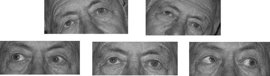
FIGURE 12.1 The three-step test in a patient with a right fourth NP. Note the RHT is greater with left gaze and with right head tilt.
G. Other examination features. It is important to look for orbital signs, which are sometimes subtle. These include proptosis, chemosis, conjunctival injection, and globe retraction with attempted eye movement, the latter best observed from the side. Careful inspection for pupil asymmetry or abnormal function is important, particularly in cases of mild or partial third NP. It is also helpful to look specifically and separately for evidence of aberrant regeneration causing third nerve misdirection. Lid abnormalities may be helpful, including both retraction (usually indicative of thyroid eye disease) and ptosis (common in myasthenia, third NP, and oculosympathetic palsy). When eye movements are incomplete with voluntary gaze, it is sometimes helpful to also test reflex movements, either with head turning (the oculocephalic or “doll’s head” maneuver), Bell’s phenomenon (upward gaze with forced eyelid closure), or calorics.
V. LOCALIZATION AND ETIOLOGIES
Diplopia can be caused by disorders of the brainstem, cranial nerves, extraocular muscles, and orbit. In most cases, the pattern of ocular motor dysfunction and the presence of associated abnormalities will allow accurate localization.
A. Brainstem. Disorders within the brainstem can cause abnormal eye movement by causing nuclear or fascicular cranial NP, INO, and skew deviation. Most lesions that involve supranuclear structures do not produce diplopia because reflex input keeps the eyes aligned. The main exceptions to this concept are diseases that affect vergence: divergence insufficiency causes esotropia at distance, and convergence paresis produces exotropia at near. Common causes of brainstem dysfunction are stroke, demyelinating disease, hemorrhage, inflammation, trauma, congenital anomalies, and certain metabolic derangements (e.g., Wernicke’s encephalopathy).
1. Nuclear lesions cause distinctive patterns of ocular motor dysfunction.
a. Unilateral lesions of the oculomotor nucleus cause ipsilateral paresis of the EOMs innervated by the third nerve, plus bilateral ptosis and loss of upgaze. The ipsilateral superior rectus subnucleus projects to the contralateral superior rectus causing loss of upgaze in both eyes. Because the levators are innervated by a single midline subnucleus, a unilateral nuclear palsy causes bilateral ptosis.
b. The trochlear nucleus innervates the contralateral superior oblique muscle. Head trauma, midbrain tumors, and hydrocephalus may damage both fourth nerves because they decussate in the anterior medullary velum.
c. A lesion of the abducens nucleus produces ipsilateral horizontal gaze palsy rather than a sixth NP because, in addition to motor neurons for abduction, the nucleus contains interneurons that supply the contralateral medial rectus subnucleus.
2. Fascicular lesions usually affect adjacent brainstem structures and can be localized accordingly. These syndromes are characterized by an ipsilateral ocular motor NP and contralateral hemisensory or hemimotor deficit and/or ipsilateral cerebellar dysfunction.
3. Damage to the medial longitudinal fasciculus produces a disconnection between the ipsilateral abducens nucleus and the contralateral medial rectus subnucleus, resulting in an INO. In addition to a variable degree of ipsilateral adduction deficit, there is slowing of medial rectus saccades and overshoot of contralateral abducting saccades with abduction nystagmus. Despite limitation of adduction, the eyes are usually aligned in primary position. In young patients, the most common etiology is demyelination and INO is commonly bilateral; in older individuals, the cause is most often stroke, and lesions are more often unilateral.
4. Loss of otolith input causes vertical strabismus termed skew deviation. Unlike misalignment due to cranial NP, the muscle imbalance in skew is typically comitant, that is, the same in all directions of gaze.
B. Cranial nerves. Common causes of cranial neuropathy are ischemia, compression, meningitis (inflammatory or neoplastic), trauma, and congenital. The most common cause of an isolated ocular motor palsy in older adults is microvascular disease, termed a vasculopathic palsy. Most patients have one or more vascular risk factors (diabetes mellitus, hypertension, and hypercholesterolemia). Onset is acute, usually with ipsilateral pain, which resolves spontaneously within 7 to 10 days. Resolution of the motility disturbance takes place within 6 months.
1. Oculomotor NP (third NP), when complete, produces an eye that is exotropic and hypotropic with absent adduction and vertical movements, profound ptosis, and a large poorly reactive pupil. Partial forms are often seen and the pattern is sometimes helpful (Fig. 12.2). Loss of upgaze plus ptosis indicates a lesion of the superior division. Pupil-sparing third NP is typical of a vasculopathic palsy. In contrast, third NP secondary to a posterior communicating (pCOM) artery aneurysm usually includes early pupil involvement. When third NP is partial, however, the presence of pupil sparing is less reassuring because this pattern is occasionally seen with pCOM aneurysm. An otherwise complete but pupil-sparing third NP is never due to a pCOM aneurysm. Pupil sparing is also common in third NP due to intracavernous lesions.
2. Trochlear NP (fourth NP) causes limitation of infraduction when the eye is adducted. Patients report vertical and torsional diplopia. The most common cause is trauma, which frequently produces bilateral fourth NP. Congenital fourth NP is also common and may present at any age due to decompensation of fusion.
3. Abducens NP causes esotropia and loss of abduction with slowing of lateral rectus saccades (Fig. 12.3). In young adults, multiple sclerosis and tumors are important considerations. Chronic sixth NP is often due to a compressive lesion at the skull base, such as meningioma. Sixth NP is sometimes a “false localizing sign” of increased ICP.
4. Combined CN palsy involving third, fourth, and sixth nerves localizes to the cavernous sinus or superior orbital fissure. The oculosympathetics and first division of the trigeminal nerve are commonly involved. Etiologies include tumor (primary and metastatic), vascular conditions (cavernous sinus thrombosis, fistula, and aneurysm), pituitary apoplexy, and inflammation.
C. Extraocular muscles.
1. Neuromuscular junction disease is most commonly due to myasthenia, which causes painless, pupil-sparing ptosis and diplopia. Other etiologies include paraneoplastic disease, botulism, and tick paralysis. Myasthenia is characterized by variability and prominent fatigability, often evident in the history and the examination. Symptoms are typically absent upon awakening. Ptosis increases with prolonged upgaze. Myasthenia may affect just a solitary muscle, two or more EOMs or all muscles diffusely. Because of this enormous variability, the disease may mimic a number of different ocular motor conditions, including CN palsies, gaze palsy, and INO. Weakness of eyelid closure as well as eyelid opening is a very helpful finding when present because the levators and the orbicularis oculi muscles are innervated by different cranial nerves (third and seventh, respectively).
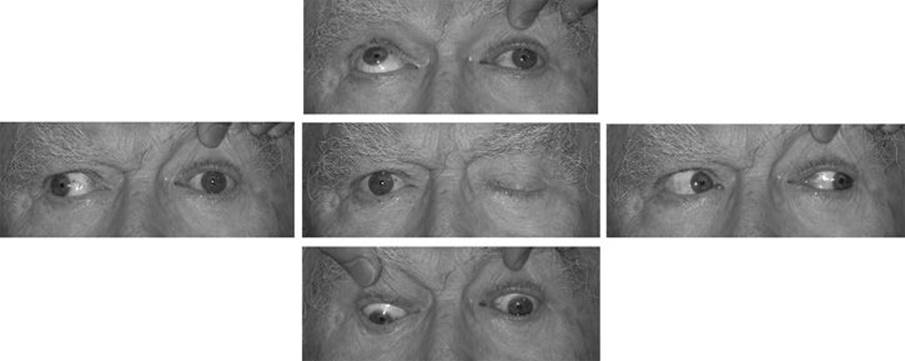
FIGURE 12.2 A 65-year-old man with a diabetic (vasculopathic) left third NP. There is loss of all third nerve function with the exception of the pupil.
2. Chronic progressive external ophthalmoplegia represents a group of hereditary disorders that causes limitation of eye movements with marked slowing of saccades and ptosis. Most are due to mitochondrial mutations, including Kearn–Sayre, which is sporadic and includes cardiac conduction abnormalities, atypical retinitis pigmentosa, and spongiform CNS changes.
3. Orbital myositis is occasionally due to a systemic granulomatous or vasculitic disorder but most commonly occurs as a form of idiopathic orbital inflammatory disease. Acute onset of diplopia is accompanied by pain with eye movement and the diagnosis can be confirmed on neuroimaging.
4. Graves’s ophthalmopathy causes restriction of eye movements due to inflammatory infiltrates, proliferation of fibroblasts, and edema. The inferior rectus is most commonly affected, producing loss of supraduction. Similarly, involvement of the medial rectus causes loss of abduction, mimicking a sixth NP (Fig. 12.4).
5. Giant cell arteritis is an important cause of diplopia in older individuals. Ischemia of extraocular muscles can produce a variety of patterns, sometimes mimicking CN palsy.
D. Orbit.
1. Masses in the orbit may displace the globe, mechanically interfere with EOMs, or cause cranial NP. Specific etiologies include primary tumors, vascular lesion, and inflammation including lesions of the paranasal sinuses. Pain, proptosis, chemosis, and conjunctival injection are common.
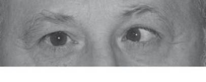
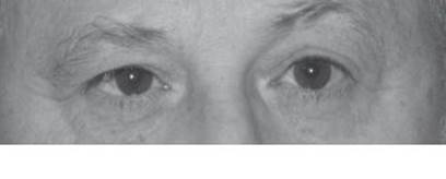
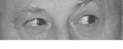
FIGURE 12.3 A 35-year-old man with acute onset of a left sixth NP secondary to multiple sclerosis. There is a moderate esotropia in primary position that increases on left gaze.

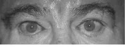
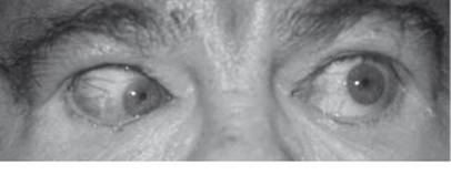
FIGURE 12.4 Esotropia and bilateral abduction deficit in a patient with thyroid eye disease. The presence of bilateral lid retraction and conjunctival injection are helpful signs indicating orbital restrictive disease rather than bilateral sixth NP.
2. Lesions at the orbital apex cause a distinctive combination of ipsilateral optic neuropathy and ocular motor disturbance. Because structures are crowded at the back of the orbit, a relatively small lesion can produce severe dysfunction. Small lesions in this area may not be appreciated on neuroimaging, but the clinical findings should point to the correct localization.
3. Orbital trauma often causes fracture of delicate orbital bones. A blowout fracture of the orbital floor can cause entrapment of the inferior rectus muscle, producing loss of supraduction. Fracture of the medial wall can entrap the medial rectus, producing an abduction deficit and thus mimicking a sixth NP.
VI. EVALUATION
A. Clinical diagnosis. It is often helpful to start by asking if the diplopia fits the pattern of a cranial NP. In the case of third NP, it is helpful to be able to recognize partial forms; however, it is important to note that isolated weakness of one-third nerve muscle is exceedingly rare. Cases that have this appearance more likely represent myasthenia, INO, or orbital restriction.
In cases in which the diplopia is not consistent with a single cranial NP, pattern recognition becomes extremely important. For instance, loss of supraduction when the eye is abducted is typical of inferior rectus restriction, such as from Graves’s disease or orbital floor fracture. Limitation of eye movement and an ipsilateral optic neuropathy constitute the orbital apex syndrome. Any combination of third, fourth, and sixth nerve palsies points to a lesion in the cavernous sinus or superior orbital fissure; there is no other location where these nerves come together. Certain brainstem disorders create distinctive ocular motor patterns. The one-and-a-half syndrome, for example, causes complete loss of conjugate gaze toward the side of the lesion and loss of adduction on the opposite side, leaving intact only abduction away from the lesion. In any patient with painless, pupil-sparing diplopia, the possibility of myasthenia should be considered.
B. Ancillary testing.
1. Radiographic testing should be directed to the area of interest determined from the clinical findings. MRI generally provides more information than CT; however, if an orbital process is suspected, it is important to include dedicated fat-suppressed orbit views with gadolinium. Alternatively, CT is an equally effective modality for imaging most orbital structures with the exception of the optic nerve. If optic neuritis is suspected, an MRI is the study of choice.
2. The ideal endpoint for an edrophonium chloride (Tensilon) test is a ductional deficit. A small phoria or a subjective judgment by the patient (e.g., “my eyes feel less tired”) is unreliable and should be avoided. False negative results are not uncommon; false positives are rare.
3. Blood tests. In all elderly patients with diplopia, GCA should be considered. Testing includes CBC, ESR, and CRP. Testing for acetylcholine receptor antibodies is appropriate if myasthenia is suspected, but antibodies are only positive in 30% of patients with the ocular form of the disease. Similarly, thyroid function tests may be normal in patients with thyroid orbitopathy (termed euthyroid Graves’s disease).
VII. URGENCY OF EVALUATION
In certain conditions, the clinical outcome depends on timely and appropriate treatment. Prompt recognition of these syndromes is therefore of paramount importance.
A. Aneurysmal third NP. Third NP due to a posterior communicating artery (pCOM) aneurysm usually presents with acute onset of ipsilateral pain and pupil involvement. Most such aneurysms can be identified on good quality MRA or CTA; however, these techniques are not yet 100% accurate, and the degree of accuracy varies among different institutions. If there is a high clinical suspicion of an aneurysm, catheter angiography should be obtained despite a negative MRA or CTA. This would include patients without vascular risk factors and those with a history suggestive of subarachnoid hemorrhage.
B. Pituitary apoplexy. Hemorrhage or infarction of a pituitary tumor usually causes acute onset of severe headache with symptoms and signs related to meningeal irritation. Visual loss, usually with a bitemporal pattern, and diplopia are common, the latter most often due to third nerve involvement, which may be unilateral or bilateral. In the large majority of cases of pituitary apoplexy, the presence of a tumor was not suspected prior to hemorrhage. The diagnosis is usually apparent on MRI scanning but may be missed with CT, which is usually the most easily accessed radiographic study in an emergency room setting. Prompt diagnosis is crucial because of the potential for acute adrenal insufficiency. Emergency management should include the administration of systemic corticosteroids in stress dosages (e.g., hydrocortisone 100 mg IV every 6 to 8 hours) with careful monitoring of electrolyte balance. Surgical decompression is usually indicated, although occasional patients do well with conservative management.
C. Giant cell arteritis. In any elderly patient with diplopia, the possibility of GCA should be entertained. In addition to inquiring about typical symptoms, an ESR and CRP should be obtained. High-dose steroid treatment should be started upon suspicion of the diagnosis and temporal artery biopsy obtained thereafter.
VIII. TREATMENT
Treatment of diplopia is generally directed toward the underlying condition. In some cases, it is helpful to provide symptomatic relief with prism correction. For diplopia that is expected to be temporary or to change over time, a paste-on (Fresnel) prism is most appropriate; for long-term treatment, a ground-in prism is generally preferred. In cases in which the deviation is too large and/or incomitant to treat with prisms, eye muscle surgery may be undertaken after ocular motility has been stable for a sufficient time period. Diplopia can always be dealt with by patching one eye. In young children, the patch should be alternated to prevent the development of amblyopia; in adults, this is not a concern, and patients are usually most comfortable with the nondominant eye covered.
![]()
Recommended Readings
Biousse V, Newman NJ, Oyesiku NM. Precipitating factors in pituitary apoplexy. J Neurol Neurosurg Psychiatry. 2001;71:542–545.
Borchert MS. Principles and techniques of the examination of ocular motility and alignment. In: Miller NR, Newman NJ, eds. Walsh and Hoyt’s Clinical Neuro-ophthalmology. Vol 1, 5th ed. Baltimore, MD: Lippincott Williams & Wilkins; 1998.
Brazis PW, Lee AG. Binocular vertical diplopia. Mayo Clin Proc. 1998;73:55–66.
Crane TB, Yee RD, Baloh RW, et al. Analysis of characteristic eye movement abnormalities in internuclear ophthalmoplegia. Arch Ophthalmol. 1983;101:206–210.
Jacobson DM, Trobe JD. The emerging role of magnetic resonance angiography in the management of patients with third cranial nerve palsy. Am J Ophthalmol. 1999;128:94–96.
Lee AG, Hayman LA, Brazis PW. The evaluation of isolated third nerve palsy revisited: an update on the evolving role of magnetic resonance, computed tomography and catheter angiography. Surv Ophthalmol. 2002;47:137–157.
Leigh RJ, Zee DS. The Neurology of Eye Movements. Oxford: Oxford University Press; 1999.
Richards BW, Jones FR Jr, Younge BR. Causes and prognosis in 4278 cases of paralysis of the oculomotor, trochlear and abducens cranial nerves. Am J Ophthalmol. 1992;113:489.
Weinberg DA, Lesser RL, Vollmer TL. Ocular myasthenia: a protean disorder. Surv Ophthalmol. 1994;39:169–210.
Yee RD, Whitcup SM, Williams IM, et al. Saccadic eye movements in myasthenia gravis. Ophthalmology. 1987;94:219–225.