
I. BACTERIAL MENINGITIS
The initial signs and symptoms of bacterial meningitis are fever, stiff neck, headache, lethargy, confusion or coma, nausea and vomiting, and photophobia. Examination of the CSF shows an elevated opening pressure (>180 mm H2O), a decreased glucose concentration (<40 mg per dl), polymorphonuclear pleocytosis, and an elevated protein concentration. The diagnosis is made by demonstrating the organism with Gram’s stain or in culture. Polymerase chain reaction (PCR) to detect bacterial nucleic acid in CSF is available at some centers. Bacterial meningitis is a neurologic emergency, and initial treatment is empiric until a specific organism is identified.
A. Therapeutic approach
1. Dexamethasone therapy. The American Academy of Pediatrics recommends consideration of dexamethasone in the treatment of infants and children 2 months of age and older with proven or suspected bacterial meningitis on the basis of findings on CSF examination, a Gram’s-stained smear of the CSF, or antigen test results. Dexamethasone is also recommended in adults with suspected bacterial meningitis and in proven pneumococcal meningitis (based on CSF gram-positive diplococci or blood or CSF cultures that are positive for Streptococcus pneumoniae). In clinical trials, dexamethasone improves the outcome of meningitis. In experimental models of bacterial meningitis, dexamethasone inhibits synthesis of the inflammatory cytokines, decreases leakage of serum proteins into the CSF, minimizes damage to the blood–brain barrier (BBB), and decreases CSF outflow resistance. Dexamethasone also decreases brain edema and intracranial pressure (ICP).
The recommended dosage of dexamethasone is 0.15 mg per kg intravenous (IV) every 6 hours for the first 4 days of therapy. The initial dose of dexamethasone should be given before or at least with the initial dose of antimicrobial therapy for maximum benefit. Dexamethasone is not likely to be of much benefit if started >24 hours or more after antimicrobial therapy has been initiated. The concomitant use of an histamine-2 receptor antagonist is recommended with dexamethasone to avoid gastrointestinal bleeding.
2. Antimicrobial therapy. If bacterial meningitis is suspected, antimicrobial therapy must be initiated immediately. This should be done before the performance of CT or lumbar puncture. Initial antimicrobial therapy is empiric and is determined by the most likely meningeal pathogen according to the patient’s age and underlying condition or predisposing factors.
a. The most likely etiologic organisms of bacterial meningitis in neonates are group B streptococci, enteric gram-negative bacilli (Escherichia coli), and Listeria monocytogenes. Empiric therapy for bacterial meningitis in a neonate should include a combination of ampicillin and either a third or fourth generation cephalosporin (cefotaxime or cefepime).
b. Empiric therapy for community-acquired bacterial meningitis in infants and children should include coverage for S.pneumoniae and Neisseria meningitidis. A third or fourth generation cephalosporin (ceftriaxone, cefotaxime, or cefepime) and vancomycin are recommended as initial therapy for bacterial meningitis in children in whom the etiologic agent has not been identified. Cefuroxime, also a third-generation cephalosporin, is not recommended for therapy for bacterial meningitis in children because of reports of delayed sterilization of CSF cultures associated with hearing loss in children treated with cefuroxime.
c. Empiric therapy for community-acquired bacterial meningitis in adults (15 to 50 years of age) should include coverage for S. pneumoniae and N. meningitidis. A third-generation cephalosporin (ceftriaxone or cefotaxime) or a fourth-generation cephalosporin (cefepime) plus vancomycin is recommended for empiric therapy. All CSF isolates of pneumococci and meningococci should be tested for antimicrobial susceptibility. Cefotaxime, ceftriaxone, or cefepime is recommended for relatively resistant strains of pneumococci (penicillin minimal inhibitory concentrations [MIC], 0.1 to 1.0 µg per ml and MICs of cefotaxime or cefepime ≤0.5 µg per ml). For highly penicillin-resistant pneumococcal meningitis (MIC > 1.0 µg per ml), a combination of vancomycin and a third-generation or fourth-generation cephalosporin is recommended. Penicillin G or ampicillin can be used for meningococcal meningitis.
d. Initial therapy for meningitis in postneurosurgical patients should be directed against gram-negative bacilli, Pseudomonas aeruginosa, and Staphylococcus aureus. Ceftazidime or meropenem is recommended for management of gram-negative bacillary meningitis in neurosurgical patients. Ceftazidime is the only cephalosporin with sufficient activity against P. aeruginosa in the CNS. Vancomycin should be added until infection with staphylococci is excluded.
e. In infants, children, and adults with CSF ventriculoperitoneal shunt infections, initial therapy for meningitis should include coverage for coagulase-negative staphylococci and S. aureus. The assumption can be made that the organism will be resistant to methicillin; therefore, initial therapy for a shunt infection should include IV vancomycin. Intrashunt or intraventricular vancomycin may also be needed to eradicate the infection.
f. In immunocompromised patients, the infecting organism can be predicted on the basis of the type of immune abnormality. In patients with neutropenia, initial therapy for bacterial meningitis should include coverage for L. monocytogenes, staphylococci, and enteric gram-negative bacilli. Patients with defective humoral immunity and those who have undergone splenectomy are unable to mount an antibody response to a bacterial infection or to control an infection caused by encapsulated bacteria. These patients are at particular risk of meningitis caused by S. pneumoniae, Haemophilus influenzae type b (Hib), and N. meningitidis.
g. The most common organisms causing meningitis in the older adult (50 years or older) are S. pneumoniae and enteric gram-negative bacilli; however, meningitis caused by Listeria organisms and Hib are increasingly recognized. The recommended initial therapy for meningitis in the older adult is either ceftriaxone, cefotaxime, or cefepime in combination with vancomycin and ampicillin. Table 43.1 lists empiric antimicrobial therapy for bacterial meningitis by age group. Tables 43.2 and 43.3 list the recommended antimicrobial therapy for bacterial meningitis in neonates, infants and children, and adults by meningeal pathogen.
TABLE 43.1 Empiric Antimicrobial Therapy for Bacterial Meningitis
|
Age Group |
Antimicrobial Agent |
|
Neonates |
Ampicillin plus cefotaxime or cefepime |
|
Infants and children |
Ceftriaxone, cefotaxime, or cefepime plus vancomycin |
|
Adults (15–50 y) |
|
|
Community acquired |
Ceftriaxone, cefotaxime, or cefepime plus vancomycin |
|
Otitis, mastoiditis, sinusitis |
Ceftriaxone plus vancomycin plus metronidazole |
|
Postneurosurgical |
Ceftazidime or meropenem plus vancomycin |
|
Immunocompromised |
Ceftazidime or meropenem plus ampicillin |
|
Older adults |
Ceftriaxone, cefotaxime, or cefepime plus vancomycin plus ampicillin |
TABLE 43.2 Recommended Antimicrobial Therapy for Bacterial Meningitis in Neonates, Infants and Children by Organism
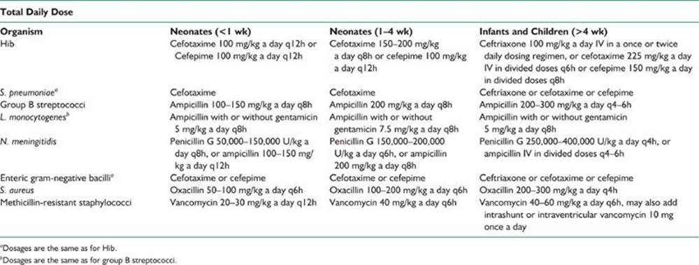
TABLE 43.3 Recommended Antimicrobial Therapy for Bacterial Meningitis in Adults by Organism
|
Organism |
Antimicrobial Agent |
|
S. pneumoniae |
Ceftriaxone 4 g/d (q12h) or cefotaxime 12 g/d (q4h) or cefepime 6 g/d (q8h) plus vancomycin 45–60 mg/kg/d (q6–12h) |
|
N. meningitidis |
Penicillin G 20–24 miU/kg a day (q4h) or ampicillin 12 g/d (q4h) |
|
Gram-negative bacilli (except P. aeruginosa) |
Ceftriaxone 4 g/d (q12h) or cefotaxime 12 g/d (q4h) or cefepime 6 g/d (q8h) |
|
P. aeruginosa |
Ceftazidime 8 g/d (q8h) or meropenem 6 g/d (q8h) |
|
H. influenzae type b |
Ceftriaxone or cefotaxime or cefepime |
|
S. aureus (methicillin-sensitive) |
Oxacillin 9–12 g/d (q4h) or nafcillin 12 g/d (q4h) |
|
Staphylococcus aureus (methicillin-resistant) |
Vancomycin 45–60 mg/kg/d (q6–12h) |
|
L. monocytogenes |
Ampicillin 12 g/d (q4h) with or without gentamicin |
|
Enterobacteriaceae |
Ceftriaxone or cefotaxime or cefepime |
3. Management of increased ICP. Increased ICP is an expected complication of bacterial meningitis and should be anticipated.
a. Elevation of the head of the bed 30˚.
b. Hyperventilation to maintain PaCO2 between 30 and 35 mm Hg.
c. Mannitol.
(1) Children. 0.5 to 2.0 g per kg infused over 30 minutes and repeated as necessary.
(2) Adults. 1.0 g per kg bolus injection and then 0.25 g per kg every 2 to 3 hours. A dose of 0.25 g per kg appears as effective as a dose of 1.0 g per kg in lowering ICP. The main exception is that the higher dose has a longer duration of action. Serum osmolarity should not be allowed to rise above 320 mOsm per kg.
d. Pentobarbital.
(1) Loading dose. 10 mg per kg over 30 minutes.
(2) 5 mg/kg/hour for 3 hours, supplemented with 200-mg IV boluses until a burst-suppression pattern is obtained on an EEG.
(3) Maintenance dosage. 1 mg/kg/hour by constant IV infusion.
4. Seizure activity is such a common complication of bacterial meningitis in adults, especially pneumococcal meningitis, which prophylactic anticonvulsant therapy is not unreasonable.
a. Prophylactic therapy. Phenytoin is administered at a dosage of 18 to 20 mg per kg at a rate no faster than 50 mg per minute. Propylene glycol, the diluent of IV phenytoin, can cause bradycardia, hypotension, and cardiac dysrhythmias. IV phenytoin should be administered via central access while the ECG and BP are monitored. If these side effects are observed, the rate of administration should be decreased. It is recommended that phenytoin be administered no faster than 25 mg per minute in the elderly.
Fosphenytoin, the prodrug to phenytoin, is administered at a dosage of 18 to 20 mg pulmonary embolism (PE) per kg at a rate no faster than 150 mg per minute. IV fosphenytoin does not contain propylene glycol and thus can be administered at a faster rate. A standard maintenance dosage is 100 mg of phenytoin or 100 mg PE of fosphenytoin every 8 hours. A serum concentration of 10 to 20 µg per ml should be maintained.
Levetiracetam is an alternative option for parenteral seizure prophylaxis therapy. The loading dose is 1,000 to 1,500 mg intravenously, followed by a maintenance dose of 500 mg every 12 hours. Levetiracetam dosing should be adjusted for creatinine clearance.
b. Status epilepticus.
(1) Lorazepam (0.1 mg/kg for adults; 0.05 mg/kg/dose for children) or diazepam (5 to 10 mg for adults; 0.2 to 0.3 mg/kg/dose for children) is administered IV.
(2) Phenytoin is administered in a dose of 18 to 20 mg per kg as described in I.A.4.a. Fosphenytoin is administered in a dose of 20 mg PE per kg as described in I.A.4.a. If seizures are not controlled, a repeat bolus of 10 mg PE per kg fosphenytoin or 500 mg phenytoin can be given.
(3) If phenytoin fails to control seizure activity, phenobarbital is administered intravenously at a rate of 100 mg per minute to a loading dose of 20 mg per kg. The loading dose of phenobarbital for children is also 20 mg per kg. The most common adverse effects of phenobarbital loading are hypotension and respiratory depression. Before phenobarbital loading, ensure that an endotracheal tube has been placed and mechanical ventilation begun. The primary reason for failure to control seizure activity is either that anticonvulsants are administered in subtherapeutic dosages or, as is the case for phenobarbital, the rate of administration is too slow.
(4) For more information on the management of refractory status epilepticus see Chapters 38, 39, and 58.
5. Fluid management. Most children with bacterial meningitis have hyponatremia (serum sodium concentration <135 mEq per L) at the time of admission. For this reason, fluid restriction to correct the serum sodium level is important, but this must be done taking into consideration the adverse effects of hypovolemia on cerebral perfusion pressure. The recommended initial rate of IV fluid administration is approximately three-fourths normal maintenance requirements, or approximately 1,000 to 2,000 ml/m2/day. A 5% dextrose solution with one-fourth to one-half normal saline solution and 20 to 40 mEq per L potassium is recommended. The volume of fluids administered can be gradually increased when serum sodium concentration increases to >135 mEq per L.
B. Expected outcome. Despite appropriate antimicrobial therapy, patients with bacterial meningitis are very sick. Prognosis depends on age, underlying or associated conditions, time from onset of illness to institution of appropriate antimicrobial therapy, the infecting organism, and the development of brain edema, coma, arterial and venous cerebrovascular complications, hydrocephalus or seizures. Pneumococcal meningitis has the worst prognosis, and a poor prognosis is associated with the extremes of age.
C. Prevention.
1. Rifampin or ciprofloxacin is recommended for all close contacts with a patient who has meningococcal meningitis. Rifampin is given in divided doses at 12-hour intervals for 2 days as follows: adults, 600 mg; children, 10 mg per kg; neonates (younger than 1 month), 5 mg per kg. It should not be given during pregnancy. Pregnant and lactating women may be given intravenous or IV ceftriaxone (a single injection of 250 mg).
The Advisory Committee on Immunization Practices recommends that adolescents and college freshmen be vaccinated against meningococcal meningitis with the tetravalent (Men A,C,W135,Y) meningococcal polysaccharide vaccine.
2. The vaccination of infants with the Hib conjugate vaccine has greatly decreased the incidence of Hib. Rifampin prophylaxis should be given to children younger than 4 years of age who have not been fully vaccinated against Hib disease who come in contact with a patient with Hib meningitis. It is recommended for all adults who have close contact with the patient and for the patient, because the organism usually is not eradicated from the nasopharynx with systemic antimicrobial therapy. Rifampin in the following dosages is recommended: adults, 20 mg per kg a day orally for 4 days; children, 20 mg per kg a day orally (maximum 600 mg per day) for 4 days; and neonates (younger than 1 month), 10 mg per kg a day for 4 days. Rifampin is not recommended for pregnant women.
II. HERPES ENCEPHALITIS
Encephalitis is inflammation of the brain parenchyma. Herpes simplex virus (HSV-1) is the principal cause of herpes encephalitis. Initial infection occurs either after exposure to infected saliva or respiratory secretions, the virus gaining access to the CNS along the olfactory nerve and tract into the limbic lobe, or as a result of reactivation of latent virus from the trigeminal ganglion. Virus is transmitted from infected persons to other persons only through close personal contact. The typical clinical presentation is a several-day history of fever and headache followed by memory loss, confusion, olfactory hallucinations, and seizures. The hallmark sign is a focal neurologic deficit suggestive of a structural lesion in the frontotemporal area. The EEG often is abnormal, demonstrating periodic sharp-wave complexes from one or both temporal regions. The abnormalities on the EEG arise from one temporal lobe initially but typically spread to the contralateral temporal lobe over a period of 6 to 10 days. On CT scans, there is a low-density lesion within the temporal lobe with mass effect. On MRI, the infection appears as an area of high signal intensity on T2-weighted (Fig. 43.1) and fluid-attenuated inversion recovery images. Examination of the CSF shows a lymphocytic pleocytosis (with an average WBC count of 50 to 500 cells per mm3), elevation in protein concentration, and normal or mildly decreased glucose concentration. CSF should be sent for the PCR assay to detect nucleic acid of HSV-1 and it should be examined for HSV-1 antibodies. Antibodies to HSV-1 do not appear in the CSF until approximately 8 to 12 days after symptom onset, and increase significantly during the first 2 to 4 weeks of infection. In order to determine if there is an intrathecal synthesis of antibodies against HSV-1, send CSF and serum samples. A serum:CSF ratio of <20:1 is considered diagnostic of HSV-1 infection. Because this infection produces areas of hemorrhagic necrosis, the CSF may contain RBC or xanthochromia. RBC in the CSF may inhibit the PCR, giving a false-negative result.
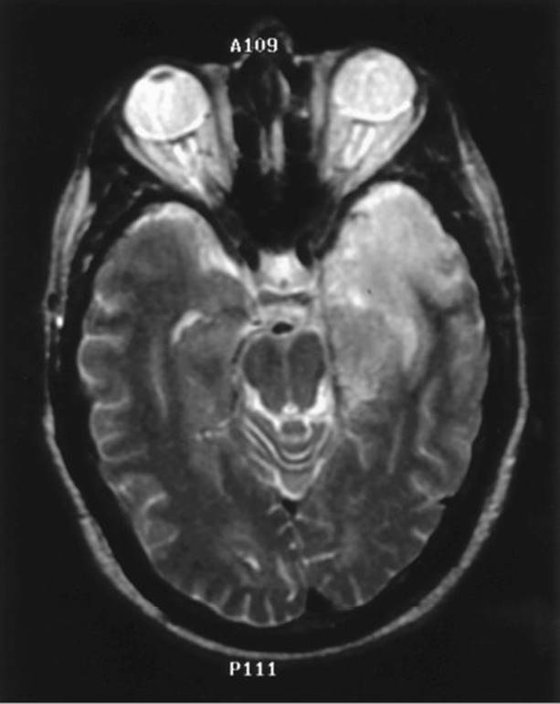
FIGURE 43.1 T2-weighted MRI shows classic high signal intensity lesion in left temporal lobe in herpes encephalitis.
A. Therapeutic approach.
1. Antiviral activity. Acyclovir is the antiviral drug of choice for HSV-1 encephalitis. It is given at a dosage of 10 mg per kg every 8 hours (30 mg per kg a day) IV, each infusion lasting >1 hour, for a period of 3 weeks. IV acyclovir can cause transient renal insufficiency secondary to crystallization of the drug in renal epithelial cells. For this reason, it is recommended that acyclovir be infused slowly over a period of 1 hour and that attention be paid to adequate IV hydration of the patient.
2. Anticonvulsant therapy. Seizure activity, either focal or focal with secondary generalization, occurs in two-thirds of patients with HSV-1 encephalitis. Anticonvulsant therapy is indicated if seizure activity develops, and the following drugs are recommended.
a. Lorazepam at dosages of 0.1 mg/kg for adults and 0.05 mg/kg for children, or diazepam at 5 to 10 mg for adults and 0.2 to 0.3 mg/kg/dose for children.
b. Phenytoin at a dosage of 18 to 20 mg per kg at a rate no faster than 50 mg per minute, or fosphenytoin at a dose of 18 to 20 mg PE per kg no faster than 150 mg per minute. The daily maintenance dosage of phenytoin should be determined by serum levels.
3. Therapy for increased ICP. Increased ICP is a common complication of herpes encephalitis and is associated with a poor outcome. Increased ICP should be aggressively managed as outlined in I.A.3.
B. Expected outcome. Among untreated patients with HSV-1 encephalitis, the mortality is higher than 70%, and only 2.5% of patients return to normal function after recovery. Patients treated with acyclovir have a significantly lower mortality of 19%, and 38% of these patients return to normal function.
C. Referrals. Because the clinical diagnosis of herpes encephalitis typically requires interpretation of the neurologic presentation, the EEG, neuroimaging studies, and CSF, the diagnosis of this severe and devastating neurologic illness should be made in consultation with a neurologist.
III. HERPES ZOSTER (SHINGLES)
A. Therapeutic approach. Oral valacyclovir 1,000 mg three times a day has been found superior to acyclovir in reducing zoster-associated pain. Oral acyclovir 800 mg five times a day and valacyclovir accelerate the rate of cutaneous healing and reduce the severity of acute neuritis and are most beneficial if treatment is initiated within 48 hours. Neither valacyclovir nor acyclovir reduces the incidence or severity of postherpetic neuralgia. For immunosuppressed patients, and for those with zoster ophthalmicus, many experts recommend the use of IV acyclovir. Ganciclovir can be considered an alternative agent for treatment. Corticosteroids have been proposed as adjunctive therapy in immunocompetent patients with varicella-zoster virus encephalitis.
B. Side effects. Oral acyclovir therapy has not been associated with renal dysfunction, but IV acyclovir can cause renal insufficiency. The risk of this complication is decreased by a slow rate of infusion.
C. Prevention. A varicella-zoster vaccine is available to decrease the risk of herpes zoster. Individuals with primary or acquired immunosuppression should not receive the vaccine.
IV. LYME’S DISEASE
Lyme’s disease is caused by the spirochete Borrelia burgdorferi, which is transmitted by the bite of an infected tick. It is endemic in the coastal northeast from Massachusetts to Maryland (particularly in New York), in the upper midwest in Minnesota and Wisconsin, and on the Pacific coast in California and southern Oregon.
In the majority of patients, initial infection is manifested by the appearance of an annular erythematous cutaneous lesion with central clearing of at least 5 cm in diameter, called erythema migrans. This lesion appears within 3 days to 1 month after a tick bite. Early disseminated Lyme’s disease is characterized by cardiac conduction abnormalities, arthritis, myalgia, fatigue, fever, meningitis, and cranial and peripheral neuropathy and radiculopathy. The most common neurologic abnormality during early disseminated Lyme’s disease is meningitis. The clinical features are typical of viral meningitis with symptoms of headache, mild neck stiffness, nausea, vomiting, low-grade fever, and photophobia. These symptoms may be associated with unilateral or bilateral facial nerve palsy or with symptoms of radiculitis (paresthesia and hyperesthesia) with or without focal weakness, transverse myelitis, mononeuritis multiplex or cognitive difficulties.
The majority of patients with Lyme’s disease have or have had an erythema migrans lesion. The diagnosis of Lyme’s disease begins with a serologic test for antibodies against B. burgdorferi. Most laboratories use an enzyme-linked immunosorbent assay technique. False-positive serologic results are a problem with this test for two reasons:
1. Tests can be performed on identical sera in different laboratories with different results. Because these tests are not well standardized, it is important to use a laboratory that is reliable in performing this test. A positive test result may indicate only exposure to B. burgdorferi rather than active infection. Persons who live in high risk areas may have measurable antibodies without having Lyme’s disease.
2. False-positive serologic results can occur with rheumatoid arthritis, Rocky Mountain spotted fever, infectious mononucleosis, syphilis, tuberculous meningitis, and leptospirosis.
When the ELISA is positive, then a Western blot should be obtained. The CSF is generally abnormal in neurologic Lyme’s disease. The typical spinal fluid abnormalities include lymphocytic pleocytosis (100 to 200 cells per mm3), an elevated protein concentration, and a normal glucose concentration. The CSF should be examined for intrathecal production of anti-B. burgdorferi antibodies, and an antibody index should be calculated. The antibody index is determined as the ratio of (anti-Borrelia IgG in CSF/anti-Borrelia IgG in serum) to (total IgG in CSF/total IgG in serum) and is defined as positive when the result is ≥2. The use of the antibody index to determine that there is an intrathecal production of antibodies is important as antibodies can be passively transferred from serum to CSF giving a false positive result, and Lyme’s antibodies may persist in CSF for years.
A. Therapeutic approach.
1. Patients with facial nerve palsy without other neurologic manifestations can be treated with doxycycline 100 mg by mouth twice a day for 14 to 28 days. Doxycycline should not be given to pregnant women.
2. Parenteral ceftriaxone is recommended for patients with neurologic complications of Lyme’s disease, although oral doxycycline may be equally effective in the absence of brain or spinal cord involvement. The adult dosage of ceftriaxone is 2 g per day, which may be given in a single daily dose, and the dosage for children is 75 to 100 mg per kg a day (up to 2 g per day). Treatment is given for at least 2 weeks and should be continued for an additional 2 weeks if the response to treatment is slow or there is severe infection.
3. Alternatives to ceftriaxone are penicillin G and cefotaxime. Penicillin G is administered at an adult dosage of 3 to 4 million units (miU) every 4 hours for 10 to 14 days or at a child dosage of 250,000 U per kg a day in divided doses. The major side effect of penicillin G is hypersensitivity reaction. Cefotaxime is given at dosages of 2 g three times a day for adults and 150 to 200 mg per kg a day (every 6 hours) for children.
B. Expected outcome. The condition of patients with neurologic complications of early disseminated Lyme’s disease (meningitis, cranial neuropathy, and peripheral neuropathy) should improve clinically within days, although improvement of facial weakness and radicular symptoms can take weeks. Prolonged antimicrobial therapy does not improve symptoms of post-Lyme’s syndrome and is not recommended.
C. Prevention. The deer tick is the usual vector of Lyme’s disease in the northeastern and the midwestern United States. Wearing protective clothing can help decrease the risk of infection. Transmission of infection is unlikely if the tick has been attached for <24 hours.
V. CRYPTOCOCCAL MENINGITIS
The diagnosis of cryptococcal meningitis is made when examination of the CSF shows lymphocytic pleocytosis, a decreased glucose concentration, and a positive result of a CSF cryptococcal antigen assay, fungal smear, or culture.
A. Therapeutic approach. The management of cryptococcal meningitis is divided into induction, consolidation and maintenance (suppressive therapy). Induction therapy includes amphotericin B 0.7 to 1 mg per kg a day in combination with flucytosine 100 mg per kg a day divided into four daily doses for at least 4 weeks. Treatment then is switched to consolidation therapy with fluconazole 400 mg per day for a minimum of 8 weeks. Present guidelines recommend slight modifications for induction and consolidation therapy in HIV infected patients and in organ transplant recipients. In the treatment of patients with HIV infection, fluconazole 200 mg per day is continued for lifelong suppressive therapy.
B. Side effects. The most important adverse effect of amphotericin B is renal dysfunction, which occurs in 80% of patients. Renal function, hemoglobin concentration, and electrolytes should be monitored closely. Renal toxicity appears to be reduced or prevented by means of careful attention to serum sodium concentration at the time of administration of amphotericin B. If renal insufficiency develops, AmBisome (Fujisawa, Deerfield, IL, USA) 5 mg per kg a day or amphotericin B lipid complex 5 mg per kg a day can be substituted for amphotericin B. Flucytosine is generally well-tolerated; however, bone marrow suppression with anemia, leukopenia, or thrombocytopenia can develop. These hematologic abnormalities occur more often when serum concentrations of the drug exceed 100 mg per ml; therefore serum concentrations of flucytosine should be monitored and the peak serum concentration kept well below 100 mg per ml.
VI. NEUROSYPHILIS IN IMMUNOCOMPETENT PATIENTS
In immunocompetent patients, the clinical presentation of neurosyphilis falls into one or more of the following categories: (1) asymptomatic neurosyphilis, (2) meningitis, (3) meningovascular syphilis, (4) dementia paralytica, and (5) tabes dorsalis. The diagnosis of neurosyphilis is based on a reactive serum treponemal test and CSF abnormalities. In neurosyphilis, there is often a mild CSF mononuclear pleocytosis with a mild elevation in the CSF protein concentration or a reactive CSF venereal disease research laboratory (VDRL) test. A positive CSF VDRL test establishes the diagnosis of neurosyphilis. A nonreactive CSF VDRL result does not exclude neurosyphilis. The CSF VDRL test is nonreactive in 30% to 57% of patients with neurosyphilis. A nonreactive CSF fluorescent treponemal antibody absorption test (FTA-ABS) excludes the diagnosis of neurosyphilis in all cases except early syphilis. A reactive CSF FTA-ABS is nonspecific and cannot be used to make a diagnosis of neurosyphilis.
A. Therapeutic approach. The regimen recommended by the CDC for management of neurosyphilis is IV aqueous penicillin G at 18 to 24 miU per day (3 to 4 miU every 4 hours) for 10 to 14 days. An alternative regimen is intramuscular procaine penicillin at 2.4 miU per day and oral probenecid at 500 mg four times a day, both for 10 to 14 days.
B. Expected outcome. The serum VDRL titer should decrease after successful therapy for neurosyphilis. The serum fluorescent treponemal antibody absorption test and the microhemagglutination—Treponema pallidum test remains reactive for life. The CSF WBC count should be normal 6 months after therapy is completed. If on reexamination of the CSF, the WBC count remains elevated, repetition of treatment is indicated.
VII. TUBERCULOUS MENINGITIS
A. Clinical presentation. Tuberculous meningitis manifests as either subacute or chronic meningitis, as a slowly progressive dementing illness, or as fulminant meningoencephalitis. The intradermal tuberculin skin test is helpful when the result is positive. Radiographic evidence of pulmonary tuberculosis is found more often in children with tuberculous meningitis than in adults with tuberculous meningitis. The classic abnormalities at CSF examination are decreased glucose concentration, elevated protein concentration, and polymorphonuclear or lymphocytic pleocytosis. The CSF pleocytosis is typically neutrophilic initially but then becomes mononuclear or lymphocytic within several weeks. Acid-fast bacilli are difficult to find in smears of CSF. Culture of CSF is the standard for diagnosis but is insensitive. Cultures are reported to be positive in 25% to 75% of cases of tuberculous meningitis, requiring 3 to 6 weeks for growth to be detectable. PCR assays that detect Mycobacterium tuberculosis rRNA in CSF have shown good results in clinical trials.
B. Therapeutic approach. Current recommendations for the management of tuberculous meningitis in children and adults include a combination of isoniazid (5 to 10 mg per kg a day up to 300 mg per day), rifampin (10 to 20 mg per kg a day up to 600 mg per day), and pyrazinamide (25 to 35 mg per kg a day up to 2 g per day). If the clinical response is good, pyrazinamide is discontinued after 8 weeks, and isoniazid and rifampin are continued for an additional 10 months. Ethambutol is added, and the course of treatment is extended to 1 to 2 years for immunocompromised patients. The American Academy of Pediatrics recommends addition of streptomycin at 20 to 40 mg per kg a day to the foregoing regimen for the first 2 months. Pyridoxine may be administered at a dosage of 25 to 50 mg per day to prevent the peripheral neuropathy that can result from use of isoniazid. Corticosteroid therapy is recommended when clinical deterioration occurs after treatment has begun. Dexamethasone can be administered at a dosage of 0.3 to 0.5 mg per kg a day for the 1st week of treatment and followed by oral prednisone.
VIII. NEUROCYSTICERCOSIS
A diagnosis of neurocysticercosis should be considered when a patient has seizures and neuroimaging evidence of cystic brain lesions. Cysticercosis is acquired by ingesting the eggs of the Taenia soliumtapeworm shed in human feces.
A. Principal forms. The lesions of neurocysticercosis can be found in the brain parenchyma, the ventricles, the subarachnoid space, or in the basilar cisterns (racemose forms). In the parenchymal form, single or multiple cysts are found in the gray matter in the cerebrum and cerebellum. The most common clinical manifestation of parenchymal neurocysticercosis is new-onset seizure activity. In the ventricular form, single or multiple cysts are adherent to the ventricular wall or free in the CSF. Cysts are most common in the area of the fourth ventricle. In subarachnoid neurocysticercosis, cysts are found in the subarachnoid space or fixed under the pia and burrowed into the cortex. In the racemose form, cysts grow, often in clusters, in the basilar cisterns and obstruct the flow of CSF. Hydrocephalus and cysticercotic encephalitis, a severe form of neurocysticercosis due to intense inflammation around cysticerci and cerebral edema, are the most common cause of increased ICP.
B. Diagnosis. The diagnosis of neurocysticercosis depends on clinical features and evidence of cystic lesions demonstrating the scolex at CT or MRI, or other neuroimaging abnormalities (ring enhancing cystic lesions and intraparenchymal calcifications) (Fig. 43.2) suggestive of neurocysticercosis in addition to a positive result of a serologic assay for the detection of antibodies to T. solium. Biopsy for histologic demonstration of the parasite is rarely done.
C. Therapeutic approach.
1. Cysticidal therapy consists of praziquantel at a dose of 50 mg/kg/day for 15 days, or praziquantel 100 mg/kg in three divided doses at 2-hour intervals (single day course) or albendazole at a dosage of 15 mg/kg a day for 8 days.
2. Corticosteroids. Cysticidal therapy frequently causes an inflammatory response with an increase in CSF protein concentration and CSF pleocytosis. This may result in an exacerbation of signs and symptoms. The incidence of an inflammatory response is reduced by the concomitant use of corticosteroids, and their use is recommended both before and during treatment with anticysticidal therapy. Plasma levels of albendazole are increased by dexamethasone; plasma levels of praziquantel are decreased by dexamethasone therapy. This should be taken into consideration when corticosteroid therapy is used to decrease the headaches and vomiting induced by the destruction of parasites in cysticidal therapy. Dexamethasone 24 to 32 mg per day is recommended for patients with subarachnoid cysts, encephalitis, angiitis, or arachnoiditis.
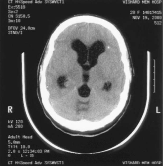
FIGURE 43.2 CT scan shows parenchymal brain calcification and hydrocephalus due to neurocysticercosis.
3. Side effects. Phenytoin and carbamazepine decrease serum praziquantel levels due to their induction of the cytochrome P-450 liver enzyme system. If one of these anticonvulsants is used with praziquantel, it is recommended that oral cimetidine be added at a dosage of 800 mg twice a day. Cimetidine inhibits the cytochrome P-450 enzyme system, and in this way increases serum levels of praziquantel.
4. Surgical therapy. Intraventricular cysts necessitate surgical therapy, and when they obstruct the flow of CSF with resulting hydrocephalus, an intraventricular shunting device is indicated.
D. Expected outcome. The prognosis of patients with parenchymal neurocysticercosis is very good with cysticidal therapy. Cystic lesions should disappear within 3 months of treatment. The mortality is higher among patients with increased ICP, hydrocephalus, or the racemose form of the disease.
E. Prevention. Humans can acquire cysticercosis by eating food handled and contaminated by T. solium tapeworm carriers. Persons at high risk of tapeworm infestation who are employed as food handlers should be screened for intestinal parasites. Improved sanitation can decrease the incidence of cysticercosis from contaminated food or drinking water.

Recommended Readings
Brouwer MC, Heckenberg SGB, de Gans J, et al. Nationwide implementation of adjunctive dexamethasone therapy for pneumococcal meningitis. Neurology. 2010;75:1533–1539.
Cadavid D. Spirochetal infection. In: Roos KL, Tunkel AR, eds. Handbook of Clinical Neurology. Bacterial Infections of the Central Nervous System. Edinburgh: Elsevier; 2010:179–219.
Corona T, Lugo M, Medina R, et al. Single day praziquantel therapy for neurocysticercosis. N Engl J Med. 1996;334:125.
Del Brutto OH, Wadia NH, Dumas M, et al. Proposal of diagnostic criteria for human cysticercosis and neurocysticercosis. J Neurol Sci. 1996;142:1–6.
Halperin JJ, Shapiro ED, Logigian E, et al. Practice parameter: treatment of nervous system Lyme disease (an evidence-based review). Neurology. 2007;69:91–102.
Kent SJ, Crowe SM, Yung A, et al. Tuberculous meningitis: a 30-year review. Clin Infect Dis. 1993;17:987–994.
Marra CM. Syphilis and human immunodeficiency virus infection. Semin Neurol. 1992;12:43–50.
Newton RW. Tuberculous meningitis. Arch Dis Child. 1994;70:364–366.
Perfect JR, Dismukes WE, Dromer F, et al. Clinical practice guidelines for the management of cryptococcal disease: 2010 update by the Infectious Diseases Society of America. Clin Infect Dis. 2010;50:291–322.
Pfister HW, Roos KL. Bacterial meningitis. In: Hacke W, ed. Neurocritical Care. Berlin: Springer–Verlag; 1994:377–397.
Roos KL, Tunkel AR, Scheld WM. Acute bacterial meningitis. In: Scheld WM, Whitley RJ, Marra CM, eds. Infections of the Central Nervous System. Philadelphia, PA: Lippincott Williams & Wilkins; 2004:347–422.
Skoldenberg B. Herpes simplex encephalitis. Scand J Infect Dis. 1991;78:40–46.
Tunkel AR, Hartman BJ, Kaplan SL, et al. Practice guidelines for the management of bacterial meningitis. Clin Infect Dis. 2004;39:1267–1284.
Whitley RJ, Gnann JW. Drug therapy: acyclovir—a decade later. N Engl J Med. 1992;327:782–789.
Wood MJ, Shukla S, Fiddian AP, et al. Treatment of acute herpes zoster: effect of early (<48 h) versus late (48–72 h) therapy with acyclovir and valacyclovir on prolonged pain. J Infect Dis. 1998;178(suppl 1):S81–S84.