
Sleep disturbances are prevalent in the general population, but certain groups such as older adults, women, and patients with chronic comorbid medical, neurologic, and psychiatric disorders are at particular risk. Indeed, the most recent evidence points to a bidirectional relationship between health and sleep. Sleep problems influence health-related quality of life and may contribute to the development of, or exacerbate, medical and neurologic conditions. Patients who report disturbed sleep generally describe one or more of three types of problems—insomnia, excessive daytime sleepiness (EDS), and abnormal motor activities, complex behaviors, or disturbed sensations during sleep.
The 2005 revised International Classification of Sleep Disorders (ICSD-2) lists five major categories of sleep disorders: insomnias—that is, disorders of initiating and maintaining sleep; sleep-related breathing disorders; hypersomnias—that is, disorders of excessive sleepiness; movement disorders; and circadian rhythm sleep disorders (CRSDs). To assist the clinician, in diagnosing and treating sleep disorders, a differential diagnosis-based classification adapted from the ICSD-2 is used in this chapter.
I. INSOMNIA
Insomnia is a disorder characterized by symptoms of inability to fall asleep, maintain sleep, perception of inadequate sleep, and/or nonrestorative sleep. These symptoms result in distress or impairment of daytime functioning. The three main categories of insomnia are psychophysiologic insomnia, idiopathic insomnia, and paradoxical insomnia (state misperception).
A. Psychophysiologic and idiopathic insomnias.
1. Course. For the diagnosis of psychophysiologic insomnia to be made, a patient has to have sleep difficulties that substantially affect daytime functioning and have a learning or conditioning component that typically involves one or more of the following: daily worries about not being able to fall asleep or stay asleep accompanied by intense efforts to fall asleep each night; paradoxical improvement away from the usual sleep environment (e.g., in another room of the house or away from home); and somatized tension and anxiety associated with bedtime and the subject of sleep. The most difficult differential diagnosis is with generalized anxiety disorders in which anxiety is pervasive and involves most aspects of daily life rather than exclusively the inability to sleep. Differentiation from affective disorders, such as depression, is also important. Idiopathic or primary insomnia is a lifelong inability to sleep, presumably associated with a predisposition for insomnia resulting from abnormality of the sleep–wake cycle or autonomic activity. Patients with this condition are a heterogeneous group. Most have been poor sleepers since childhood, and the insomnia, although it persists over the entire life span, can be aggravated by stress and tension. Patients with idiopathic insomnia may have atypical reactions to stimulants and sedatives. Idiopathic insomnia often is accompanied by other factors such as poor sleep hygiene or psychiatric disorders. Therefore, there tends to be some overlap between primary insomnia with insomnia that is comorbid with psychiatric disorders.
2. Treatment and outcome. A multimodal individualized approach is indicated for most patients. Optimizing the treatment of comorbid medical, neurologic, and psychiatric conditions, as well as identifying medications or behaviors that promote insomnia are essential first steps. A combined treatment approach involving good sleep hygiene, cognitive behavioral therapy (CBT), and medications is most often employed. A 4- to 8-week program of sleep hygiene counseling, cognitive/behavioral modifications, and judicious use of hypnotics is recommended. If insomnia does not improve after this period of treatment, referral to a sleep specialist should be considered for further evaluation.
a. Cognitive behavioral therapy. The most widely used behavioral therapy program in the management of insomnia includes a combined program of sleep hygiene education, relaxation techniques, stimulus-control therapy, and sleep restriction therapy. Relaxation techniques may include progressive muscle relaxation, biofeedback, deep breathing, meditation, guided imagery, and other techniques to control cognitive arousal. These techniques are first taught during training sessions and then practiced daily for 20 to 30 minutes by the patient at home, usually around bedtime. Stimulus-control therapy is useful in the management of conditioned insomnia. This technique is an attempt to break the conditioning by teaching the patient to associate the bedroom with sleep behavior. The instructions for sleep hygiene and stimulus-control behavioral therapy are listed in Tables 55.1 and 55.2. Sleep restriction therapy involves curtailment of time in bed, so that sleep efficiency (time asleep divided by time in bed) is 85% or greater. As sleep efficiency increases, time in bed is gradually lengthened. Shorter duration behavioral interventions and internet-based CBT are also available.
b. Hypnotic drugs. The most widely used prescription hypnotics are the benzodiazepine receptor agonists, which include benzodiazepines and the non-benzodiazepine receptor agonist (BZRA) hypnotics, such as eszopiclone, zaleplon, and zolpidem. Traditionally, this class of medications was indicated for short-term use. More recently, with the recognition that insomnia is often chronic and with the availability of longer term studies for up to a year, the short-term indication has been removed from the newly The U.S. Food and Drug Administration (FDA)-approved BZRA hypnotics such as eszopiclone and zolpidem MR. Ramelteon, a melatonin receptor agonist and low-dose doxepin, represent different classes of hypnotics that in the nonscheduled category.
TABLE 55.1 Sleep Hygiene Instructions
|
Homeostatic drive for sleep |
|
Avoid naps, except for a brief 10- to 15-min nap 8 hr after arising; check with your physician first, because in some sleep disorders naps can be beneficial |
|
Restrict sleep period to average number of hours you have actually slept per night in the preceding week. Quality of sleep is important. Too much time in bed can decrease quality on the subsequent night |
|
Get regular exercise every day, preferably 40 min in the afternoon. It is best to finish exercise at least 3 hr before bedtime |
|
Take a warm bath 90 to 120 min before bedtime to help lower body temperature |
|
Circadian factors |
|
Keep a regular out-of-bed time (do not deviate >1 hr) 7 d a week |
|
Do not expose yourself to bright light if you have to get up at night |
|
Expose yourself to bright light, either outdoor or artificial, during the day |
|
Drug effects |
|
Do not smoke to get yourself back to sleep |
|
Do not smoke after 7:00 p.m.; give up smoking entirely |
|
Avoid caffeine and limit caffeine use to no more than three cups no later than 10:00 a.m. |
|
Avoid alcoholic beverages after 7:00 p.m. |
|
Arousal in sleep setting |
|
Keep clock face turned away, and do not find out what time it is when you wake up at night |
|
Avoid strenuous exercise after 6:00 p.m. |
|
Do not eat or drink heavily for 3 hr before bedtime. A light bedtime snack may help |
|
If you have trouble with regurgitation, be especially careful to avoid heavy meals and spices in the evening. You may have to raise the head of your bed |
|
Keep your room dark, quiet, well ventilated, and at a comfortable temperature |
|
Use a bedtime ritual. Reading before lights-out may be helpful if it is not occupationally related |
|
Do not try too hard to sleep; instead, concentrate on the pleasant feeling of relaxation |
|
Use stress management and relaxation techniques in the daytime |
|
Be sure your mattress pillow is of the right height and firmness |
|
Use the bedroom only for sleep; do not work or do other activities that lead to prolonged wakefulness |
TABLE 55.2 Stimulus-Control Behavioral Therapy
|
Go to bed only when sleepy. Stay up until you are really sleepy, and then return to bed. If sleep still does not come easily, get out of bed again. The goal is to associate bed with falling asleep quickly |
|
Use the bed only for sleeping. Do not read, watch television, or eat in bed |
|
If unable to sleep, get up and move to another room |
|
Repeat the preceding step as often as necessary throughout the night |
|
Set the alarm and get up at the same time every morning, regardless of how much you slept during the night. This helps the body acquire a constant sleep–wake rhythm |
|
Do not nap during the day |
TABLE 55.3 FDA Approved Hypnotics for the Management of Insomnia

The choice of hypnotic may depend on the type of insomnia. For example, if the predominant problem is falling asleep, a fast-acting, short-half-life hypnotic may be preferable. If the problem is frequent awakenings and sleep maintenance insomnia, a longer acting hypnotic may be more effective. Most hypnotics approved by the FDA are indicated for the treatment of sleep onset insomnia, whereas eszopiclone, zolpidem-CR and low-dose doxepin are also indicated for the treatment of sleep maintenance insomnia. In practice, sedating antidepressants, such as the tricyclic antidepressants and heterocyclics (trazodone), are often used off label for the treatment of insomnia. However, there is limited data regarding their efficacy or long-term safety for the treatment of insomnia that is not comorbid with depression. The exception is low-dose doxepin (3 to 6 mg), which is FDA approved for insomnia.
The most widely used prescription hypnotics and their properties are listed in Table 55.3. Although patients with chronic insomnia rarely become “great” sleepers after treatment, most can manage the predisposition to insomnia by using sleep hygiene, cognitive behavioral treatment, and when indicated hypnotics.
B. Paradoxical insomnia.
1. Course. It is not uncommon for patients to overestimate sleep latency and underestimate total sleep time. In paradoxical insomnia, this tendency is extreme.
2. Diagnosis. The disorder is characterized by reports of persistent difficulty falling, staying asleep or disturbed sleep, although sleep duration and quality are objectively normal.
3. Treatment and outcome. Reassuring patients with the fact that their sleep is normal and that they sleep longer than they think they do and cognitive behavioral treatments are effective.
C. Insomnia associated with psychiatric disorders.
1. Course. Insomnia is often comorbid with psychiatric conditions. Results of epidemiologic studies suggest that as many as 57% of persons with insomnia have a psychiatric condition or will have one within 1 year. The comorbid condition usually is a mood disorder, anxiety disorder, somatoform disorder, personality disorder, schizophrenia, or substance abuse. Sleep in major depression is characterized by early morning awakening (2 to 4 hours after sleep onset) and frequent nocturnal awakening with inability to reinitiate sleep. Insomnia often precedes the diagnosis of depression. The incidence of insomnia among patients with anxiety disorders is high. The typical symptoms are difficulty with sleep initiation and, to a lesser degree, nocturnal awakenings. Fatigue is common, but napping is unusual. Patients with anxiety disorders are susceptible to conditioning factors that produce psychophysiologic insomnia.
2. Treatment and outcome. Treatment should address the comorbid psychiatric disorder as well as insomnia. For major depressive and anxiety disorders, this involves use of antidepressants or anxiolytics such as the selective serotonin reuptake inhibitors (SSRIs). An antidepressant with sedative properties is favored over a less-sedating one for patients with insomnia. Administration 30 minutes before bedtime also aids in promoting sleep. Amitriptyline, trimipramine, doxepin, trazodone, and mirtazapine are the most sedating, whereas protriptyline and SSRIs such as fluoxetine have stimulating effects that may worsen insomnia. Antidepressants with anxiolytic properties are useful in the treatment of anxious, depressed patients and facilitate psychotherapeutic or pharmacologic treatment. Anticholinergic side effects of tricyclic antidepressants (cardiotoxicity, urinary retention, erectile dysfunction, and dry mouth) limit the usefulness of these agents, particularly in the elderly.
Recent studies demonstrate that insomnia may persist despite adequate treatment of depression and that insomnia predicts future relapse of depression. Therefore, oftentimes, a parallel approach that combines treatment for both depression and insomnia is recommended. If the patient is refractory to treatment, referral to a sleep specialist or psychiatrist is recommended for further evaluation of comorbid psychiatric or other sleep disorders.
II. CIRCADIAN RHYTHM SLEEP DISORDERS
Circadian rhythms are generated by a neural clock located in the suprachiasmatic nucleus of the hypothalamus. Disruption of biologic timing results in circadian rhythm disorders that are most often associated with patients’ reports of insomnia and excessive sleepiness. CRSDs are characterized by essentially normal total sleep time that is not synchronized with conventional environmental light–dark cycles and periods of sleep. Diagnosis requires specialized assessment, including use of a sleep diary for 7 days alone, or in combination with actigraphy, physiologic markers of circadian timing such as core body temperature or melatonin onset. A careful history interview to elicit the appropriate major diagnostic criteria is a key. CRSDs include delayed sleep phase disorder (DSPD), advanced sleep phase disorder (ASPD), non-24-hour sleep–wake disorder, irregular sleep–wake rhythm disorder, (ISWR) shift work sleep disorder (SWSD), and jet lag disorder. Effective treatment for CRSDs typically require a multifaceted approach to realign circadian rhythms with the use of timed bright light exposure and low-dose melatonin, together with cognitive behavioral treatments that promote healthy sleep habits. Melatonin is not approved by the FDA for the treatment of CRSDs, and one should also be aware of potential side effects such as headaches, vivid dreams, nausea, and cardiovascular effects.
A. DSPD and ASPD.
1. Course. DSPD is characterized by a persistent inability to fall asleep until the early morning hours (1 to 3 a.m., and sometimes later) and difficulty waking up in the morning. If allowed, the patient would sleep until the late morning or early afternoon (10 a.m. to 2 p.m.). When the patient is forced to rise at 7 or 8 a.m., sleep is curtailed, and daytime sleepiness develops. Despite the daytime sleepiness, patients find that in the evening they become more alert and remain unable to fall asleep until the early morning hours. The prevalence rate is estimated to be between 1.7% in the general population to 7% of those with insomnia complaints. Onset of this disorder typically occurs during adolescence or early adulthood. ASPD is characterized by early evening sleep onset (7 to 9 p.m.) and early morning awakening (3 to 5 a.m.). Although DSPD predominates at younger ages and ASPD at older ages, both disorders can result in sleep problems throughout life. Because many features of the sleep of patients with depression resemble those of either DSPD or ASPD, depression and other psychiatric disorders must be considered in the differential diagnosis.
2. Treatment and outcome.
a. Chronotherapy is a behavioral technique in which bedtime is systematically delayed (for DSPD) or advanced (for ASPD) in 3-hour increments each day until the desired sleep phase is achieved. The patient is then instructed to maintain the newly established bedtime rigidly. Although this approach works, it is an arduous procedure, and maintenance of the effect has been difficult.
b. Bright light therapy. Light intensity >2,500 lux is considered bright. Appropriately timed bright light (white or blue/green enriched) exposure can reset the timing of circadian rhythms, and normalize circadian phase in DSPD and ASPD. Exposure to bright light in the early morning results in an advancement of circadian phase, whereas exposure to light in the evening delays circadian rhythms. For management of DSPD, exposure to light usually is scheduled for 1 to 2 hours in the morning (close to the time of habitual awakening). For ASPD, light exposure is recommended in the evening, approximately 2 to 4 hours before scheduled bedtime. Avoidance of bright light in the evening in DSPD should also be encouraged. Despite high rates of success in achieving the desired sleep phase under immediate treatment, many patients do not continue the light regimen and have a relapse. Some patients are able to maintain a normalized phase without maintenance of light exposure for as long as several months, whereas others drift back toward the pretreatment phase within a few days.
c. Melatonin has been shown to shift the phase of circadian rhythms in humans. Although not approved by the FDA, melatonin of 1 to 5 mg has been shown to be effective when taken in the early evening for patients with DSPD.
B. Free-running disorder (FRD).
1. Course. Individuals with FRD typically have a longer than 24-hour circadian rhythm, similar to those living in temporal isolation. Because these patients are unable to entrain to the external 24-hour physical, social or activity cycles, sleep and wake periods progressively drift later each day. Diagnosis of FRD includes complaints of insomnia or excessive sleepiness associated with the misalignment between the endogenous circadian rhythm and the light-dark cycle. FRD is most common in blind people, but can occur in sighted persons.
2. Treatment and outcomes. Both behavioral and pharmacologic options are available for the treatment of FRD, depending on whether the patient is sighted or blind. For blind and sighted patients, planned sleep schedules and/or low-dose melatonin (0.5 to 3 mg) approximately 1 to 2 hours before habitual bedtime are recommended. In sighted persons, the addition of timed exposure to bright light is also recommended.
C. Irregular sleep wake rhythm disorder.
1. Course. ISWR differs from the phase disorders in that there is loss of circadian rhythmicity, which results in the lack of a long, consolidated sleep period. Sleep usually is broken into three or more short sleep periods or naps during the course of 24 hours. Irregular sleep–wake patterns occur among patients with Alzheimer’s disease and among other elderly persons in nursing homes.
2. Treatment and outcomes. Management of irregular sleep–wake patterns and associated behavioral problems in this group of elderly and often cognitively impaired patients is a challenge. Treatment with sedative-hypnotics is prevalent in nursing homes. These medications have side effects that may not be well-tolerated by older patients. Some promising studies have indicated that structured activity programs, increasing exposure to bright light and evening melatonin may alleviate these sleep–wake and behavioral disorders. The effects of melatonin have been mixed, and a recent placebo-controlled multicenter study in Alzheimer’s disease failed to demonstrate its effectiveness. Light therapy units are commercially available.
E. SWSD.
1. Course. SWSD is characterized by chronic symptoms of insomnia and excessive sleepiness that are due to unconventional work schedules, resulting in circadian misalignment. Typically, sleep is curtailed by 1 to 4 hours in patients with SWSD, with most complaints associated with night and early morning work. Excessive sleepiness at work and commute poses important safety concerns.
2. Treatment and outcomes. Clinical management of SWSD is aimed at realigning circadian rhythms with the sleep and work schedules, as well as improving sleep, alertness, and safety. Nonpharmacologic treatments are basic to the management of SWSD. Optimizing the sleep environment, adherence to healthy sleep habits, and planned naps, when possible, should be encouraged for all patients.
a. Bright light therapy. Timed bright light therapy and avoidance of light at the wrong time of the day can help accelerate and maintain entrainment to the shift schedule. For night workers, circadian rhythms need to be delayed, so that the highest sleep propensity occurs during the day, rather than at night. Intermittent bright light exposure (approximately 20 minutes per hour blocks) and avoidance of bright light exposure in the morning during the commute home (using driving safe sunglasses) has being shown to accelerate circadian adaptation to night shift.
b. Melatonin. Studies on the effectiveness of melatonin for the treatment of SWSD have been mixed. When taken at bedtime after the night shift, melatonin can improve daytime sleep; it may have limited effects on alertness at work. Other pharmaceuticals often used for the treatment of sleep disturbance and excessive sleepiness in shift workers includes: hypnotics for sleep and stimulants for maintaining alertness. However, these approaches do not specifically address the issue of circadian misalignment, and thus should be used in concert with behavioral strategies as discussed above.
III. DISORDERS OF EXCESSIVE DAYTIME SLEEPINESS
Sleepiness severe enough to affect activities of daily living is estimated to be present among 30% of the population and is most commonly caused by self-imposed restriction of sleep. However, approximately 4% to 5% of the population has EDS as a result of a sleep disorder. Sleepiness is excessive and an indication of a sleep disorder when it occurs at undesirable times, such as while driving and during social activities. EDS can be divided into two types: extrinsic and intrinsic. Some extrinsic causes include environmental factors, drug dependency, sleep-disordered breathing, and movement disorders during sleep. The more common types of intrinsic hypersomnia usually associated with primary CNS includes disorders such as narcolepsy and idiopathic hypersomnia.
A. Narcolepsy.
1. Course. Narcolepsy is a manifestation of dissociation between wakefulness and sleep, particularly rapid eye movement (REM) sleep. The onset usually occurs in adolescence or young adulthood, and men are affected more often than are women. Studies have shown a strong genetic association between narcolepsy and the human leukocyte antigen (HLA) type DR2 and DQ1. A more sensitive marker for narcolepsy is the DQB1*0602 genotype, which appears to be correlated with both the frequency and severity of cataplexy (loss of muscle tone elicited by a strong emotional response).
The role of hypocretin in narcolepsy is supported by the finding that hypocretin levels are abnormally low or undetectable in the CSF of most narcoleptic patients. Values below 110 pg per ml are highly diagnostic for narcolepsy in the absence of severe brain pathology. The most consistent abnormalities were observed in the amygdala, where increased dopamine and metabolite levels were found.
2. Clinical features. Narcolepsy is a syndrome characterized by a pentad of severe unremitting EDS manifesting as sleep attacks, cataplexy, sleep paralysis, and hypnagogic/hypnopompic hallucinations and disturbed nocturnal sleep. Some patients will also have other comorbid primary sleep disorders such as restless legs and REM sleep behavior disorder (RBD; see next section). All patients must have pathologic levels of daytime sleepiness, and the presence of unequivocal cataplexy, a feature pathognomonic for narcolepsy. Cataplexy is associated with a drop in H-reflex and loss of skeletal muscle and is induced by strong emotional stimuli. Decreased quality and quantity of nocturnal sleep exacerbate the EDS even further.
B. Classification of narcolepsy.
1. Narcolepsy with cataplexy. Characterized by EDS and bona fide cataplexy. Sleepiness is maximal during monotonous activities and may appear as irresistible sleep attacks.
2. Narcolepsy without cataplexy. Narcolepsy without cataplexy is similar to narcolepsy with cataplexy in most clinical respects except for the lack of definite cataplexy.
3. Narcolepsy caused by a medical condition. Narcolepsy with and without cataplexy is found in a number of key medical and neurologic conditions including genetic disorders associated such as type Prader–Willi’s syndrome, structural lesions in the hypothalamic region, and inflammatory lesions such as multiple sclerosis and acute disseminated encephalomyelitis.
4. Diagnosis. In addition to the clinical history, nocturnal polysomnography (PSG) and multiple sleep latency testing (MSLT) are performed to establish a diagnosis of narcolepsy.
a. Sleep studies.
(1) PSG. A baseline sleep study is generally required for an accurate diagnosis of narcolepsy because of the spectrum of conditions that can cause excessive sleepiness. Most typically, the nocturnal PSG is required, followed by the MSLT. PSG features of narcolepsy include sleep disruption, repetitive awakenings, and decreased REM sleep latency. A sleep-onset REM period (SOREMP) at night is highly predictive of narcolepsy.
(2) The MSLT. The MSLT during the day following the PSG and is designed to determine a patient’s propensity to fall asleep. Current criteria for narcolepsy include a mean sleep latency (MSL) ≤8 minutes and ≥2 SOREMPs. Up to one-third of the general population may have an MSL of ≤8 minutes so the finding of a short MSL alone, without any SOREMP, should be interpreted cautiously together with the clinical picture.
If the results of sleep studies are inconclusive, results of HLA typing and CSF hypocretin (below 110 pg per ml) may provide additional aid in establishing the diagnosis.
5. Treatment and outcome. Treatment approaches to narcolepsy emphasize control of narcoleptic symptoms to allow optimal social and professional productivity by maintaining the patient’s alertness throughout the day. Choice of treatment must take into account that narcolepsy is a lifelong disorder and that patients will have to take medications for many years. Clinicians are not unanimous in their approach to management of narcolepsy.
a. The drugs commonly used to manage EDS and sleep attacks are the nonamphetamine stimulants such as armodafinil and modafinil and CNS stimulants including, methylphenidate, and dextroamphetamine. Because of frequent side effects of sympathomimetic stimulants, such as irritability, tachycardia, elevated blood pressure, and nocturnal sleep disturbance, methylphenidate and amphetamines are probably less preferred first-line treatment. Armodafinil and modafinil has several advantages over other stimulants in that it has fewer cardiovascular side effects, has longer half-life, and can be taken one daily in the morning, and the prescription can be refilled. Sodium oxybate has been recently approved for the management of symptoms of hypersomnia and cataplexy in narcolepsy. Medications used in the management of EDS and the dosages are listed in Table 55.4. Drugs with norepinephrine-releasing properties have the greatest impact on sleepiness. However, evidence shows that even at the highest recommended doses, no drug is capable of returning a person with narcolepsy to a normal baseline level of alertness.
b. The management of abnormal REM-intrusion phenomenon such as cataplexy, sleep paralysis, and hypnagogic hallucinations involves sodium oxybate and tricyclic antidepressant medications. Sodium oxybate is currently approved for the management of cataplexy and daytime sleepiness in narcolepsy. Protriptyline and clomipramine have been used widely, often with good results. Other tricyclic medications, such as imipramine, desipramine, and amitriptyline, are also effective; however, anticholinergic side effects (particularly erectile dysfunction) limit the ability of many patients to tolerate these medications, particularly if high doses are needed to control cataplexy. Fluoxetine is somewhat less effective for cataplexy, but it has the advantage of being a mild stimulant (Table 55.4). An example of an initial regimen for narcolepsy among adults is provided in Table 55.5.
c. A third approach to the management of narcolepsy is to improve the nocturnal sleep of persons with narcolepsy. Improvement of nocturnal sleep not only decreases EDS but also may help cataplexy. Nocturnal sleep disturbances may be related to periodic limb movements of sleep (PLMS), which frequently occur among patients with narcolepsy. They may, however, also be a complication of treatment with stimulants and tricyclic medications. Management of PLMS with dopamine agonist drug (ropinirole and pramipexole) may be helpful.
d. Nonpharmacologic treatment. Scheduled short “power” naps and support therapy must be emphasized. Short naps of 15 to 20 minutes three times during the day help maintain alertness and have been shown to have a recuperative power in narcoleptic subjects.
(1) Drug holiday. In cases of tolerance, switching to a different class of medication or providing a drug holiday for 1 to 2 days can be useful.
(2) Psychosocial considerations. Patients with narcolepsy often experience social and professional difficulties owing to sleepiness and cataplexy. Narcolepsy can result in unemployment, rejection by friends, and depression. For these reasons, it is important to encourage patients with narcolepsy to join support groups, as the Narcolepsy Network (http://www.narcolepsynetwork.org/) and to provide referral for psychotherapy when needed.
6. Side effects of stimulant medications. The amphetamine-like medications are typically associated with side effects such as hypertension, alterations in mood, and psychosis. Moreover, tolerance and, less frequently, addiction may be observed with drugs such as amphetamines. Interestingly, with high dosages of amphetamines (100 mg per day), a paradoxic effect of increased sleepiness may result. This paradoxic effect disappears with reduction of the daily dosage. Other common side effects include increased jitteriness, verbal aggressiveness, “racing thoughts,” increased heart rate, tremor, and involuntary movements. The most commonly reported side effects of the nonamphetamine stimulants, armodafinil, and modafinil includes headache, gastrointestinal (GI) irritability, nausea, and the potential to interact and lower the efficacy of oral contraceptives. Side effects associated with sodium oxybate include disorientation in the middle of the night and morning grogginess, enuresis, and nausea at the time of initiating the medication and at higher doses.
C. Hypersomnia other than narcolepsy.
1. Course. This group of disorders characterizes patients whose diagnosis does not meet that of narcolepsy but is associated with severe disabling hypersomnia without the associated cataplexy, which is unique with narcolepsy. The age at onset varies from adolescence to middle age. The symptoms are life-long, with some potential for improvement if an associated condition is identified.
2. Clinical features. Patients report sleepiness throughout the day associated with prolonged naps, which unlike narcolepsy, are not refreshing. Automatic behaviors and some features of REM sleep intrusion (such as hypnogogic hallucinations) may occur during periods of drowsiness. These behaviors often are inappropriate, and patients usually do not have any recollection of these events. Patients have severe difficulty awakening in the morning.
TABLE 55.4 Medications Used to Treat CNS Hypersomnias
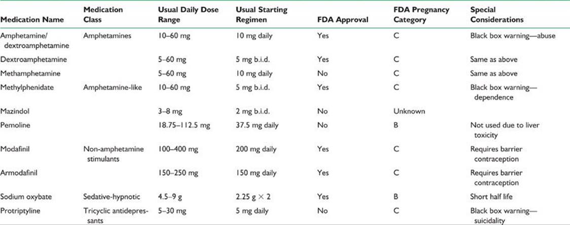

TABLE 55.5 Example of an Initial Treatment Plan for Narcolepsy in Adults
|
Avoidance of shifts in sleep schedule |
|
|
Avoidance of heavy meals and alcohol intake |
|
|
Regular timing of nocturnal sleep: 10:30 p.m. to 7:00 a.m. |
|
|
Naps. Strategically timed naps, if possible (e.g., 15 min at lunchtime and 15 min at 5:30 p.m.) |
|
|
Medication. The effects of stimulant medications vary widely among patients. The dosing and timing of medications should be individualized to optimize performance. Additional doses, as needed, may be suggested for periods of anticipated sleepiness |
|
|
Armodafinil (150–250 mg daily upon awakening), modafinil: 200 mg/d (200 mg on awakening or 100 mg b.i.d.) |
|
|
If difficulties persist: may increase modafinil to 400 mg/d |
|
|
Methylphenidate: 5 mg (three or four tablets) or 20 mg SR in morning (on empty stomach) |
|
|
If difficulties persist: |
|
|
methylphenidate (SR): |
20 mg in morning |
|
5 mg after noon nap |
|
|
5 mg at 4:00 p.m. |
|
|
If no response: |
|
|
Dexedrine spansule (SR): |
15 mg at awakening |
|
5 mg after noon nap |
|
|
5 mg at 3:30 or 4:00 p.m. (or 15 mg at awakening and 15 mg after noon nap) |
|
3. Examples of hypersomnia other than narcolepsy.
(a) Recurrent hypersomnia. The recurrent hypersomnias are very rare conditions in which patients experience prolonged episodes of severe sleepiness separated by periods of normal alertness and function. Recurrent hypersomnia presents as two distinct clinical forms: With the Kleine–Levin’s syndrome (KLS), which usually affects adolescent males, patients may sleep for all but a few hours daily for periods lasting from days to weeks. The hypersomnia in KLS is often accompanied by variable disturbances of mood, cognition, and temperament, often including increased appetite and significantly aggressive or hypersexual behavior. Menstrual-associated hypersomnia is a poorly characterized condition in which episodic sleepiness coincides with the menstrual cycle; it is postulated to be secondary to hormonal influences.
(b) Hypersomnia caused by a medical condition. Hypersomnia here may be diagnosed when sleepiness is thought to be the direct result of a medical or neurologic condition, but the patient does not meet clinical or laboratory criteria for a diagnosis of narcolepsy. A variety of conditions may underlie this disorder including associated neurologic disorders such as encephalitis, cerebrovascular accidents, brain tumor, head trauma, and Parkinson’s disease. Common genetic conditions associated with sleepiness include Prader–Willi’s syndrome and myotonic dystrophy.
4. Diagnosis. The differential diagnosis includes narcolepsy and primary sleep disorders such as sleep-disordered breathing or PLMS, which may also be associated with significant daytime sleepiness. Therefore, the diagnosis is made by means of elimination of other causes of daytime sleepiness. PSG should be performed to further assess these possibilities, and MSLT should be performed to document the level of objective daytime sleepiness.
Mean sleep latencies are often <8 minutes but unlike narcolepsy, which is diagnosed electrographically when the MSL is <8 minutes and when two or more SOREMP are present, the criteria for the latter must include less than two SOREMS and an equally short sleep latency.
5. Treatment and outcome. Because multiple etiologic factors and because of the relative lack of understanding of the underlying pathophysiologic mechanism, treatment is symptomatic and the response is variable. Behavioral therapies and sleep hygiene instructions should be recommended but have only modest positive effect. The only medications that provide partial relief of excessive sleepiness are stimulant-like drugs. The most commonly suggested medications are armodafinil, modafinil, sodium oxybate, methylphenidate, and dextroamphetamine. Tricyclic antidepressants, SSRIs, clonidine, bromocriptine, amantadine, and methysergide have been used with varying success. Sometimes combinations of these drugs yield better control of sleepiness. Even with the highest recommended dose, complete control of daytime sleepiness is seldom achieved in this group of patients. Therefore, prescribing >400 mg of modafinil, >60 mg of methylphenidate, or 40 mg of dextroamphetamine does not provide significant additional symptomatic relief. The patient should be advised not to drive or engage in potentially dangerous activities that require high levels of alertness. Pemoline was recently withdrawn in the United States due to its potential hepatotoxicity (see Table 55.4). Treatment for patients with KLS includes the amphetamine and nonamphetamine stimulants, and mood stabilizers such as lithium, valproic acid, and carbamazepine.
IV. PARASOMNIA
Parasomnia is a group of disorders that occur during sleep, are associated with wake-to-sleep transition, or are associated with arousal from sleep. These conditions with important consideration in neurology include RBD, sleepwalking (somnambulism), night terrors, nightmares, confusional attaches, and nocturnal frontal lobe epilepsy. The ones that are most often encountered in adult clinical practice are discussed.
A. Non-REM parasomnia. Sleepwalking and sleep terrors are episodic behaviors that occur as arousals from non-REM stages of sleep, usually when the patient is coming out of slow-wave sleep. Because sleepwalking (somnambulism) and sleep terrors (pavor nocturnus) among adults are most often associated with each other, the key features are discussed together.
1. Course. The prevalence of sleepwalking and sleep terrors is estimated at approximately 6% of the population. These types of parasomnia are most frequent among children and often disappear by adolescence. These behaviors may be considered normal among children, but for a large number of persons, they persist into adulthood. Patients may report a family history of parasomnia.
2. Clinical features. During these episodes, patients will exhibit polymorphic motor behaviors, such as talking, sitting up, and getting up to walk. These episodes have the potential to become dangerous because patients may bump into walls and windows or fall down stairs. With sleep terrors, extreme autonomic discharge culminating with screaming is a unique feature. Patients usually have only vague recollections of these events and are confused or agitated if awakened.
3. Diagnosis. For adults, thorough evaluation of abnormal nocturnal behavior should be performed to differentiate non-REM parasomnias from other pathologic entities, particularly nocturnal seizures.
4. Treatment and outcome. Therapy for non-REM parasomnia includes several approaches consisting of preventive measures, psychological interventions, and medications (Table 55.6).
a. Preventive measures and psychological intervention. Preventive measures are taken to avoid serious injury during episodes of sleepwalking. The patient should be advised to locate the bedroom on the first floor, lock windows and doors, cover windows and glass doors with heavy draperies, and remove hazardous objects from the house.
Hypnosis and psychotherapy have also been used in the management of parasomnia. Hypnosis has been shown helpful, at least for a short time, to young adults. The need for psychotherapy depends on the association of psychological factors with the parasomnia. Psychotherapy has been used most widely to treat young adults for sleep terrors. Most cases of parasomnia increase in severity and frequency with psychological stress. Therefore, in addition to psychotherapy, relaxation programs, such as progressive muscle relaxation and biofeedback, may be beneficial. Anticipatory awakening has been reported as a treatment modality for sleepwalking and perhaps other disorders of arousal. The technique involves waking the patients between 15 and 30 minutes prior to the time of the typical episodes.
TABLE 55.6 Treatment for Most Common Non-REM Parasomnias
|
Treatment |
|
|
Confusional arousal |
Reassurance when benign in nature |
|
Avoid precipitants such as: |
|
|
–Sleep deprivation |
|
|
–Alcohol |
|
|
–CNS depressants |
|
|
Escitalopram (10 mg)—for sexsomnia |
|
|
Somnambulism (sleep walking) |
Safeguard the sleep environment and protect the patient |
|
Avoid precipitants: |
|
|
–Sleep deprivation |
|
|
–Lithium |
|
|
–Nonbenzodiazepines receptor agonists (i.e., zolpidem) |
|
|
Anticipatory awakenings |
|
|
Benzodiazepines |
|
|
–Clonazepam (0.5–1 mg) |
|
|
–Diazepam (10 mg) |
|
|
–Triazolam (0.25 mg) |
|
|
Imipramine (50–300 mg) |
|
|
Sleep terrors |
Reassurance when benign in nature |
|
CBT |
|
|
Progressive muscle relaxation |
|
|
Biofeedback |
|
|
Hypnosis |
|
|
Psychotherapy |
|
|
Pharmacotherapy: |
|
|
–Paroxetine (20–40 mg) |
|
|
–Clonazepam (0.5–1 mg) |
b. Medications. The benzodiazepines—most commonly clonazepam, alprazolam, and diazepam—have been used. In the management of sleep terrors, tricyclic antidepressants (particularly imipramine) have been used either alone or in combination with benzodiazepines to provide control of symptoms. In addition, several studies have shown that treatment with carbamazepine may be beneficial. An example of an initial therapeutic approach is to start with clonazepam (0.25 to 1.0 mg) approximately 30 minutes before bedtime. If the response is inadequate, the dose should be increased by balancing the side effects, which include confusion and daytime drowsiness, particularly in the care of the older adult. A secondary line of treatment includes initiation of low doses of tricyclic antidepressant drugs or carbamazepine at bedtime.
Results of management of non-REM parasomnia are poorly documented. However, the little information in the literature indicates that response to combinations of pharmacologic and nonpharmacologic therapies is excellent. After various lengths of time, as many as 70% of adult patients report disappearance of the symptoms.
B. RBD.
1. Course. REM sleep and dreaming is normally accompanied by muscle atonia. RBD is characterized by the loss of REM-sleep atonia or excessive motor activation during sleep. Patients with this disorder most commonly report vigorous sleep behaviors that are accompanied by vivid dreams. These behaviors may be quite violent and can result in serious injury. RBD occurs in both acute and chronic forms. The acute form usually is associated with toxic–metabolic etiologic factors, most commonly, drug withdrawal states, particularly delirium tremens. Loss of REM atonia may also occur among patients taking medications that suppress REM sleep, such as tricyclic antidepressants and fluoxetine, and substances such as caffeine. The chronic form usually occurs among older adults. It has been seen in association with various brainstem abnormalities, extrapyramidal neurologic disorders, and medical conditions (α-synucleopathies such as Parkinson’s disease and Parkinson’s plus syndrome [Shy–Drager’s syndrome, multiple systems atrophy, Lewy’s body dementia], brainstem stroke, brainstem tumor, demyelinating disease, and medication toxicity or withdrawal [i.e., SSRI, alcohol]) or is idiopathic. The differential diagnosis of RBD includes non-REM parasomnias, severe obstructive sleep apnea (OSA; “pseudo RBD”), periodic movements of sleep, nocturnal seizures, and nocturnal rhythmic movements. It is important to recognize this condition and differentiate it from other nocturnal behaviors because RBD can be managed effectively. The condition may precede the onset of the neurodegeneration by a few years-to-decades and may be a predictor of an evolving synucleinopathy.
2. Treatment and outcome. Management of RBD involves validation of the condition and counseling light of the possible association with dementia, pharmacologic therapy and interventions that address issues concerning environmental safety.
a. The most commonly prescribed drug therapy is clonazepam at a dosage of 0.5 to 1.0 mg at bedtime. Clonazepam may be taken earlier (1 to 2 hours before bedtime) by patients who report sleep-onset insomnia or morning drowsiness as a result of the medication. If patients have significant OSA associated with RBD, they should be treated with continuous positive airway pressure (CPAP) because Clonazepam may potentially exacerbate OSA. Clonazepam is effective in 90% of cases, and there is little evidence of abuse and infrequent in tolerance in this group of patients. Beneficial effects are observed within the first week of treatment. Typically, treatment with clonazepam results in control of vigorous, violent sleep behaviors, but mild to moderate limb movement, sleep talking, and other complex behaviors may persist. Discontinuation of treatment usually results in recurrence of symptoms. More recently, melatonin (3 to 12 mg per q.h.s.) has been shown to be useful in improving dream enactment behavior and even restoring muscle atonia. This treatment has the advantage of relative lack of sedation, cognitive impairment, and lack of respiratory suppression. There have been few reported cases of successful treatment with dopamine agonists; pramipexole was 0.5 to 1 mg, dopamine precursors, and antiepileptic agents. Table 55.7 depicts a list of treatment options for RBD with the respective level of evidence based on a recent literature review. Both melatonin and clonazepam appear to have the strongest level of evidence in the management of this condition.
b. Environmental safety is an important issue in the management of RBD. Patients should be advised to remove potentially dangerous objects from the house, to pad hard and sharp surfaces around the bed, to cover windows with heavy draperies, and even to place the mattress on the floor to avoid falling out of bed. The combination of drug therapy and implementation of safety precautions offers safe and effective management of RBD.
c. Nocturnal seizures should be considered in the differential diagnosis of many forms of parasomnias. If the history suggests a seizure disorder or if symptoms are not controlled, referral to a neurologist or sleep specialist for further evaluation is recommended. Sometimes, a referral to a movement disorders/cognitive specialist may be indicated if patient present with motor findings, dementia, and behavioral disturbances.
TABLE 55.7 Treatment of RBD

V. MOVEMENT DISORDERS OF SLEEP
A. Restless legs syndrome (RLS) and PLMS.
1. Course. RLS is characterized by creeping, crawling, and disagreeable sensations in the lower and occasionally in the upper extremities associated with irresistible movements of the extremities. The symptoms are present at rest (quiesogenic) and are relieved by movements such as stretching, rubbing, and walking. Lying down in bed and falling asleep is a major problem for patients with RLS. Dysesthesia and the need to move the lower extremities are the most severe at bedtime and are often associated with sleep-initiation insomnia, which is indeed the most common reason for presentation. Many patients also report severe dysesthesia and leg jerks in the middle of the night with difficulty returning to sleep.
Up to 80% of patients with RLS also have periodic limb movement disorder (PLMD). However, PLMD can occur without RLS and has its own diagnostic category. Unlike RLS, which is a clinical diagnosis, PLMD is suspected when a patient or bed partner reports repeated leg kicks and is confirmed by periodic limb movements in the PSG. The typical PSG findings consist of stereotyped repetitive rhythmic movements. The leg movement must last for 0.5-to-10 seconds, and candidate leg movements are considered “periodic” if three or more occur with their onsets separated by 5 to 90 seconds. The legs movements consist of dorsiflexion of the foot, and occasionally may also involve the upper extremities. PLMD usually is more frequent during the first half of the night but can be present throughout sleep. Movements may be associated with sleep disruption. If numerous, the movements can result in nocturnal awakenings and EDS. The prevalence of PLMD increases with age, from 5% among those younger than 50 years to 44% among those 65 years and older.
For most patients with RLS or PLMD, the cause is unknown and therefore termed idiopathic. In many cases, RLS is familial and has an autosomal dominant inheritance with a significant genome-wide association with a common variant in an intron of BTBD9 on chromosome 6p21.2. Both RLS and PLMD have recently been shown to be associated with anemia resulting from iron deficiency, particularly when the ferritin level is <45 mcg per L. Folic acid, vitamin B12 deficiency, neuropathy, myelopathy, rheumatoid arthritis, thyroid dysfunction, and uremia have also been shown to have an association. Results of studies suggest that RLS is associated with alteration in CSF ferritin levels. Furthermore, PLMD may be induced or exacerbated by SSRIs and tricyclic antidepressants as well as by withdrawal from a variety of hypnotic agents. The existence of these conditions should be entertained in the differential diagnosis of PLMD so that patients receive the appropriate therapy. If these conditions are suspected, referral to the appropriate specialist is recommended.
2. Treatment. The four major classes of drugs that have been shown to be effective in the management of RLS are dopaminergic drugs, benzodiazepines, anticonvulsants, and opioids. Common medications used in the treatment of RLS are shown in Table 55.8.
a. The first approach for patients with symptoms consistent with RLS symptoms is to draw a serum ferritin level. Patients with levels <45 mg per L should begin iron replacement therapy with iron sulfate along with vitamin C to improve absorption.
b. When iron stores are normal, nonergotamine dopamine (D2 D3) agonists such as ropinirole and pramipexole may be started. Dopamine agonists are preferred because they are specifically FDA approved for the treatment of RLS. Major side effects include unpredictable sleep attached, GI side effects, postural orthostatic hypotension, and at higher doses, very rare cases of compulsive behaviors such as compulsive gambling.
TABLE 55.8 Pharmacotherapy for RLS

c. Historically carbidopa/levodopa (at a dose of 25 per 100 mg q.h.s.) was initially widely used. However, its wide use is limited due to the risk of rebound symptoms as well as augmentation decreased. Carbidopa/levodopa is given at bedtime, and the dosage is increased progressively until a therapeutic effect is obtained. Usually a dose of carbidopa/levodopa (50 per 200 mg) is sufficient to control RLS and PLMD. A second administration during the day may be necessary if patients report an increase in leg movements in the morning. Treatment with controlled-release carbidopa/levodopa (Sinemet CR) may also alleviate the rebound effect. Additional side effects include dysesthesia or leg movements during the day. Dyskinesia associated with long-term levodopa treatment, as observed among patients with Parkinson’s disease, are uncommon in this group of patients.
d. The anticonvulsant gabapentin (starting with 100 to 300 mg), either alone or in combination with the dopamine agonist medications, also relieves the symptoms of RLS and PLMD. The FDA has very recently approved gabapentin enacarbil extended-release. Tablets for the treatment of moderate-to-severe primary RLS in adults.
e. Several benzodiazepines, including clonazepam, nitrazepam, lorazepam, and temazepam, have been found to improve the nocturnal sleep of patients with RLS and PLMS. Of these, clonazepam is the most widely used. The therapeutic action of clonazepam most likely results from its ability to decrease the number of arousals caused by leg movements. The usual starting dosage of clonazepam is 0.5 to 1.0 mg at bedtime for management of PLMS. Management of RLS may require additional doses to control symptoms during the day. Because benzodiazepines are CNS depressants, they may aggravate sleep apnea, particularly among older persons.
f. Finally, opioids are highly effective in the management of RLS and PLMS. In severe cases refractory to other treatments, intermittent therapy with opioids provides good relief. Other proposed treatments include carbamazepine, clonidine, and baclofen.
VI. SLEEP-DISORDERED BREATHING
The most commonly encountered types of abnormal nocturnal breathing are the sleep apnea and hypopnea. Sleep apnea is cessation of breathing for at least 10 seconds caused by obstruction of the upper airway (OSA), loss of respiratory effort or rhythmicity (central apnea), or a combination of the two (mixed apnea). Hypopnea is a decrease in airflow, which can be obstructive or central in origin. Many patients with sleep apnea have combinations of the central and obstructive types, which suggest that the mechanisms of the different types of sleep apnea may overlap.
A. Central sleep apnea (CSA).
1. Course. Patients with CSA constitute <10% of all patients with sleep apnea who undergo studies in sleep laboratories. Therefore, only a few studies have been reported, which limits knowledge of this disorder. Little information is available regarding the cardiovascular sequelae of CSA. The most common finding is sinus arrhythmia with bradycardia. Oxygen desaturation in patients with CSA tends to be generally mild to moderate compared with that in patients with OSA. Although the cause of CSA in most cases is unknown, it has been associated with certain diseases that should be considered in the differential diagnosis and management of this disorder. These diseases include central alveolar hypoventilation (Ondine’s curse), obesity hypoventilation (Pickwickian) syndrome, congestive heart failure (Cheyne–Stokes’ breathing pattern), autonomic dysfunction (Shy–Drager’s syndrome, familial dysautonomia, and diabetes mellitus), neuromuscular disorders (muscular dystrophy, myasthenia gravis, and motor neuron disease), and brainstem lesions.
2. Treatment of patients with CSA is limited and not satisfactory. Studies regarding treatment usually have involved small numbers of patients, and very few have addressed the long-term efficacy of the proposed treatments.
a. One approach is noninvasive nocturnal ventilation delivered by means of a nasal mask with a volume- or pressure-cycled ventilator. This approach is used to manage only the most severe cases of central alveolar hypoventilation or in the care of patients with neuromuscular disorders.
b. For patients with medical or neurologic conditions known to be associated with CSA, the condition should be managed specifically and the central apnea reassessed. However, if the problem persists or if a cause is not found, several pharmacologic agents can be used. Acetazolamide, a carbonic anhydrase inhibitor, has been shown to improve CSA. In a small number of patients, acetazolamide has been shown to reduce substantially the number of episodes of central apnea. The recommended dosage is 250 mg four times a day. Even fewer studies address the long-term efficacy of this treatment. The side effects associated with mild metabolic acidosis usually are well-tolerated by this group of patients.
c. Other medications, such as theophylline, naloxone, and medroxyprogesterone acetate, have been used with varying degrees of success. Tricyclic antidepressants, particularly clomipramine, have been used successfully to treat a small number of patients. Because none of these medications has been studied systematically, more precise recommendations regarding their use are currently not available.
d. Some patients with CSA have been shown to benefit from therapy with nasal CPAP. This type of therapy is most beneficial to obese patients who also have signs of upper airway obstruction with predominantly central apnea. Nasal CPAP has also been shown effective in the treatment of patients with congestive heart failure in whom central apnea and periodic breathing are observed during sleep. Finally, oxygen therapy has been useful in managing central apnea.
Adaptive seroventilation (ASV). It provides a relatively low baseline pressure and variable ventilatory support to establish a preset level of ventilation for each breath. If the patient’s effort decreases, the ASV’s inspiratory support increases to maintain a steady level of ventilation. This treatment is indicated in patients with CSA, but also OSA patients are refractory to standard CPAP. Some sleep experts have established the term complex sleep apnea referring to patients with OSA who develop CSA on initiation of CPAP. These patients with so-called treatment-emergent central apneas often experience spontaneous resolution of their disease with ongoing therapy.
B. OSA.
1. Course. The initial symptoms of OSA syndrome are loud snoring, excessive sleepiness, fatigue, morning headaches, memory problems, alterations in mood, and episodes of apnea witnessed by the bed partner. OSA is associated with considerable morbidity, including sleep fragmentation, daytime sleepiness that may lead to vehicular and industrial accidents, nocturnal hypoxemia, and cardiovascular as well as cerebrovascular sequelae (e.g., stroke, right heart failure, and hypertension). OSA is generally caused by upper airway obstruction resulting from obesity and skeletal and soft-tissue abnormalities. Examination of the nose and throat may indicate a possible cause. However, some patients with OSA may have normal findings at physical examination.
If OSA is suspected, PSG should be performed to ascertain the severity of the breathing disorder, which will determine the appropriate therapy. Some patients who have symptoms indistinguishable from those of OSA may have predominantly sleep hypopnea. Sleep hypopnea syndrome should be managed in the same manner as sleep apnea syndromes.
The apnea–hypopnea index (AHI) (number of respiratory events per hour of sleep) is used to measure sleep-disordered breathing. An index of 5 is generally accepted as the upper limit of the normal range. An AHI >20 has been shown to result in increased mortality. Therefore, all patients with indexes >20 should be treated. Patients who have milder indexes but whose respiratory events are accompanied by more significant oxygen desaturations and who have additional cardiovascular risk factors such as hypertension, history of heart disease, high cholesterol level, and cigarette smoking also should be treated.
2. Therapy. The approach to management of OSA and hypopnea syndromes involves both general measures and interventions that address specific abnormalities. For most patients, nasal CPAP is the most effective medical therapy for control of sleep apnea.
a. General measures for identifying and addressing coexistent lifestyle issues that exacerbate OSA should be part of treatment of all patients. Although difficult to achieve, weight loss is an important factor in the treatment of obese persons with apnea. Sleep apnea generally improves with weight loss and may even be improved with weight loss of 40 to 50 pounds (18 to 23 kg). In addition to dietary control, this approach requires an exercise program and psychological counseling for long-lasting results. Unfortunately, results indicate that most patients regain the weight within 2 years. If sleep-disordered breathing is more prominent in the supine position, positional therapy to avoid sleep in the supine position is very useful. Alcohol, hypnotic drugs, and other CNS depressant drugs interfere with the arousal response that terminates apneic episodes. Therefore, patients should avoid alcohol use and should not take hypnotics or sedatives. If a specific cause for upper-airway obstruction is found, an otorhinolaryngologic or maxillofacial evaluation is recommended for possible surgical intervention and trials of orthodontic devices, including tonsillectomy or adenoidectomy for enlarged tonsils or adenoids and correction of retrognathia or micrognathia. Results indicate that dental devices may be useful to those patients with mild-to-moderate sleep apnea with some degree of retrognathia or micrognathia. If chronic rhinitis is found, nasal steroid sprays may be beneficial.
b. Nasal CPAP. If no specific cause of upper airway obstruction is found, nasal CPAP is the treatment of choice. This treatment is effective for most patients with obstructive apnea and hypopnea. The level of CPAP should be determined by means of titration of the therapeutic pressure in a sleep laboratory, respiratory data being obtained in all sleep stages. Nasal CPAP requires patency of the nasal airway. Therefore, this procedure may not be effective for patients with severe nasal obstruction. The most common causes of intolerance of nasal CPAP are nasal symptoms, dryness, discomfort from the mask, and social and psychological factors of having to use the mask during sleep (due to claustrophobia). Added humidification often alleviates dryness and associated nasal congestion. With higher pressures, bilevel positive airway pressure (BiPAP) may be a more comfortable alternative to CPAP. Most home care companies provide both nasal CPAP and BiPAP services. If a patient with sleep apnea also has low baseline oxygen saturation during the day or during sleep, referral to an internist or pulmonologist is recommended. Although improvement of symptoms, including daytime sleepiness, may be observed within 1 or 2 days of treatment with nasal CPAP, maximal improvement may not occur for several weeks. Follow-up studies indicate that long-term compliance with nasal CPAP is a substantial problem for many patients not using CPAP throughout the night and on a daily basis. Compliance increases with close follow-up care. Follow-up visits should be scheduled 1 month after the start of CPAP and every 6 months thereafter. The Centers for Medicare & Medicaid Services (CMS) has recently issued a memo that authorized payment for CPAP may take place only if formal PSG was performed and was diagnostic for OSA, and that CMS will be pay for CPAP therapy for 3 months (and subsequently if OSA improves) for adults diagnosed with either PSG or with unattended home sleep monitoring devices. The use of portable home monitoring devices may improve access to diagnosis and treatment of OSA. However, these devices must be used as part of a comprehensive sleep evaluation program that includes access to board-certified sleep specialists, PSG facilities, and therapists experienced in fitting and troubleshooting CPAP devices.
c. Oxygen therapy. Oxygen has been previously reviewed for the treatment of OSA, but the data are quite limited, including limited population. An American Academy of Sleep Medicine (AASM) practice parameter review from 2006 did not recommend oxygen as a primary treatment for OSA. In contrast, in some cases, oxygen may be utilized as a supplement to positive airway pressure therapy in cases of refractory hypoxemia and may, in some circumstances, be an option for individuals who fail or refuse all other OSA treatments and have significant nocturnal hypoxia associated with their sleep apnea.
d. Oral appliances. Custom made oral appliances improve upper airway size during sleep by enlarging the upper airway and/or by decreasing upper airway collapsibility. One specific type of an oral appliance, the mandibular repositioning appliances covers the upper and lower teeth and hold the mandible in a relatively advanced position with respect to the resting position, improving the air space. Oral appliances may not be as efficacious as CPAP in treating sleep apnea, but are indicated for use in patients with mild-to-moderate OSA who do tolerate or respond to CPAP, or fail behavioral interventions to improve compliance. Oral appliances are appropriate for first line therapy in patients with primary snoring who do not respond to weight loss or positional therapy.
e. Uvulopalatopharyngoplasty (UPPP) is a surgical procedure in which excess soft tissue of the soft palate, uvula, and sometimes the tonsils and adenoids are removed. A recent advance in this type of approach is laser-assisted uvuloplasty. The advantages are that the laser procedure is office based and that the amount of tissue removed can be titrated to effect. However, the efficacy of these procedures in the management of sleep apnea is variable. It is estimated that these surgical approaches are effective approximately 50% of the time for amelioration of sleep apnea but are more effective for snoring. Thus patients may continue to have silent obstructive apnea after surgery.
A 2010 AASM’s practice parameter on surgical treatment options for adult OSA patients reviewed the literature regarding the following specific surgical procedures: tracheostomy, maxillo-mandibular advancement, laser-assisted uvulopalatoplasty (LAUP), UPPP, tongue base submucosal radiofrequency ablation, and palatal implants.
Establishing a diagnosis of OSA and its severity by PSG prior to any surgical intervention was considered a standard recommendation by this position paper. In addition, so that patients can make an informed decision regarding therapy, the standard proposed that all patients be advised of the anticipated success rates and potential complications, related to surgical intervention as compared with the alternative treatment options for their OSA (namely CPAP and OA). If patients chose to have surgery, clinical follow-up including a nocturnal polysomnogram is a standard recommendation in order to demonstrate resolution of OSA as measures by the AHI oxygen saturation, and sleep architecture. As for the specific surgical procedure, none, with the exception of LAUP, received more than a recommendation of option as an intervention for the management of OSA. The standard recommendation was not in favor of using LAUP as a treatment for OSA. A multidisciplinary approach is recommended to identify appropriate patients for surgical interventions.
f. Drug therapy. When nasal CPAP is not an option, patients with mild-to-moderate OSA may benefit from drug therapy. Protriptyline at a dosage of 10 mg at bedtime with upward adjustment depending on response and side effects may be an alternative treatment. Drug therapy is generally unsatisfactory for the management of OSA. Recently, the FDA has approved the use of modafinil to improve wakefulness in patients with EDS associated with OSA if CPAP is used with adequate compliance and when total sleep time is adequate.

Recommended Readings
Allen R. Dopamine and iron in the pathophysiology of restless legs syndrome (RLS). Sleep Med. 2004;5:385–391.
Allen R, Becker PM, Bogan R, et al. Ropinirole decreases periodic leg movements and improves sleep parameters in patients with restless legs syndrome. Sleep. 2004;27:907–914.
Aurora RN, Casey KR, Kristo D, et al. Practice parameters for the surgical modifications of the upper airway for obstructive sleep apnea in adults. Sleep. 2010;33(10):1408–1413.
Aurora RN, Zak RS, Maganti RK, et al. Best practice guide for the treatment of REM sleep behavior disorder (RBD). J Clin Sleep Med. 2010;6(1):85–95.
Avidan AY. Parasomnias and movement disorders of sleep. Semin Neurol. 2009;29(4):372–392.
Avidan AY, Kaplish N. The parasomnias: epidemiology, clinical features, and diagnostic approach. Clin Chest Med. 2010;31(2):353–370.
Avidan AY, Zee PC. Handbook of Sleep Medicine. 2nd ed. Philadelphia, PA: Wolters Kluwer Health/Lippincott Williams & Wilkins; 2011.
Dodson ER, Zee PC. Therapeutics for circadian rhythm sleep disorders. Sleep Med Clin. 2010;5(4):701–715.
Earley CJ, Silber MH. Restless legs syndrome: understanding its consequences and the need for better treatment. Sleep Med. 2010;11(9):807–815.
Guilleminault C, Faull KF. Sleepiness in non-narcoleptic, nonsleep apneic EDS patients: the idiopathic CNS hypersomnolence. Sleep. 1982;5:S175–S181.
Harsora P, Kessmann J. Nonpharmacologic management of chronic insomnia. Am Fam Physician. 2009;79(2):125–130.
Holty JC, Guilleminault C. Surgical options for the treatment of obstructive sleep apnea. Med Clin North Am. 2010;94:479–515.
The International Classification of Sleep Disorders: Diagnostic and Coding Manual. 2nd ed. Westchester, IL: AASM; 2005.
Kapur V, Maganti R, Owens J, et al.; Standards of Practice Committee of the American Academy of Sleep Medicine. Practice parameters for the clinical evaluation and treatment of circadian rhythm sleep disorders. An American Academy of Sleep Medicine report. Sleep. 2007;30(11):1445–1459.
Kryger MH, Roth T, Dement WC. Principles and Practice of Sleep Medicine. 5th ed. Philadelphia, PA: Saunders/Elsevier; 2011.
Loube DI, Gay PC, Strohl KP, et al. Indications for positive airway pressure treatment of adult obstructive sleep apnea patients: consensus statement. Chest. 1999;115:863–866.
Manthena P, Zee PC. Neurobiology of circadian rhythm sleep disorders. Curr Neurol Neurosci Rep. 2006;6(2):163–168.
Mignot E. Narcolepsy: pharmacology, pathophysiology and genetics. In: Kryger M, Roth T, Dement WC, eds. Principles and Practice of Sleep Medicine. Philadelphia, PA: Elsevier Saunders; 2005:761–779.
Mitler M, Erman M, Hajdukovic R. The treatment of excessive somnolence with stimulant drugs. Sleep. 1993;16:203–206.
Morgenthaler TI, Lee-Chiong T, Alessi C, et al. Practice parameters for the psychological and behavioral treatment of insomnia: an update. An American Academy of Sleep Medicine report.
The National Institutes of Health Consensus Development Program. NIH State-of-the-Science Conference statement on manifestations and management of chronic insomnia in adults. Sleep. 2005;28:1049–1057.
Neubauer DN, Flaherty KN. Chronic insomnia. Semin Neurol. 2009;29(4):340–353.
Nishino S, Mignot E. Narcolepsy and cataplexy. In: Vinken PJ, Bruyn GW, eds. Handbook of Clinical Neurology. New York, NY: Elsevier, Vol. 99. 2011:783–814.
NIH State-of-the-Science Conference Statement on manifestations and management of chronic insomnia in adults. NIH Consens State Sci Statements. 2005;22(2):1–30.
Overeem S, Mignot E, van Dijk JG, et al. Narcolepsy: clinical features, new pathophysiologic insights, and future perspectives. J Clin Neurophysiol. 2001;18(2):78–105.
Overeem S, Reading P. Sleep Disorders in Neurology: A Practical Approach. Chichester, West Sussex: Wiley-Blackwell; 2009.
Pack AI, Pien GW. Update on sleep and its disorders. Annu Rev Med. 2011;62:447–460.
Reid KJ, Zee PC. Circadian rhythm sleep disorders. Med Clin North Am. 2004;(3):631–651.
Reid KJ, Zee PC. Circadian rhythm sleep disorders. In: Vinken PJ, Bruyn GW, eds. Handbook of Clinical Neurology. New York, NY: Elsevier, Vol. 99. 2011:963–977.
Riemann D, Perlis ML. The treatments of chronic insomnia: a review of benzodiazepine receptor agonists and psychological and behavioral therapies. Sleep Med Rev. 2009;13(3):205–214.
Robinson A, Guilleminault C. Obstructive sleep apnea syndrome. In: Chokroverty S, ed. Sleep Disorders Medicine: Basic Science, Technical Considerations, and Clinical Aspects. 2nd ed. Boston, MA: Butterworth-Heineman; 1999:331–354.
Rubins JB, Kunisaki KM. Contemporary issues in the diagnosis and treatment of obstructive sleep apnea. Postgrad Med. 2008;120(2):46–52.
Silber MH, Krahn LE, Morgenthaler TI. Sleep Medicine in Clinical Practice. 2nd ed. New York, NY: Informa Healthcare; 2010.
Wise MS, Arand DL, Arger RR, et al. Treatment of narcolepsy and other hypersomnias of central origin. Sleep. 2007;30(12):1712–1727.
Zee PC, Goldstein CA. Treatment of shift work disorder and jet lag. Curr Treat Options Neurol. 2010;12(5):396–411.