
1. An 85-year-old man is being evaluated for gait difficulties. He says that he frequently trips walking up stairs or on uneven surfaces. On examination, it is found that joint proprioception is absent in his toes. People with impaired position sense will usually fall if they simultaneously stand with their feet together and do which of the following?
a. Flex the neck
b. Extend their arms in front of them
c. Flex the knees
d. Turn the head
e. Close their eyes
2. A 62-year-old right-handed man has “involuntary twitches” of his left hand. He first noticed between 6 months and 1 year ago that when he is at rest, his left hand shakes. He can stop the shaking by looking at his hand and concentrating. The shaking does not impair his activities in any way. He has no trouble holding a glass of water. There is no tremor in his right hand, and the lower extremities are not affected. He has had no trouble walking. There have been no behavioral or language changes. On examination, a left hand tremor is evident when he is distracted. Handwriting is mildly tremulous. He is very mildly bradykinetic on the left. The most likely examination finding would be which of the following?
a. Upper motor neuron pattern of weakness on the left
b. Lower motor neuron pattern of weakness on the left
c. Bilateral upper motor neuron pattern of weakness
d. Mild cogwheel rigidity on the left only with distraction
e. Bilateral severe cogwheel rigidity
3. A 42-year-old man notices that his right pupil is smaller than the left. His wife has also commented that the right eye is “droopy looking.” The only remarkable recent history is that he was tackled a little hard while playing football the day before. An axial T1-weighted magnetic resonance image (MRI) is shown below. Which of the following is present?
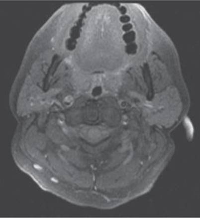
(Courtesy of David Panasci, MD)
a. Increased T2 signal in a periventricular distribution
b. Contrast enhancement along the tentorial margin
c. Increased T1 signal in the wall of the right carotid artery
d. Enlarged optic nerve in the orbit
e. Thrombosed cavernous sinus aneurysm
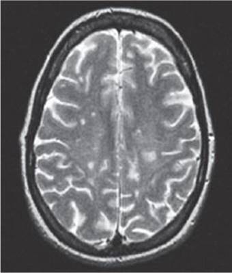
4. A 21-year-old, generally healthy college student presents with loss of sensation in the right arm that has been progressive over a few days. She says that she had flu for about a week and noticed the symptoms started afterward. It began in her hand, and then very slowly progressed over 36 hours to involve the entire right upper extremity. Her physician ordered blood tests and an MRI. A T2 image is pictured below. Which of the following is the most likely process?
a. Ischemic
b. Demyelinating
c. Neoplastic
d. Hemorrhagic
e. Psychogenic
5. A 42-year-old attorney presents with a tremor in her hands that is most obvious when she is awake and trying to perform an action. She had first noticed it several years ago, but is concerned that it may be very slowly worsening. A tremor of this type is most likely caused by disease in which of the following structures?
a. Thalamus
b. Cerebellum
c. Substantia nigra
d. Spinal cord
e. Internal capsule
6. A 65-year-old man was forced to retire from iron working because of a neurological condition which has progressed over the past several years. It is characterized by tremor, rigidity, and bradykinesia worse on the left side. The symptoms are somewhat alleviated by treatment with L-dopa/carbidopa. This patient’s resting tremor is most likely to do which of the following upon falling asleep?
a. It becomes more rapid.
b. Its amplitude increases.
c. It generalizes to limbs that were uninvolved when the patient was awake.
d. It disappears.
e. It transforms into choreiform movements.
7. A 25-year-old woman with a history of epilepsy presents to the emergency room with impaired attention and unsteadiness of gait. Her phenytoin level is 37 (normal therapeutic range 10-20). She has white blood cells (WBCs) in her urine and has a mildly elevated thyroid-stimulating hormone (TSH) level. Examination of the eyes would be most likely to show which of the following?
a. Weakness of abduction of the left eye
b. Lateral beating movements of the eyes
c. Impaired convergence
d. Papilledema
e. Impaired upward gaze
8. A 75-year-old retired journalist is generally healthy but has noticed worsening problems maneuvering over the past 4 months. He has particular trouble getting out of low seats and off toilets. He most likely has which of the following?
a. Poor fine finger movements
b. Electromyography results indicating widespread denervation
c. Distal muscle weakness
d. Elevated creatine kinase
e. Gait apraxia
9. A 50-year-old right-handed man has presented to a neurologist because of gradually progressive hearing loss of the right ear. He denies worsening balance or ringing in his ears. There is no associated pain. A vibrating tuning fork is applied to the center of his forehead. The sound is louder in his left ear. This finding suggests which of the following?
a. Bilateral sensorineural hearing loss.
b. Bilateral conductive hearing loss.
c. Right ear conductive hearing loss.
d. Left ear sensorineural hearing loss.
e. Right ear sensorineural hearing loss.
10. A 38-year-old woman says that she is “dizzy.” A more careful history reveals that she has an abnormal sensation of movement intermittently. Examination reveals several beats of horizontal rhythmic eye movements on leftward gaze. A T1 MRI image from this patient is shown. Dix-Hallpike test is positive. Which is the most likely diagnosis?

a. Ocular bobbing
b. Pontine hemorrhage
c. Cervicomedullary junction glioma
d. Benign positional vertigo
e. Brainstem stroke
11. A 48-year-old left-handed man develops increased sensitivity to sound in his left ear. A brain MRI reveals a posterior fossa mass. This symptom may develop in one ear with damage to which of the following ipsilateral CNs?
a. V
b. VII
c. VIII
d. IX
e. X
12. A 42-year-old woman is being evaluated for gait difficulties. On examination, it is found that her ability to walk along a straight line touching the heel of one foot to the toe of the other is impaired. This finding is most common with which of the following?
a. Cerebellar dysfunction
b. Parietal lobe damage
c. Temporal lobe damage
d. Ocular motor disturbances
e. Dysesthesias in the feet
13. A 55-year-old woman is being examined because of difficulty walking. The clinician notices the presence of fine twitching movements beneath the surface of the tongue and wasting of one side of the tongue. This finding suggests which of the following?
a. Pseudobulbar affect
b. Aberrant reinnervation of muscles from CN X
c. Aberrant reinnervation of muscles from CN XII
d. Denervation of muscles from CN X
e. Denervation of muscles from CN XII
14. A 46-year-old longshoreman has lower back pain radiating down the posterior aspect of his left leg and paresthesias in the lateral aspect of his left foot. This has been present for 6 months. Strength and bowel and bladder function have been normal. Examination would be most likely to show which of the following?
a. Left Babinski sign
b. Loss of pinprick sensation over the web space between the first and second digits of the left foot
c. Hyperreflexia at the left knee jerk
d. Hyporeflexia in the left Achilles tendon reflex
e. Decreased rectal tone
15. A 28-year-old graduate student presents with confusion and mild right hemiparesis developing over the course of an evening. His girlfriend relates that he has been having severe headaches each morning for the past 2 weeks. While being evaluated in the emergency room, he has a generalized tonic-clonic seizure. When examined 2 hours later, he is lethargic and unable to recall recent events, has difficulty naming, and has a right pronator drift. There is mild weakness of abduction of the eyes bilaterally. Funduscopic examination might be expected to show which of the following?
a. Pigmentary degeneration of the retina
b. Hollenhorst plaques
c. Retinal venous pulsations
d. Blurring of the margins of the optic disc
e. Pallor of the optic disc
16. A 42-year-old man sustained multiple injuries in an automobile accident. After orthopedic surgery, he is difficult to arouse. Assuming that his brainstem function is intact, when he is lying supine with his head slightly elevated (30°) and one external auditory meatus is irrigated with warm water, which of the following would be expected?
a. Tonic deviation of the eyes toward the ear that is stimulated
b. Nystagmus in both eyes toward the ear that is stimulated
c. Tonic deviation of the ipsilateral eye toward the ear that is stimulated
d. Nystagmus in both eyes away from the ear that is stimulated
e. Tonic deviation of both eyes away from the ear that is stimulated
17. A 33-year-old woman has an acute onset of right orbital pain after a tennis match. The following morning, her 10-year-old son comments that her right eye looks funny. On examination, she has a mild right ptosis and anisocoria. The right pupil is 2 mm smaller than the left, but both react normally to direct light stimulation. Visual acuity, visual fields, and eye movements are normal. The site of injury is caused by interruption of fibers from which of the following structures?
a. Optic tract
b. Optic chiasm
c. CN III
d. T1 nerve root
e. Superior cervical ganglion
18. An 81-year-old woman with a history of type 2 diabetes mellitus and atrial fibrillation presents with right body weakness and slurred speech. She realized that there was a problem on awakening in the morning, and her husband called emergency medical services (EMS), who brought her to the emergency room. There are no word-finding difficulties, dysesthesia, or headaches. She is taking warfarin. Physical examination findings include blood pressure of 210/95 and irregularly irregular heartbeat. There is leftside neglect with slurred speech. There is a corticospinal pattern of weakness of the right body, with the face and upper extremity worse than the lower extremity. Routine chemistries and cell counts are normal. Her INR is 1.7. Computed tomography (CT) of the head reveals a large right-sided subdural hematoma. The intracranial material appearing most dense on CT is which of the following?
a. Blood clot
b. White matter
c. Gray matter
d. Cerebrospinal fluid (CSF)
e. Pia mater
19. A 15-year-old boy developed a left Bell palsy over the course of 1 week. He was treated with acyclovir and prednisone. Over the next 3 months he seemed to recover almost fully. However, he has noticed involuntary twitching at the left corner of the mouth each time he tries to blink the left eye. This is most likely caused by which of the following?
a. A habit spasm
b. Cerebellar damage producing impaired coordination
c. Aberrant regeneration of the facial nerve
d. Trigeminal neuralgia
e. Focal seizures
20. You are working in the emergency room when a 30-year-old man presents with a headache that started yesterday. As he was shoveling snow, he felt a sudden pain in the front of his head. The pain does not throb and has been relatively constant since. He says that now his neck also has become a little stiff. He carries a diagnosis of migraine headaches, but says that this is different than his usual headaches. He is afebrile and has a normal examination except for slight photophobia and mild discomfort with neck flexion. Which of the following is the most appropriate next step in management?
a. Obtain a brain MRI
b. Obtain a brain CT
c. Obtain a cerebral angiogram
d. Obtain an electroencephalogram (EEG)
e. Obtain a psychiatric consult
21. A 56-year-old right-handed woman presents to the emergency room with a sudden-onset, severe, left-sided headache. The pain began when she stood up from her couch while watching TV. A head CT is normal. Which of the following is the most appropriate next step in management of this patient?
a. Begin intravenous heparin
b. Perform a lumbar puncture
c. Obtain a brain MRI
d. Obtain a cerebral angiogram
e. Give the patient a prescription for zolmitriptan and send her home
22. A 60-year-old man is clinically suspected to have had a subarachnoid hemorrhage. A lumbar puncture shows 7000 red blood cells (RBCs) in tube 1 and 7200 in tube 4. There are nine WBCs in each. The fluid is xanthochromic. The opening pressure is 22 cm H2O. Which of the following is the next best step in managing this case?
a. Arrange for a cerebral angiogram and call a neurosurgical consult
b. Give the patient a prescription for sumatriptan and send him home
c. Immediately give 2 g of intravenous ceftriaxone
d. Immediately start intravenous acyclovir
e. Repeat the lumbar puncture
23. A 28-year-old man presents to the emergency room with a severe headache. It is different than any that he has ever had before. It is in the right posterior region and is not throbbing. The headache started suddenly, about 5 hours ago, while he was watching television and eating pizza. He is now noticing some mild neck stiffness and blurry vision. Examination is significant for weakness of abduction of the right eye. Which of the following is the most definitive test for identifying intracranial aneurysms?
a. MRI scanning
b. CT scanning
c. Single photon emission computed tomography (SPECT)
d. Positron emission tomography (PET)
e. Cerebral angiography
24. In this MRI scan, the site most likely to produce a noncommunicating hydrocephalus when it is obstructed is identified by which of the following?

a. Arrow A
b. Arrow B
c. Arrow C
d. Arrow D
e. Arrow E
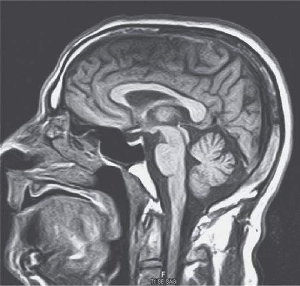
25. An 18-year-old woman tells her physician that she has throbbing right-sided headaches. They are most common just prior to her menstruation and are associated with nausea and photophobia. Examination at the time of the office visit is normal. A T1 sagittal image from her MRI is shown below. The location of the cerebellar tonsil in the MRI scan suggests which of the following?
a. Arnold-Chiari type 1 malformation
b. Arnold-Chiari type 2 malformation
c. Giant cisterna magna
d. Dandy-Walker syndrome
e. Normal posterior fossa
26. A 46-year-old woman with depression has a brain CT performed at the request of her psychiatrist. There is the incidental finding of a dense mass that appears to originate from the tentorium cerebelli. The tentorium cerebelli separates the superior cerebellum from the cerebrum and is a common site of origin for which of the following?
a. Meningiomas
b. Ependymomas
c. Hemangioblastomas
d. Medulloblastomas
e. Astrocytomas
27. A 35-year-old woman has noticed that over the past 3-to-5 months she has had some difficulties with balance, particularly when she closes her eyes. On examination, she has decreased hearing in her left ear and also left body dysdiadochokinesia. Her physician orders a head CT, shown below. Given this CT scan, which was obtained without contrast enhancement, the physician must assume that the posterior fossa mass at the arrow is which of the following?

a. Normal
b. Calcified
c. Highly vascular
d. Granulomatous
e. Highly cystic
28. A 35-year-old woman presents with slowly evolving left arm ataxia, left-sided head tilt, dysarthria, and left facial weakness. The patient denies vertigo, tinnitus, or hearing loss. MRI reveals a posterior fossa mass that lies close to the bone and enhances with contrast. Which of the following is the most likely explanation for this lesion?
a. Cerebellar infarction
b. Cerebellar hemorrhage
c. Meningioma
d. Schwannoma
e. Astrocytoma
29. A 45-year-old woman presents with worsening right-sided headaches. Examination is significant for a left-sided drift. Brain MRI and CT scans reveal a homogeneously enhancing, round, dural-based, calcified lesion compressing the right frontal lobe. Which of the following is the most appropriate course of action for the management of this case?
a. Anticoagulation
b. Triple therapy with isoniazid, rifampin, and ethambutol
c. Surgical resection
d. Chemotherapy
e. Craniospinal axis irradiation
30. A patient with bilateral posterior fossa masses has café au lait spots and reports a family history of bilateral hearing loss at a relatively young age. A gene abnormality should be suspected on which chromosome?
a. 5
b. 13
c. 17
d. 21
e. 22
31. A 65-year-old diabetic man has a history of a cerebellar stroke. The stroke occurred 5 years ago, and he says that he has now fully recovered. He cannot recall the symptoms, but his medical records state that he presented with left-sided dysdiadochokinesia. Which of the following was most likely impaired?
a. Successive finger movements
b. Heel-to-toe walking
c. Rapid alternating movements
d. Tremor suppression
e. Conjugate eye movements
32. A 27-year-old healthy normal woman is having a routine EEG examination. The study begins with a 5-minute recording of her sleeping. Then she is awakened and given photic stimulation. Next she is alert and awake, lying with her eyes closed in a quiet room. At this point she will exhibit what frequency of EEG activity over the occipital and parietal areas bilaterally?
a. 0-to-3 Hz
b. 4-to-7 Hz
c. 8-to-13 Hz
d. 14-to-25 Hz
e. 26-to-45 Hz
33. A 38-year-old right-handed woman presented with early morning headaches. There was papilledema on examination. Below is shown a T1-weighted postcontrast brain MRI of the patient. Which of the following symptoms is most likely to also be present?

a. Aphasia
b. Neglect
c. Left hemiparesis
d. Left homonymous hemianopia
e. Alexia without agraphia
Questions 34 to 38
Match each of the following vignettes with the appropriate diagnostic study. Each lettered option may be used once, more than once, or not at all.
a. MRI of the brain
b. Brain biopsy
c. Nerve conduction studies (NCS)
d. CSF analysis
e. EEG
f. Serum creatine phosphokinase (CPK)
g. Cerebral angiography
h. Myelography of the spinal canal
i. Cranial computerized axial tomography (head CT)
j. Skull x-ray
k. Visual evoked responses (VER)
l. Nerve biopsy
34. A 65-year-old man with a history of atrial fibrillation is brought into the emergency room at 1:00 PM because of the acute onset of right-sided weakness and inability to speak beginning at noon. On examination, he is alert but unable to speak. He follows simple one-step commands. There is left gaze deviation and impaired rightward gaze. Flaccid paresis of the right face and arm is present, but he is able to lift his right leg off the bed. Reflexes are decreased on the right side.
35. A 50-year-old homeless woman is admitted to the hospital with ulcerations on her feet. She complains of burning in her feet and lower legs, but does not localize the pain to where her skin is ulcerated. She is unable to stand with her eyes closed and her feet together. Her deep tendon reflexes are diffusely hypoactive. On systematic testing of her strength and sensation, decreased pain, position, and vibration sense are evident in her hands and feet. She also has weakness on dorsiflexion of the ankles and wrists.
36. A 7-year-old boy has recurrent staring episodes while at school. His school performance is poor. The episodes never last more than 30 seconds, and afterward he immediately resumes normal attention. There are no lip-smacking movements or other automatisms. He never falls down during the episodes. If he is walking or eating during the episode, he merely stops. He is unaware of this behavior.
37. A previously healthy 7-month-old infant is brought to the emergency room after having had three generalized convulsions. The infant has a stiff neck, is poorly responsive to the examiner, and has a rectal temperature of 38.9°C (102°F). The parents report that 1 day of diarrhea preceded this episode.
38. A 70-year-old right-handed woman with a history of polio describes 1 month of increasing difficulty rising from a chair and walking. She also has trouble combing her hair and cooking, and there is mild swallowing trouble, but only with solids. Her legs and upper arms are painful and mildly swollen. Periungual telangiectasias are seen. Erythrocyte sedimentation rate (ESR) is 61.
Questions 39 to 45
For each clinical scenario, choose the CSF pattern most likely to be found.

39. A 26-year-old woman with a 7-year history of epilepsy develops a generalized convulsion while shopping. She is taken to an emergency room, but no one accompanying her is aware of a previous history of epilepsy. Because she has a protracted postictal period, numerous investigations are performed over the course of the next hour. CT scan is completely normal, but her arterial blood gases reveal a mild acidosis.
40. A 72-year-old man is brought to the emergency room in a coma. He has a fever and was observed to have a generalized tonic-clonic seizure just prior to arriving in the emergency room. His family reports that he had lethargy and cough about 1 week prior to the acute deterioration. On the day of his seizure, he developed a headache and blurred vision. He had some vomiting early in the day and became more stuporous as the day progressed. There is no evidence of alcohol or drug use.
41. A 19-year-old man notices discomfort in his ankles within a few days of recovering from an upper respiratory infection. Over the next 7 days, he develops progressive weakness in both of his legs and subsequently in his arms. He has no loss of sensation in his limbs, despite the progressive loss of strength. He does not lose bladder or bowel control, but on the tenth day of his weakness he develops problems with breathing and requires ventilatory assistance.
42. A 40-year-old man was involved in an automobile accident. There is an obvious laceration on his head, and he has neck pain. Police at the scene report that he was unconscious when they arrived, but the patient cannot recall this loss of consciousness. In fact, he cannot remember the accident or events within 10 minutes prior to the accident. On examination, he has obvious neck stiffness and photophobia. Within a few hours of his arrival at the emergency room, he develops vomiting. Lumbar puncture is delayed until after an MRI can be obtained. The tap is performed 2 days after the accident because the patient is still confused and irritable.
43. A 22-year-old woman is brought to the hospital in a coma. She has had changes in her behavior characterized by excessive suspiciousness and facetiousness over the month prior to her hospitalization. One week prior to her hospitalization, she had visual and auditory hallucinations. Drug testing reveals no apparent illicit drug use. On the day of admission, she had a generalized seizure and lapsed into a coma. MRI shows unilateral changes in the temporal lobe.
44. A 26-year-old man develops bed wetting and transient sexual dysfunction that resolves over the course of 6 weeks. One month later, he notices a pins-and-needles sensation in his right leg that never clears completely. On examination, he has hyperreflexia in both of his legs and past-pointing in his right arm. His gait is slightly ataxic, and he is unable to perform tandem gait.
45. A 26-year-old woman weighing in excess of 300 lb has headache and blurred vision that began 2 weeks prior to consulting a physician. She has no vomiting or diplopia. Examination of her eyes reveals florid papilledema but without hemorrhages. Her neurologic examination is otherwise entirely normal. She had a similar problem while pregnant with her fourth child.
1. The answer is e. (Ropper, p 113.) Standing with the feet together and the eyes closed is the Romberg test. The person with poor position sense needs visual cues to remain standing. This test was introduced as a helpful way to check for deficits associated with tabes dorsalis, a form of neurosyphilis. In tabes dorsalis, the dorsal columns of the spinal cord are damaged. These dorsal or posterior columns carry the nerve fibers that transmit vibration and position sense to the brain. With the widespread use of penicillin, tabes dorsalis has become rare in industrialized nations, and impaired position sense is much more likely to be a consequence of diabetes mellitus, spondylosis, or alcoholism. In performing the test, it is important to remember that even normal people will tend to sway slightly with their eyes closed, and that those with loss of cerebellar function or vestibulopathy will also sway more with loss of visual cues.
2. The answer is d. (Ropper, pp 1033-1045.) This patient gives a typical history for early Parkinson disease. The classic triad is asymmetric resting tremor, rigidity, and bradykinesia. The rigidity is generally severe later, not early in the disease. Answers a-c are incorrect as weakness is not a feature of Parkinson disease, and there is no suggestion of any type of weakness in the history.
3. The answer is c. (Osborn, pp 72-73.) The most likely cause of this patient’s symptoms and signs is a carotid dissection. Dissections are caused by a tear in the intimal lining of the vessel with penetration of blood beneath the intimal surface, forming an intramural hematoma (blood clot within the wall of the vessel). Carotid artery dissections can occur spontaneously, post-traumatically, or in the setting of underlying disease process that weakens the endothelial intima. They may appear as an area of increased signal within the lining of the vessel on T1-weighted images, on which a blood clot appears bright. Typically, this takes a crescentic pattern, and the lumen of the vessel is displaced eccentrically. An increased T2 signal in a periventricular distribution is typical of the plaques of demyelinating disease seen in multiple sclerosis. Contrast enhancement along the tentorial margin might be seen in inflammatory diseases of the dura mater or meninges. An enlarged optic nerve might be seen in the setting of optic neuritis or infiltration of the nerve by a tumor or other process, and this would be expected to cause visual impairment. A cavernous sinus aneurysm that is sufficient to produce oculosympathetic palsy would be expected to cause other deficits in ocular motility, because the CNs subserving oculomotor function course through the cavernous sinus.
4. The answer is b. (Ropper, pp 896-899.) The history and MRI are consistent with a demyelinating process, such as acute disseminated encephalomyelitis (ADEM) or multiple sclerosis. The MRI shown is a T2-weighted image. Several areas of abnormal high signal are apparent. MRI depends primarily on the water content of tissues, but it can be customized to look at more specific properties of tissues. A variety of methods are available for affecting the information produced by and analyzing the information generated by MRI. The T2-weighted image is not specific for demyelination, but it is useful in following changes in plaques of demyelination, an application that has been used in studies of agents useful in the management of multiple sclerosis. The MRI appearance and history are not highly consistent with ischemic, neoplastic, or hemorrhagic processes. Ischemic or hemorrhagic processes will typically present more acutely, whereas a neoplasm would either present very acutely or much more subacutely.
5. The answer is b. (Ropper, pp 93-95.) Intention or kinetic tremors are most characteristic of damage to the cerebellum. Kinetic tremors of the hand or arms are most common with disease of the cerebellar hemispheres, but they may also develop with damage to the spinocerebellar tracts of the spinal cord. Damage to the substantia nigra, such as that occurring in Parkinson disease, produces a resting tremor that abates when the patient moves the involved limb intentionally. Damage to the thalamus is more likely to produce a sensory disturbance. Tremors may develop with spinal cord damage, but they do not follow a typical pattern and do not suggest a spinal cord origin. Internal capsule lesions typically cause weakness.
6. The answer is d. (Watts, p 235.) Patients with Parkinson disease often have a characteristic pill-rolling tremor of the hand while they are awake. The tremors associated with Parkinson disease are worse when the patient is at rest and not moving the affected limb. Paradoxically, this resting tremor ceases when relaxation progresses to sleep. In fact, most tremors and other types of movement disorders caused by disease of the caudate, putamen, and globus pallidus (ie, the basal ganglia) and of the substantia nigra remit during sleep. Choreiform movements are jumping or dance-like movements and occur with Wilson disease (hepatolenticular degeneration) and Huntington disease, a hereditary degenerative disease of the basal ganglia.
7. The answer is b. (Ropper, p 331.) Most rhythmic to-and-fro movements of the eyes are called nystagmus. Nystagmus has a fast component in one direction and a slow component in the opposite direction. Nystagmus with a fast component to the right is called right-beating nystagmus. Phenytoin may evoke nystagmus at levels of 20-to-30 mg/dL. The eye movements typically appear as a laterally beating nystagmus on gaze to either side; this type of nystagmus is called gaze-evoked. If the patient has nystagmus on looking directly forward, he or she is said to have nystagmus in the position of primary gaze. Therapeutic levels for phenytoin are usually 10-to-20 mg/dL, and some patients develop asymptomatic nystagmus even within that range. Ataxia, dysarthria, impaired judgment, and lethargy may also occur at toxic levels of phenytoin. Many other drugs, such as alcohol, barbiturates, and other sedatives, also evoke nystagmus. Weakness of abduction of the left eye, or abducens palsy, is due either to injury to the sixth CN or to increased ICP. Impaired convergence can occur normally with age or may be a sign of injury to the midbrain. Papilledema is a sign of increased ICP. Impaired upward gaze may occur in many conditions, but would not be expected to occur due to a toxic phenytoin level.
8. The answer is d. (Ropper, pp 1355-1361.) With primary muscle diseases, such as polymyositis, weakness usually develops in proximal muscle groups much more than in distal groups. This means that weakness will be most obvious in the hip girdle and shoulder girdle muscles. The hip girdle is usually affected before the shoulder girdle. To get out of a low seat, the affected person may need to pull him- or herself up using both arms. Persons with more generalized weakness or problems with coordination are less likely to report problems with standing from a seated position. Poor rapid alternating movements and poor fine finger movements usually develop with impaired coordination, such as that caused by cerebellar damage. With severe weakness in the limbs, patients will do poorly on these tests of function as well. With proximal muscle weakness, the affected person will usually perform relatively well on these tests of distal limb coordination.
9. The answer is e. (Ropper, p 281.) The vibrations from a tuning fork placed on top of the head are transmitted through the skull to both ears. Bone conduction of sound through the skull should be equal in both ears. With sensorineural hearing loss, the patient will hear the midline fork more loudly in the unaffected ear. Sensorineural hearing loss is the deafness that develops with injury to the receptor cells in the cochlea or to the cochlear division of the auditory nerve. In conductive hearing loss, the vibrations of the tuning fork are perceived as louder in the affected ear. With this type of hearing loss, the injury is in the system of membranes and ossicles designed to focus the sound on the cochlea. Impairment of the conductive system causes the vibrations of the tuning fork to be transmitted to the cochlea directly through the skull. Much like a person with cotton stuffed into the external auditory meati, the patient with the conductive hearing loss has impaired perception of sound coming from around him or her but an enhanced perception of his or her own voice. This type of tuning fork test is called the Weber test.
10. The answer is d. (Ropper, pp 293-296.) Abnormal patterns of eye movement may help localize disease in the central or peripheral nervous system in patients with vertigo. The retina is negatively charged in comparison with the cornea, which creates a dipole that may be monitored during electronystagmography studies with electrodes placed on the skin about the eyes. Movement of the most posterior elements of the retina toward an electrode is registered as a negative voltage change at that electrode. Damage to the pons may produce characteristic conjugate deviations of the eyes. The conjugate eye movements are rhythmic and directed downward, but they lack the rapid component characteristic of nystagmus. This type of abnormal eye movement is called ocular bobbing. A lesion at the cervicomedullary junction, such as a meningioma at the foramen magnum, will produce a down-beating nystagmus with both eyes rhythmically deviating downward, with the rapid component of this nystagmus directed downward as well. Cervicomedullary refers to the cervical spinal cord and the medulla oblongata. Damage to the midbrain, thalamus, or hypothalamus may disturb eye movements, but down-beating nystagmus would not ordinarily develop with damage to these structures. The pictured MRI is entirely normal. Benign positional vertigo is thought to be caused by otolithic debris within the semicircular canals. In addition to facilitating acclimation to the vertigo, the Dix-Hallpike maneuver may be able to clear the debris from the canal.
11. The answer is b. (Ropper, pp 1329-1333.) The facial nerve innervates the stapedius muscle of the middle ear. With paralysis of this muscle, undamped transmission of acoustic signals across the stapedius bone of the middle ear produces hyperacusis. Hyperacusis is an indication that the damage to the facial nerve is close to its origin from the brainstem, because the nerve to the stapedius muscle is one of the first branches of the facial nerve. The tensor tympani is controlled by the motor fibers in the fifth CN. With damage to this nerve, the tympanic membrane has some inappropriate slack, but the patient does not usually comment on increased sensitivity to sound in the affected ear. Damage to CN VIII may cause a loss of hearing, but not hyperacusis. CNs IX and X are not involved in hearing.
12. The answer is a. (Ropper, pp 113-114.) Walking along a straight line with the heel of one foot touching the toe of the other foot is called heel-to-toe walking, or tandem gait. It is a routine test for ethanol intoxication because alcohol exposure impairs the coordination of gait as governed by the cerebellum. Tandem gait will be abnormal with many other problems, including weakness, poor position sense, vertigo, and leg tremors, but such abnormality in the absence of these other problems suggests a cerebellar basis for the problem.
13. The answer is e. (Ropper, pp 1336-1337.) The hypoglossal nerve provides motor innervation to the tongue. The fine movements noted under the surface of the tongue with injury to the hypoglossal nerve are called fasciculations and are an indication of denervation. They are presumed to occur through hypersensitivity to acetylcholine acting at the denervated neuromuscular junction. Atrophy and fasciculations are likely to occur together and are highly suggestive of denervation of the tongue. This is most often seen with brainstem disease, such as stroke or bulbar amyotrophic lateral sclerosis (ALS), or with transection of the hypoglossal nerve.
14. The answer is d. (Ropper, pp 196-200.) This patient has a history most consistent with a herniated lumbar disk. The most common locations for lumbar disk herniation are between the fifth lumbar and first sacral vertebrae (producing S1 nerve root compression) and between the fourth and fifth lumbar vertebrae (producing L5 root compression). S1 nerve root compression, or radiculopathy, is associated with pain in the lower back or buttock region, often radiating down the posterior thigh and calf to the lateral and plantar surfaces of the foot and affecting the fourth and fifth digits of the foot. Motor function of the foot and toe flexors, toe abductors, and hamstring muscles may be impaired, but more often it is not. Bowel and bladder function are usually preserved. The loss of the ankle jerk, or Achilles tendon reflex, is often the only objective sign of S1 radiculopathy. Deep tendon reflexes such as the ankle jerk are diminished or lost when there is damage to the sensory fibers from the tendon stretch organs.
A Babinski sign is an indication of upper motor neuron damage, which is not expected in this case with preserved autonomic and motor function. Loss of pinprick sensation over the web space between first and second toes is found in association with injury to the fifth lumbar nerve root or to the peroneal nerve. Hyperreflexia of the knee jerk is another sign of an upper motor neuron lesion and would not be expected in this case. Straight-leg raising (Lasègue maneuver) is used to determine whether symptoms are caused by nerve root compression by stretching the nerve root. With compression, lifting the leg passively may be limited to between 20° and 30°. This test may be positive on the contralateral side (crossed straight-leg-raising sign), but it is usually more prominent on the affected side.
15. The answer is d. (Ropper, pp 236-238.) The presentation with sub-acute onset of morning headaches culminating in confusion, right hemiparesis, and seizure in a young person suggests an expanding mass lesion, most likely a tumor. The weakness of eye abduction bilaterally is what may be referred to as a false localizing sign. Although this suggests injury to the sixth CN bilaterally, the injury is not restricted to the sixth CN. The increase in intracranial pressure (ICP) from the mass causes stretching of the sixth nerve fibers, which consequently leads to their dysfunction. Diplopia may be appreciated only on lateral gaze, which requires full function of the sixth nerve. Funduscopic examination in such a case would most likely reflect changes of increased ICP. The first sign of this is usually blurring of the margins of the optic disc and elevation of the disc because of swelling. Changes in the optic disc—the area in which all the nerve fibers from the retina come together and exit as the optic nerve—occur with problems other than increased ICP (such as optic neuritis), but blurring of the margins should be routinely considered a sign of increased ICP. This is especially true if the appearance of the disc has changed in association with the development of headache, obtundation, and vomiting. Pigmentary degeneration of the retina may occur with some infections, such as congenital toxoplasmosis or cytomegalovirus, or as part of a hereditary metabolic disorder, as in retinitis pigmentosa. Hollenhorst plaques are cholesterol and calcific deposits seen in the retinal arterioles in the setting of atheroembolism to the eye, along with visual loss. Retinal venous pulsations are typically not present when there is increased ICP, although they may also be absent in up to 15% of normal individuals.
16. The answer is b. (Ropper, pp 291-292.) Caloric stimulation of the ear drives the endolymphatic fluid in the inner ear up or down, depending on whether warm or cold water is used. By tilting the head of a supine patient up 30° from the horizontal, the semicircular canal responsible for detecting horizontal head movements is placed in a vertical plane, and caloric stimulation drives the endolymph in that canal more effectively than it will the endolymph in the other semicircular canals. The vestibular organ exposed to warm water sends impulses to the brainstem that indicate that the head is moving to the side that is being warmed. The eyes deviate to the opposite side to maintain fixation on their targets, but the eye movement actually breaks fixation. A reflex nystagmus toward the ear that is being stimulated develops as the brain tries to establish refixation while the vestibular signals repeatedly prompt deviation of the eyes contralateral to the warm stimulus. The mnemonic COWS (Cold Opposite, Warm Same) may be useful for the direction of nystagmus.
17. The answer is e. (Ropper, p 271.) The presence of ptosis and miosis indicate oculosympathetic palsy, or Horner syndrome. This indicates injury to the sympathetic supply to the eye. This pathway begins in the hypothalamus, travels down through the lateral aspect of the brainstem, synapses in the intermediolateral cell column of the spinal cord, exits the spinal cord at the level of T1, and synapses again in the superior cervical ganglion. From there, postganglionic fibers travel along the surface of the common carotid and internal carotid arteries until branches leave along the ophthalmic artery to the eye. Fibers of the sympathetic nervous system, which are destined to serve the sudomotor function of the forehead, travel with the external carotid artery. Thus diseases affecting the internal carotid artery and the overlying sympathetic plexus do not produce anhidrosis, the third element of Horner syndrome. In this case, the occurrence of painful Horner syndrome acutely after vigorous activity is virtually diagnostic of carotid artery dissection. Dissections may occur more frequently in migraineurs. The preservation of visual fields and acuity excludes significant disease of the optic tract and chiasm, which also would not be expected to cause ptosis. Lesions of cranial nerve (CN) III do cause ptosis, but they would also be expected to cause ipsilateral mydriasis, or pupillary enlargement, not miosis. The degree of ptosis is usually much more severe in third nerve palsy than in Horner syndrome; this is because CN III supplies the levator palpebrae, the primary levator of the lid, whereas the sympathetics supply Müller muscle, which plays an accessory role. The sympathetic pathway does exit the spinal cord at T1, but injury at this location would not cause orbital pain, which is typical of carotid arterial dissection.
18. The answer is a. (Ropper, p 19.) CT scanning measures the density of intracranial as well as extracranial structures. Bone appears much denser than blood, but blood is obvious on the unenhanced (precontrast) CT scan precisely because it is much denser than white matter, gray matter, and CSF. The resolution of the CT scan is generally not sufficient to differentiate the pia mater from the gray matter on which it lies. Other meningeal structures, such as the dura mater, may appear denser than brain, especially if there is some calcification in the membranes.
19. The answer is c. (Ropper, pp 1330-1331.) After injury to the facial nerve, regenerating fibers may be misdirected. This is especially common with Bell palsy (idiopathic facial weakness). Aberrant regeneration is possible only if the nerve cell bodies survive the injury and produce axons that find their way to neuromuscular junctions. Fibers intended for the periorbital muscles end up at the perioral muscles, and signals for eye closure induce mouth retraction. With a habit spasm or idiopathic tic, similar movements may occur, but the movement disorder would not be linked to facial weakness. Trigeminal neuralgia is painful and rare in this age group. Focal seizures may present as involuntary mouth movements, but it would be unlikely that they would be triggered by attempting to blink the eye.
20. The answer is b. (Ropper, pp 747-748, 808-816.) A sudden-onset new type of headache is a typical presentation of subarachnoid hemorrhage. As blood begins to irritate meninges, a stiff neck may occur. A CT scan will detect blood locally or diffusely in the subarachnoid spaces or within the brain or ventricular system in more than 90% of cases. It is more sensitive than an MRI in most cases and can be obtained more quickly. A cerebral angiogram could diagnose the etiology of a subarachnoid hemorrhage, such as an aneurysm. However, it is an invasive test and should not be done without first attempting to confirm the diagnosis with less risky tests. If you suspect seizures, an EEG might be useful. Because this patient did not have any loss of consciousness, personality changes, or hallucinations, there is no indication for a psychiatric consult.
21. The answer is b. (Ropper, pp 747-748, 808-816.) The history of a sudden-onset severe headache is very concerning for subarachnoid hemorrhage. Additionally, this patient is in the peak age range for a ruptured cerebral aneurysm. In suspected subarachnoid hemorrhage, CT will detect blood locally or diffusely in more than 90% of cases. However, if no blood is seen, the physician should proceed to a lumbar puncture. Elevated CSF red blood cells, xanthochromia, and increased opening pressure all may be caused by subarachnoid hemorrhage. A cerebral angiogram could diagnose the etiology of a subarachnoid hemorrhage, such as an aneurysm; however, it is a more invasive test and should not be done without first attempting to confirm the diagnosis with less risky tests. An MRI is unlikely to give new useful information in this case. Zolmitriptan is a treatment for migraines. This patient’s history is not typical for a migraine. Heparin should be given in some cases of embolic stroke; in this case the anticoagulant may lead to uncontrollable bleeding from the aneurysm and likely death.
22. The answer is a. (Ropper, pp 747-748, 808-816.) This patient probably has a subarachnoid hemorrhage and must be evaluated for an aneurysm. This does not appear to be bacterial meningitis. It is not emergent that ceftriaxone be given in this case. Sumatriptan is a treatment for migraine, and this patient’s history and CSF results do not support a diagnosis of migraine. Repeating the lumbar puncture will not help with the diagnosis or treatment. Intravenous acyclovir would be used to treat herpes encephalitis. Although there are often RBCs in the spinal fluid of such patients, the overall history makes herpes encephalitis unlikely. If this had been a traumatic lumbar puncture, the number of RBCs would have declined from tube 1 to tube 4.
23. The answer is e. (Ropper, p 19.) Computed tomographic scanning is especially sensitive to intracerebral hemorrhage, but not to unruptured aneurysms unless they are more than 5 mm across. Even such relatively large aneurysms may not be revealed by CT scanning unless there is bleeding from the aneurysm or distortion of adjacent structures by the aneurysm. Microscopic aneurysms may be localizable on CT only because of the high signal left near the aneurysm by telltale blood. In most cases of aneurysmal bleeding, angiography is needed to characterize and localize the lesion. The resolution of PET, MRI, SPECT, and CT of intracranial aneurysms is insufficient to enable surgical correction of the lesion to proceed without demonstration of the aneurysm on angiography.
24. The answer is a. (Osborn, pp 14-27.) Arrow A denotes the aqueduct of Sylvius, which connects the third ventricle with the fourth ventricle. This sagittal view of the lower part of the brain provides a high-resolution view of the posterior fossa. Noncommunicating hydrocephalus is characterized by a blockage of the ventricular system, which may be congenital or acquired. A communicating hydrocephalus is due to a mismatch between CSF formation and absorption rather than a blockage of flow.
What appears to be a connection between the most inferior aspect of the fourth ventricle and the cisterna magna (Arrow E) is an artifact. This is the obex of the fourth ventricle, and there is a complete roof over this ventricle, which communicates with the subarachnoid space through the foramens of Luschka and Magendie.
25. The answer is e. (Osborn, pp I[1]:11.) Bone has low signal on this T1-weighted image of the head, and the cerebellar tonsil is sitting above the opening of the foramen magnum. The bone marrow in the occipital bone may be seen as high signal (white). With Arnold-Chiari malformations, the tonsil would be expected to sit below the foramen magnum. With Dandy-Walker syndrome or giant cisterna magna, the tonsil would be inapparent or at least sitting much more cephalad. The history provided by the patient is typical of common migraine.
26. The answer is a. (Osborn, pp II[4]:72-77.) All five of the tumors listed are common in the posterior fossa. The tentorium cerebelli is a fold of meninges. Consequently, it is a relatively common site for the development of meningiomas. A tumor arising on the tentorium may extend either superiorly or inferiorly. Inferior extension of the tumor may damage CNs and make complete extirpation of this benign neoplasm impossible.
An ependymoma is a type of brain tumor that begins in the choroid plexus. A hemangioblastoma is a usually benign vascular tumor. An astrocytoma is composed of glial cells and is intermixed with healthy brain tissue. Medulloblastomas are a type of primitive neuroectodermal tumor (PNET) that occurs in the posterior fossa.
27. The answer is b. (Ropper, pp 19, 25.) Calcified masses appear hyper-dense without contrast enhancement, whereas highly vascular lesions may appear dense on CT scanning after the patient has received intravenous contrast material. Tumors, granulomas, and other intracranial lesions enhance because of a breakdown in the blood–brain barrier. More cystic lesions may exhibit enhancement limited to the periphery of the cyst.
28. The answer is c. (Ropper, pp 628-630.) Any type of stroke in the cerebellum would be expected to evolve over the course of hours, rather than days or weeks. With signs and symptoms that evolve slowly, a neoplasm is more likely. Because there was no involvement of the eighth CN, the most probable neoplasm is a meningioma. This tumor also appears to arise from bone, another indication that it is most likely a meningioma. An astrocytoma is unlikely to occur in the posterior fossa, especially in adults, and would also more clearly arise from brain parenchyma rather than lying close to bone.
29. The answer is c. (Ropper, pp 628-630.) The diagnosis is almost certainly a meningioma. Intraoperative confirmation of diagnosis and complete resection would be the treatment of choice. Chemotherapy is not helpful because these tumors are notoriously insensitive. Stereotactic radiosurgery may be considered in some cases, but whole craniospinal axis irradiation would not be indicated. Isoniazid, rifampin, and ethambutol may be used to treat tuberculosis. Anticoagulation is indicated for stroke prevention.
30. The answer is e. (Ropper, pp 983-984.) Meningiomas and bilateral acoustic neuromas occur with increased frequency in type 2 neurofibromatosis, a dominantly inherited disorder arising with a deletion on the long arm of chromosome 22. Women with breast cancer and other gynecologic cancers are also at increased risk of developing meningiomas, perhaps because of sex steroid receptors on these tumors that enhance their growth when gynecologic disturbances occur. Estrogen or progesterone antagonists may be useful in the management of these tumors, but tamoxifen, an estrogen inhibitor, paradoxically stimulates the growth of meningioma cells. Chromosome 17 is associated with NF1. People with NF1 (“peripheral” NF) are commonly affected by cutaneous manifestations, but would not be expected to have a family history of bilateral hearing loss at a young age.
31. The answer is c. (Ropper, pp 83-84.) Dysdiadochokinesia is usually apparent with cerebellar damage. It is most evident when strength and sensation are intact. Alternately tapping one side of the hand and then the other, or tapping the heel and then alternating with the toe of the foot, is the test usually employed to check this aspect of coordination. Multiple sclerosis in adults and cerebellar tumors in children are two of many causes of problems with this part of the neurologic examination. Focal lesions in the nervous system may produce highly asymmetric dysdiadochokinesia. A variety of movement disorders, such as Parkinsonism and choreoathetosis, may interfere with rapid alternating movements and give the false impression that the patient has a lesion in systems solely responsible for coordination.
32. The answer is c. (Ropper, pp 27-33.) The relaxed adult exhibits α-wave activity at a frequency of 8-to-13 Hz over the posterior aspects of the head; α-activity disappears with eye opening and with concentration on mathematical activities. This brain wave activity should be equally well developed over both sides of the head. As the subject becomes drowsy, the α-activity becomes less obvious. The α-wave is the most prominent EEG finding in most normal individuals and is so named because it was the first EEG-recorded brain rhythm.
33. The answer is a. (Ropper, pp 466-470.) The patient has a tumor in the left frontal-parietal-temporal region. This area is critical for language. This particular patient presented with Wernicke-type aphasia (impaired naming, comprehension, and repetition with fluent speech). Neglect and left hemiparesis would be more likely with right brain lesions. A left homonymous hemianopia would be caused by an occipital lesion on the right side. Alexia without agraphia is a disconnection syndrome associated with lesions involving the left occipital lobe and splenium of the corpus callosum.
34. The answer is i. (Fauci, pp 2489-2491.) This man has suffered an acute cerebrovascular event. The most important test in the immediate period is a CT scan of the head to establish whether the lesion is hemorrhagic or ischemic. If there is no evidence of hemorrhage on head CT, then the patient is within the 3-hour time window permitting therapy with intravenous recombinant tissue plasminogen activator (r-TPA), which has been shown to improve functional outcome after stroke.
35. The answer is c. (Ropper, pp 1258-1261, 1308-1309.) A nerve conduction study should confirm that this woman has a peripheral neuropathy—that is, a disturbance of sensory and motor nerve function in the limbs. Given her social condition, she is at high risk for a nutritional neuropathy. A glove-and-stocking pattern of sensory disturbance is usually seen with lesions that involve peripheral nerves, specifically the nerves extending out into the limbs. The meaning of glove-and-stocking is self-evident: Sensation is disturbed over the hands and the feet, with extension up the arms and the legs being quite variable. With severe neuropathy of the hands or the feet, ulcerations and pressure sores will develop over the skin that is innervated by the damaged nerves. The most severe sensory deficit affects the most terminal elements of the limbs. Metabolic or nutritional problems are the usual causes of a glove-and-stocking pattern of sensory disturbance. Diabetes mellitus, thiamine deficiency, and neurotoxin damage (eg, that caused by some insecticides) are the commonest causes of these sensory disturbances. Affected persons usually report the sensation of pins and needles in the hands and feet, but with some neuropathies, severe pain may develop along with the loss of sensory acuity.
36. The answer is e. (Ropper, pp 307-308.) This boy is probably having generalized absence seizures. The EEG will probably show the typical 3-Hz spike-and-wave pattern characteristic of this type of seizure disorder. Even if this patient does not have the typical pattern, the character of his episodes suggests epilepsy (ie, a tendency to have recurrent seizures), and the EEG should help characterize the type of epilepsy. Because a structural lesion could cause seizures, although not typically generalized absence seizures, an MRI or CT of the head should be performed to establish that no correctable lesion is present.
37. The answer is d. (Swaiman, pp 1573-1576.) Seizures associated with fever at this age are worrisome and must be aggressively investigated. The neck stiffness, fever, and recurrent seizures necessitate a spinal tap to allow examination of the CSF. The fluid should be checked for the opening pressure, Gram staining, protein and glucose content, cell count, bacterial and fungal cultures, acid-fast bacillus (AFB) staining, and flagellated treponemal antigen (FTA-ABS). Antibiotic treatment should be started immediately if there is any indication of an infection. Haemophilus influenzae was commonly responsible for meningitis at this early age prior to the availability of vaccination against it, but infections with a variety of bacteria, including gram-negative bacteria, are also possible causes of the clinical scenario described.
38. The answer is f. (Ropper, pp 1355-1361.) This patient probably has a myopathy. The elevated ESR and periungual telangiectasias suggest an inflammatory myopathy, and specifically dermatomyositis. Creatine phosphokinase would help to confirm that this is a primary muscle disease. Additional tests that will be of use are electromyography (EMG) and muscle biopsy. EMG may show brief, low-voltage action potentials, fibrillation potentials, positive sharp waves, polyphasic units, and some evidence of denervation. Muscle biopsy may show changes characteristic of the particular inflammatory myopathies. In polymyositis, extensive necrosis of muscle fiber segments is seen with macrophage and lymphocyte infiltration. In dermatomyositis, the picture is quite different: There is perifascicular muscle fiber atrophy, and the inflammatory infiltrate occurs in the perimysial connective tissue rather than throughout the muscle fibers themselves. In addition, electron microscopy shows characteristic tubular aggregates. In the rarer inclusion body myositis, the appearance is similar to that of polymyositis except that rimmed vacuoles are also seen.
39. The answer is f. (Ropper, pp 13-19.) This CSF profile is essentially normal. With idiopathic seizures, the CSF should be normal. Seizure activity does not ordinarily drive up the CSF protein content or significantly change the cellular content of the fluid. Occasionally, there is a mild pleocytosis of up to 80 cells/μL, which peaks 1 day postictally. The acidosis that is observed in this patient is inconsequential and is routinely found during the early postictal period after a generalized tonic-clonic seizure.
40. The answer is d. (Ropper, pp 667-678.) This man with fever, generalized seizure, lethargy, cough, headache, blurred vision, and progressive stupor probably has acute bacterial meningitis. Given his age of 72 and history of probable upper respiratory infection, a pneumococcal meningitis is highly probable. In bacterial meningitis, the CSF typically exhibits elevated protein content, no or few RBCs, an elevated opening pressure, milky or xanthochromic fluid, and a normal or slightly elevated gamma globulin content. If there are relatively few white cells and the CSF protein is not greatly elevated, the fluid may appear clear and colorless. The WBC count will be elevated, and the WBCs in the CSF will consist of both polymorpho-nuclear cells and lymphocytes. A very low CSF glucose content supports the diagnosis of bacterial meningitis. Tuberculous meningitis, however, produces an atypical pattern of CSF changes distinct from that caused by other bacterial pathogens and reminiscent of that caused by fungi.
41. The answer is b. (Ropper, pp 1261-1270.) This young man with ascending paralysis with preserved sensation and sphincter control has Guillain-Barré syndrome. His CSF is largely normal except for its markedly high protein. The CSF is xanthochromic (ie, yellow) because of the high protein content of the fluid. Despite the pattern of weakness, which suggests an ascending myelitis, his CSF reveals a normal cell count. The CSF protein with Guillain-Barré syndrome may exceed 1 g, becoming so viscous that normal CSF flow patterns are disturbed.
42. The answer is g. (Ropper, pp 811-812.) This man, involved in an automobile accident, probably has subarachnoid blood associated with his head trauma. This is suggested by his neck stiffness, photophobia, and vomiting. That he had transient loss of consciousness and that there was obvious trauma to his head support the notion that he sustained enough of a blow to his head to produce intracranial bleeding of some sort. Even if the neuroimaging studies do not reveal any contusion, he could still have a substantial accumulation of blood in the subarachnoid space from damage to vessels in the arachnoid itself. A high CSF protein content and xanthochromia suggest that much of the blood in the CSF has already broken down by the time of the tap. Many RBCs will persist for days with a substantial subarachnoid hemorrhage. The WBC count will be elevated because the subarachnoid blood is irritating and produces chemical meningitis. The opening pressure may be slightly elevated if there has been much bleeding into the subarachnoid space.
43. The answer is e. (Ropper, pp 724-725.) This young woman with progressive behavioral disturbances, hallucinations, seizures, and obtundation probably has a herpes simplex type 1 encephalitis. The CSF with herpes simplex encephalitis often has some RBCs in addition to the primarily mononuclear increase in WBCs. The CSF protein is elevated, but the glucose content is relatively normal with this viral infection. As the CSF protein increases, the percentage that is gamma globulin generally increases. This is not an indication that the problem is an infection, but this increase in total protein and gamma globulin component does occur with infections. The opening pressure may be markedly elevated, but the fluid may remain clear or be only slightly cloudy if the WBC count does not increase substantially.
44. The answer is c. (Ropper, pp 883, 888.) This young man has signs and symptoms of multiple sclerosis that are largely referable to the spinal cord. Gait ataxia is an especially common presenting complaint. Impotence is troublesome and common. The CSF fluid picture is distinctive in its elevation of the gamma globulin content. Oligoclonal banding studies of the fluid would most likely be positive.
45. The answer is a. (Ropper, pp 601-605.) This woman with headaches, papilledema, and slightly blurred vision probably has pseudotumor cerebri. This idiopathic increase in ICP usually occurs in obese young women, during pregnancy, or with hypervitaminosis. The extraordinarily high CSF opening pressure associated with pseudotumor cerebri does not produce herniation of the brain, and performing a spinal tap does not place the patient at increased risk for transforaminal herniation. In order to accurately measure pressure, the lumbar puncture must be performed with the patient in the lateral recumbent position or the patient placed in that position after the CSF is accessed. The CSF glucose content, protein content, cell count, and gamma globulin studies in persons with pseudotumor cerebri should all be unremarkable. Treatment most commonly involves weight loss, diuretics, and serial lumbar punctures. Brain MRI will be normal.