

Figure A-1. Cranial and spinal nerves.
(Used, with permission, from JS White. USMLE Road Map: Neuroscience. 2nd ed. New York, NY: McGraw-Hill; 2008.)
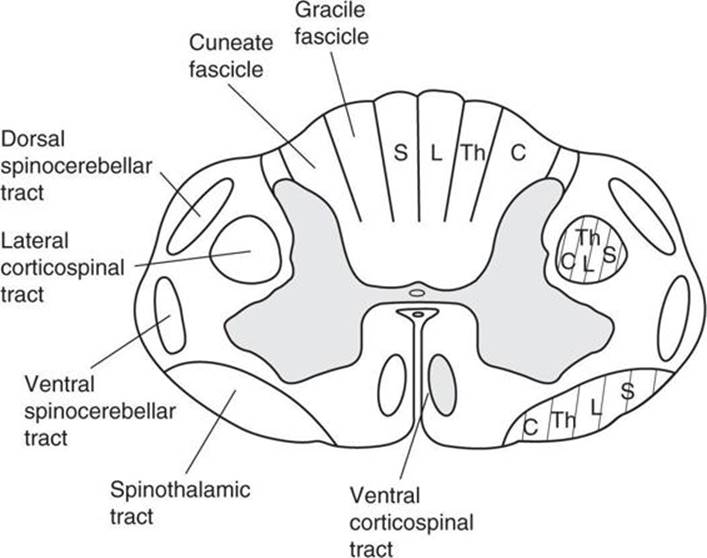
Figure A-2. Schematic diagram of the spinal cord. S, sacral; L, lumbar; Th, thoracic; C, cervical
(Modified, with permission, from JH Martin. Neuroanatomy: Text and Atlas. 3rd ed. New York, NY: McGraw-Hill; 2003.)

Figure A-3. The dermatomes of the body have a segmental organization. Note the correspondence between the spinal cord divisions (shown on a ventral view of the central nervous system) and dermatome locations. The inset illustrates dermatomal overlap.
(Used, with permission, from JH Martin. Neuroanatomy: Text and Atlas. 3rd ed. New York, NY: McGraw-Hill; 2003.)
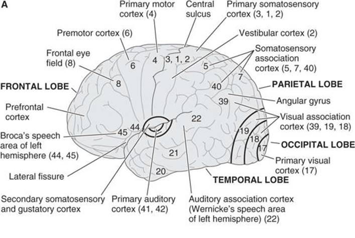
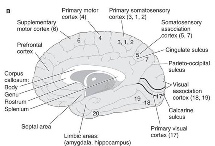
Figure A-4. Functional anatomy of the cerebral cortex.
(Used, with permission, from JS White. USMLE Road Map: Neuroscience. 2nd ed. New York, NY: McGraw-Hill; 2008.)

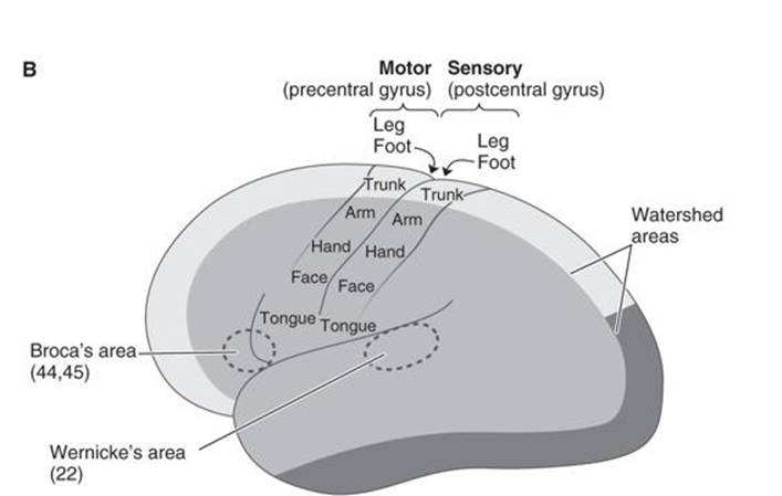
Figure A-5. (A) Coronal and (B) lateral views of the hemisphere showing the regions served by the anterior, middle, and posterior cerebral arteries.
(Used, with permission, from JS White. USMLE Road Map: Neuroscience. 2nd ed. New York, NY: McGraw-Hill; 2008.)
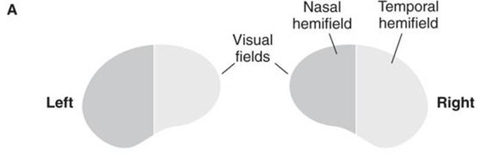
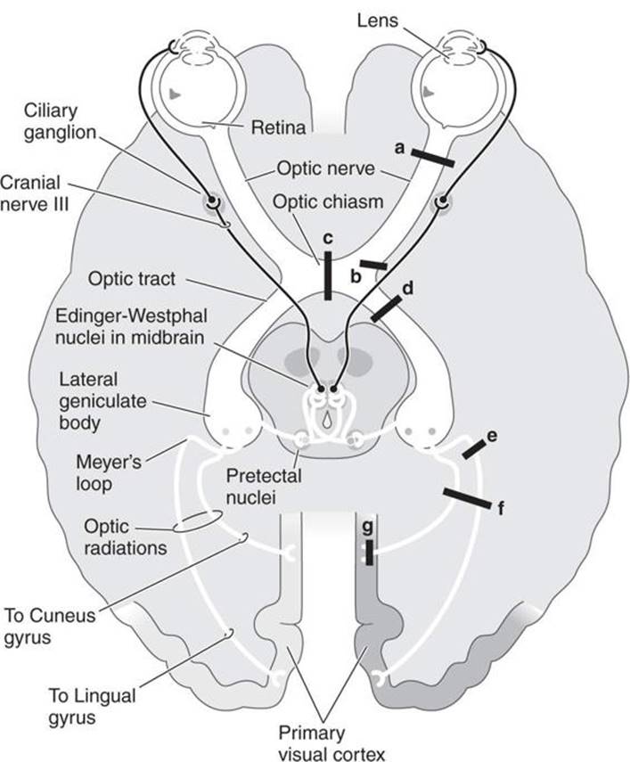
Figure A-6. A: Visual pathways.


Figure A-6. B: Laminae of the lateral geniculate body. C: Common visual field deficits.
(Used, with permission, from JS White. USMLE Road Map: Neuroscience. 2nd ed. New York, NY: McGraw-Hill; 2008.)
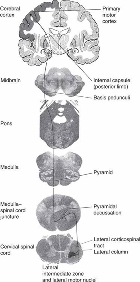
Figure A-7. Lateral corticospinal tract. The lateral corticospinal tract originates from neurons located in area 6 and the parietal lobe.
(Used, with permission, from JH Martin. Neuroanatomy: Text and Atlas. 3rd ed. New York, NY: McGraw-Hill; 2003.)
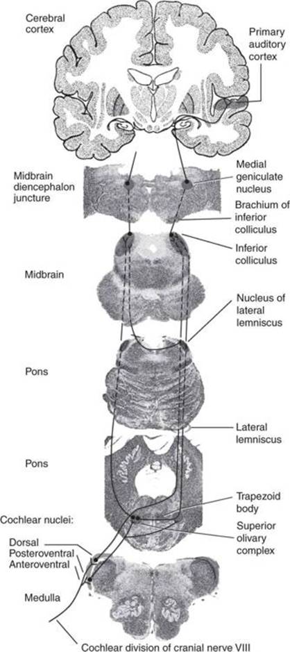
Figure A-8. Organization of the auditory system revealed in cross section of different levels through the brain stem and in coronal section through the diencephalon and cerebral hemispheres.
(Used, with permission, from JH Martin. Neuroanatomy: Text and Atlas. 3rd ed. New York, NY: McGraw-Hill; 2003.)

Figure A-9. Arterial supply at the brain and rostral spinal cord.
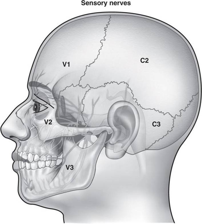
Figure A-10. Sensory innervation of the head. Middle and anterior fossa meninges are innervated by V1. Posterior fossa meninges are innervated by C2.
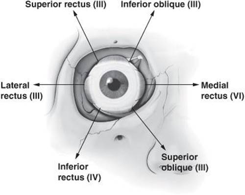
Figure A-11. Drawing of right eye indicating innervation and action of each of the muscles of extraocular movement.
(Modified, with permission, from Morton DA, Foreman KB, Albertine KH. The Big Picture: Gross Anatomy. New York: McGraw-Hill, 2011.)

Figure A-12. Brachial plexus. L, lateral; P, posterior; M, medial; s, superior; m, middle; i, inferior.
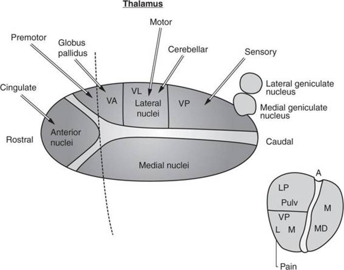
Figure A-13. Thalamic nuclei and their inputs. VA, ventral anterior nucleus; VL, ventral lateral nucleus; VP, ventral posterior nuclei (L, Lateral portion of VP; M, Medial portion of VP); Pulv, pulvinar nucleus; MD, medial dorsal nucleus (M, Medial nucleus); A, Anterior nuclei; LP, Lateral posterior nucleus.