17.1. Seizure classification
Definition of a seizure: an abnormal paroxysmal cerebral neuronal discharge that results in alteration of sensation, motor function, behavior or consciousness. Seizures may be classified by type, etiology, and by epileptic syndromes.
Classification of major seizure types1-3
1. primary generalized: bilaterally symmetrical and synchronous involving both cerebral hemispheres at the onset, no local onset, consciousness lost from the start. Represents ≈ 40% of all seizures
A. generalized tonic-clonic (GTC) (née: grand-mal seizure): generalized seizure that evolves from tonic to clonic motor activity. This is a specific type and does NOT include partial seizures that generalize secondarily
B. clonic seizures: fairly symmetric, bilateral synchronous semirhythmic jerking of the UE & LE, usually with elbow flexion and knee extension
C. tonic seizures: sudden sustained increased tone with a characteristic guttural cry or grunt as air is forced through adducted vocal cords
D. absence (née: petit-mal seizure): impaired consciousness with mild or no motor involvement (see below)
1. typical absences
2. atypical absences: more heterogeneous with more variable EEG pattern then typical absence. Seizures may last longer
E. myoclonic seizures: shock-like body jerks (1 or more in succession) with generalized EEG discharges
F. atonic seizures (AKA astatic seizures or “drop attacks”): sudden brief loss of tone that may cause falls
2. partial (née focal seizure): implies one hemisphere involved at onset. About 57% of all seizures. A new onset of partial seizure represents a structural lesion until proven otherwise
A. simple partial seizure (no impairment of consciousness)
1. with motor signs (including Jacksonian)
2. with sensory symptoms (special sensory or somatosensory)
3. with autonomic signs or symptoms
4. with psychic symptoms (disturbance of higher cerebral function)
B. complex partial seizure (many used to be classified as psychomotor seizure, often attributed to temporal lobe but they can arise from any cortical area): any alteration of consciousness, usually LOC or automatisms (including lip smacking, chewing, or picking with the fingers) with autonomic aura (usually an epigastric rising sensation)
1. simple partial onset followed by impairment of consciousness (may have premonitory aura)
a. without automatisms
b. with automatisms
2. with impairment of consciousness at onset
a. without automatisms (impairment of consciousness only)
b. with automatisms
C. partial seizure with secondary generalization
1. simple partial evolving to generalized
2. complex partial evolving to generalized
3. simple partial evolving to complex partial evolving to generalized
3. unclassified epileptic seizures: ≈ 3% of all seizures
Classification by etiology (and some epileptic syndromes)
This list is not all inclusive (see reference2, 3).
1. symptomatic (AKA “secondary”): seizures of known etiology (e.g. CVA, tumor…)
A. temporal lobe epilepsies:
1. mesial temporal sclerosis: see below
2. idiopathic (AKA “primary”): no underlying cause. Includes:
A. juvenile myoclonic epilepsy: see below
3. cryptogenic: seizures presumed to be symptomatic but with unknown etiology
A. West syndrome (infantile spasms, Blitz-Nick-Salaam Krämpfe): see below
B. Lennox-Gastaut syndrome: see below
4. special syndromes: situation-related seizures
A. febrile seizures: see page 402
B. seizures occurring only with acute metabolic or toxic event: e.g. alcohol
KEY distinctions (having therapeutic implications)
In generalized tonic-clonic seizures: primary generalized vs. partial with secondary generalization (often, local onset may not be observed).
In staring spells: absence vs. complex partial.
EPILEPSY
A disorder, not a single disease. Characterized by recurrent (2 or more), unprovoked seizures.
ABSENCE SEIZURE
Formerly called petit-mal seizure. Impaired consciousness with mild or no motor involvement (automatisms occur more commonly with bursts lasting > 7 secs). No post-ictal confusion. Aura rare. May be induced by hyperventilation x 2-3 mins. EEG shows spike and wave at exactly 3 per second.
UNCINATE SEIZURES
Obsolete term: “uncal fits”. Seizures originating in the inferior medial temporal lobe, usually in the hippocampal region. May produce olfactory hallucinations (kakosmia or cacosmia: the perception of bad odors where none exist).
MESIAL TEMPORAL SCLEROSIS4, 5
The most common cause of intractable temporal lobe epilepsy. Specific pathologic basis: hippocampal sclerosis (cell loss in hippocampus on one side). Characteristics are shown in Table 17-1. For differential diagnosis, see page 1228.
Adult seizures are initially responsive to medical therapy but become more varied and refractory, and may respond to seizure surgery.
JUVENILE MYOCLONIC EPILEPSY7
Sometimes called bilateral myoclonus. 5-10% of cases of epilepsy. An idiopathic generalized epilepsy syndrome with age-related onset consisting of 3 seizure types:
1. myoclonic jerks: predominantly after waking
2. generalized tonic-clonic seizures
3. absence
Table 17-1 Syndrome of mesial temporal-lobe epilepsy6
|
History |
|
• higher incidence of complicated febrile seizures than in other types of epilepsy • common family history of epilepsy • onset in latter half of first decade of life • auras in isolation are common • infrequent secondarily generalized seizures • seizures often remit for several years until adolescence or early adulthood • seizures often become medically refractory • common interictal behavioral disturbances (especially depression) |
|
Clinical features of seizures |
|
• most have aura (especially epigastric, emotional, olfactory or gustatory) x several secs • CPS often begin with arrest & stare; oroalimentary & complex automatisms are common. Posturing of contralateral arm may occur. Seizure usually lasts 1-2 mins • postictal disorientation, recent-memory deficit, amnesia of ictus and (in dominant hemisphere) aphasia usually lasts several mins |
|
Neurologic and laboratory features |
|
• neuro exam: normal except memory deficit • MRI: hippocampal atrophy and signal alteration with ipsilateral dilatation of temporal horn of lateral ventricle • unilateral or bilateral independent anterior temporal EEG spikes with maximal amplitude in basal electrodes • external ictal EEG activity only with CPS, usually initial or delayed focal rhythmic onset pattern of 5-7 Hz, maximal in 1 basal temporal derivation • interictal fluorodeoxyglucose PET scan: hypometabolism in temporal lobe and possibly ipsilateral thalamus and basal ganglia • neuropsychological testing: memory dysfunction specific to involved temporal lobe • Wada test: amnesia with contralateral amobarbital injection (see page 421) |
EEG → polyspike discharges. Strong family history (some studies showing linkage to the HLA region on the short arm of chromosome 6). Most responsive to depakene.
WEST SYNDROME
This term is being used less frequently as it appears not to be a homogeneous group. Classically a seizure disorder that usually appears in first year of life, and consists of recurrent, gross flexion and occasionally extension of the trunk and limbs (massive myoclonus, AKA infantile spasms, AKA salaam seizures, AKA jackknife spasms). Seizures tend to diminish with age, often abating by 5 yrs. Usually associated with mental retardation. 50% may develop complex-partial seizures, some of the rest may develop Lennox-Gastaut syndrome (see below). An associated brain lesion may be found in some.
EEG → the majority show either interictal hypsarrhythmia (huge spike/wave plus slow wave resembling muscle artifact) or modified hypsarrhythmia at some point.
Usually dramatic response of seizures and EEG findings to ACTH or corticosteroids.
LENNOX-GASTAUT SYNDROME
Rare condition that begins in childhood as atonic seizures (“drop attacks”). Often develops into tonic seizures with mental retardation. Seizures are often polymorphic, difficult to treat medically, and may occur as often as 50 per day. May also present with status epilepticus. Approximately 50% of patients have reduced seizures with valproic acid. Corpus callosotomy may reduce the number of atonic seizures.
TODD’S PARALYSIS
A post-ictal phenomena in which there is partial or total paralysis usually in areas involved in a partial seizure. More common in patients with structural lesions as the source of the seizure. The paralysis usually resolves slowly over a period of an hour or so. Thought to be due to depletion of neurons in the wake of the extensive electrical discharges of a seizure. Other similar phenomena include post-ictal aphasia and hemianopsia.
17.1.1. Factors that lower the seizure threshold
Factors that lower the seizure threshold (i.e. make it easier to provoke a seizure) in individuals with or without a prior seizure history include many items listed under Etiologies of New onset seizures (see below) as well as:
1. sleep deprivation
2. hyperventilation
3. photic stimulation (in some)
4. infection: systemic (febrile seizures, see page 402), CNS…
5. metabolic disturbances: electrolyte imbalance (especially profound hypoglycemia), pH disturbance (especially alkalosis), drugs… (see below)
6. head trauma: closed head injury, penetrating trauma (see page 398)
7. cerebral ischemia: CVA (see below)
8. “kindling”: a concept that repeated seizures may facilitate the development of later seizures
17.2. Special types of seizures
17.2.1. New onset seizures
The age adjusted incidence of new onset seizures in Rochester, Minnesota was 44 per 100,000 person years8.
Etiologies: In patients presenting with a first-time seizure, etiologies include (modified9):
1. following neurologic insult: either acutely (i.e. < 1 week) or remotely (> 1 week, and usually < 3 mos from insult)
A. cerebrovascular accident (CVA, or stroke): 4.2% had a seizure within 14 days of a CVA. Risk increased with severity of stroke10
B. head trauma: closed head injury, penetrating trauma (see page 398)
C. CNS infection: meningitis, cerebral abscess, subdural empyema
D. febrile seizures: see page 402
E. birth asphyxia
2. underlying CNS abnormality
A. congenital CNS abnormalities
B. degenerative CNS disease
C. CNS tumor: metastatic or primary
D. hydrocephalus
E. AVM
3. acute systemic metabolic disturbance
A. electrolyte disorders: uremia, hyponatremia, hypoglycemia (especially profound hypoglycemia), hypercalcemia
B. drug related, including:
1. alcohol-withdrawal: see page 399
2. cocaine toxicity: see page 276
3. opioids (narcotics), principally associated with the following:
a. propoxyphene (Darvon®)
b. meperidine (Demerol®): may also cause delerium
c. the street drug combination “T’s and blues” (pentazocine (Talwin®) + the antihistamine tripelennamine)
4. phenothiazine antiemetics
5. with administration of flumazenil (Romazicon®) to treat benzodiazepine (BDZ) overdose (especially when BDZs are taken with other seizure lowering drugs such as tricyclic antidepressants or cocaine
6. phencyclidine (PCP): originally used as an animal tranquilizer
7. cyclosporine: can affect Mg++ levels
C. eclampsia
4. idiopathic
In 166 pediatric patients presenting to an emergency department with either a chief complaint of, or a discharge diagnosis of a first-time seizure11:
1. 110 were found to actually have either a recurrent seizure or a nonictal event
2. of the 56 patients actually thought to have had a first-time seizure
A. 71% were febrile seizures
B. 21% were idiopathic
C. 7% were “symptomatic” (hyponatremia, meningitis, drug intoxication…)
In a prospective study of 244 patients with a new-onset unprovoked seizure, only 27% had further seizures during follow-up9, 12. Recurrent seizures were more common in patients with a family seizure history, spike-and-waves on EEG, or a history of a CNS insult (CVA, head injury…). No patient seizure-free for 3 years had a recurrence. Following a second seizure, the risk of further seizures was high.
EVALUATION
Adults
A new-onset seizure in an adult in the absence of obvious cause (e.g. alcohol withdrawal) should prompt a search for an underlying basis (the onset of idiopathic seizures, i.e. epilepsy, is most common before or during adolescence). A CT or MRI (without and with enhancement) should be performed. A systemic work-up should be done to identify the presence of any factors listed previously (see above). If all this is negative, then an MRI should be performed if not already done. If this is negative also, a repeat study (CT or MRI) should be done in ≈ 6 months and at 1 and possibly 2 years to rule-out a tumor which might not be evident on the initial study.
Pediatrics
Among pediatric patients with first-time seizures, laboratory and radiologic evaluations were often costly and not helpful11. A detailed history and physical exam were more helpful.
MANAGEMENT
Management of an adult with the new onset of idiopathic seizures (i.e. no abnormality found on CT or MRI, no evidence of drug withdrawal) is controversial. In one study, an EEG was performed, which if normal was followed by a sleep deprived EEG with the following observations13:
1. there is substantial interobserver variation in interpreting such EEGs
2. if both EEGs were normal, the 2-yr recurrence rate of seizures was 12%
3. if one or both EEGs showed epileptic discharges, the 2-yr recurrence rate was 83%
4. the presence of nonepileptic abnormalities in one or both EEGs had a 41% 2-yr recurrence rate
5. the recurrence rate with focal epileptic discharges (87%) was slightly higher than for generalized epileptic discharges (78%)
The conclusion is that EEGs thus obtained have moderate predictive value, and may be factored into the decision of whether or not to treat such seizures with AEDs.
17.2.2. Posttraumatic seizures
![]() Key concepts:
Key concepts:
• 2 categories: early (≤ 7 days) and late (> 7 days) after head trauma
• anticonvulsants (AEDs) may be used to prevent early posttraumatic seizures (PTS) in patients at high risk for seizures (see text)
• prophylactic AEDs do NOT reduce the frequency of late PTS
• discontinue AEDs after 1 week except for cases meeting specific criteria (see text)
Posttraumatic seizures (PTS) are often divided (arbitrarily) into: early (occurring within 1 week of injury) and late (thereafter)14. There may be justification for a third category: “immediate”, i.e. within minutes to an hour or so.
Early PTS (≤ 7 days after head trauma)
30% incidence in severe head injury (“severe” defined as: LOC > 24 hrs, amnesia > 24 hrs, focal neuro deficit, documented contusion, or intracranial hematoma) and ≈ 1% in mild to moderate injuries. Occurs in 2.6% of children < 15 yrs age with head injury causing at least brief LOC or amnesia15.
Early PTS may precipitate adverse events as a result of elevation of ICP, alterations in BP, changes in oxygenation, and excess neurotransmitter release16.
Late onset PTS (> 7 days after head trauma)
Estimated incidence 10-13% within 2 yrs after “significant” head trauma (includes LOC > 2 mins, GCS < 8 on admission, epidural hematoma…) for all age groups17, 18. Relative risk: 3.6 times control population. Incidence in severe head injury >> moderate > mild15.
The incidence of early PTS is higher in children than adults, but late seizures are much less frequent in children (in children who have PTS, 94.5% develop them within 24 hrs of the injury19). Most patients who have not had a seizure within 3 yrs of penetrating head injury will not develop seizures20. Risk of late PTS in children does not appear related to the occurrence of early PTS (in adults: only true for mild injuries). Risk of developing late PTS may be higher after repeated head injuries.
Penetrating trauma
The incidence of PTS is higher with penetrating head injuries than with closed head injuries (occurs in 50% of penetrating trauma cases followed 15 yrs21).
TREATMENT
A prospective double blind study of patients at high risk of PTS (excluding penetrating trauma) showed a 73% reduction of risk of early PTS by administering 20 mg/kg loading dose of PHT within 24 hrs of injury and maintaining high therapeutic levels; but after 1 week there was no benefit in continuing the drug (based on intention to treat)22. Carbamazepine (Tegretol®) has also been shown to be effective in reducing the risk of early PTS, and valproic acid is currently being studied16.
Phenytoin has adverse cognitive effects when given long-term as prophylaxis against PTS23.
TREATMENT GUIDELINES
Based on available information (see above) it appears that:
1. no treatment studied effectively impedes epileptogenesis
2. in high-risk patients (see Table 17-2), AEDS reduces the incidence of early PTS
3. however, no study has shown that reducing early PTS improves outcome24
4. once epilepsy has developed, continued AEDs reduces the recurrence of seizures
The following are therefore offered as guidelines.
Initiation of AEDs
AEDs may be considered for short term use especially if a seizure could be detrimentalA. Early posttraumatic seizures were effectively reduced when phenytoin was used for 2 weeks following head injury with no significant increased risk of adverse effects25.
A. acutely, seizures may elevate ICP, and may adversely affect blood pressure and oxygen delivery, and may worsen other injuries (e.g. spinal cord injury in the setting of an unstable cervical spine)16
Option: begin AEDs (usually phenytoin or carbamazepine) within 24 hrs of injury in the presence of any of the high risk criteria shown in Table 17-2 (modified16, 19, 22, 26). When using PHT, load with 20 mg/kg and maintain high therapeutic levels (see page 409). Switch to phenobarbital if PHT not tolerated.
Table 17-2 High risk criteria for PTS
|
1. acute subdural, epidural, or intracerebral hematoma 2. open-depressed skull fracture with parenchymal injury 3. seizure within the first 24 hrs after injury 4. Glasgow Coma Scale score < 10 5. penetrating brain injury 6. history of significant alcohol abuse 7. ± cortical (hemorrhagic) contusion on CT |
Discontinuation of AEDs
1. taper AEDs after 1 week of therapy except in the following:
A. penetrating brain injury
B. development of late PTS (i.e. a seizure > 7 days following head trauma)
C. prior seizure history
D. patients undergoing craniotomy27
2. for patients not meeting the criteria to discontinue AEDs after 1 week (see above):
A. maintain ≈ 6-12 mos of therapeutic AED levels
B. recommend EEG to rule-out presence of a seizure focus before discontinuing AEDs (predictive value) for the following:
1. repeated seizures
2. presence of high risk criteria shown in Table 17-2.
17.2.3. Alcohol withdrawal seizures
Also, see Alcohol withdrawal syndrome, page 273. The withdrawal syndrome may begin hours after the EtOH peak (see page 274 for prevention and treatment). Ethanol withdrawal seizures are classically seen in up to 33% of habituated drinkers within 7-30 hours of cessation or reduction of ethanol intake. They typically consist of 1-6 tonic-clonic generalized seizures without focality within a 6 hour period28. Seizures usually occur before delerium develops. They may also occur during intoxication (without withdrawal).
The seizure risk persists for 48 hrs (risk of delerium tremens (DTs) continues beyond that), thus a single loading dose of PHT is frequently adequate for prophylaxis. However, since most EtOH withdrawal seizures are single, brief, and self-limited, PHT has not been shown to be of benefit in uncomplicated cases and is thus usually not indicated. Chlordiazepoxide (Librium®) or other benzodiazepines administered during detoxification reduces the risk withdrawal seizures29 (see page 274).
Evaluation
The following patients should have a CT scan of the brain, and should be admitted for further evaluation as well as for observation for additional seizures or for DTs:
1. those with their first EtOH withdrawal seizure
2. those with focal findings
3. those having more than 6 seizures in 6 hrs
4. those with evidence of trauma
Other causes of seizure should also be considered, e.g. a febrile patient may require an LP to R/O meningitis.
Treatment
A brief single seizure may not warrant treatment, except as outlined below. A seizure that continues beyond 3-4 minutes may be treated with diazepam or lorazepam, with further measures used as in status epilepticus (see page 402) if seizures persist. Loading with phenytoin (18 mg/kg = 1200 mg/70 kg, see page 409) and long-term treatment is indicated for:
1. a history of previous alcohol withdrawal seizures
2. recurrent seizures after admission
3. history of a prior seizure disorder unrelated to alcohol
4. presence of other risk factors for seizure (e.g. subdural hematoma)
17.2.4. Nonepileptic seizures
AKA pseudoseizures (some prefer not to use this term since it may connote voluntary feigning of seizures), with the term psychogenic seizures being preferred for nonepileptic seizures (NES) with a psychologic etiology (psychogenic seizures are real events and may not be under voluntary control)30.
The hazard of NES is that patients may end up needlessly taking AEDs, which in some cases may worsen NES. Possible etiologies of NES are given in Table 17-3. Most NES are psychogenic.
DDx for seizures:
1. psychogenic: 20-90% of patients with intractable seizures referred to epilepsy centers. These patients carry the diagnosis of seizures from 5-7 years. Up to 50% of these may have legitimate seizures at some time as well31.
2. tic: can be suppressed, is not repetitive (if repetitive, may be hemifacial spasm)
3. movement disorder: myoclonus (can be epileptic or non-epileptic)
A. cataplexy: e.g. with narcolepsy often provoked by laughter or other emotional stimulus (can rarely be caught on EEG, when it is it shows REM intrusion into wakefulness)
B. parasomnia: a sleep movement disorder (occurs during sleep). Includes: night terrors (occurs in slow wave sleep, vs. nightmare which occurs in REM), sleep walking, REM behavior disorders (usually occurs in older men, and there is a high prob-ability they will go on to have degenerative brain disease (used to be called paroxysmal nocturnal PNT). Head banging is a benign parasomnia
4. syncope: 90% of the time people who faint have myoclonic jerks or shaking32
5. TIA
Table 17-3 Differential diagnosis of nonepileptic seizures30
|
1. psychologic disorders (psychogenic seizure) A. somatoform disorders: especially conversion disorder B. anxiety disorders: especially panic attack and posttraumatic stress disorder C. dissociative disorders D. psychotic disorders E. impulse control disorders F. attention-deficit disorders* G. factitious disorders: including Munchausen’s syndrome |
|
2. cardiovascular disorders A. syncope B. cardiac arrhythmias C. transient ischemic attacks D. breath-holding spells* |
|
3. migraine syndromes A. complicated migraines* B. basilar migraines |
|
4. movement disorders A. tremors B. dyskinesias C. tics*, spasms D. other (including shivering) |
|
5. parasomnias & sleep-related disorders A. night terrors*, nightmares*, somnambulism* B. narcolepsy, cataplexy C. rapid eye movement behavior disorder D. nocturnal paroxysmal dystonia |
|
6. gastrointestinal disorders A. episodic nausea or colic* B. cyclic vomiting syndrome* |
|
7. other A. malingering B. cognitive disorders with episodic behavioral or speech symptoms C. medication effects or toxicity D. daydreams* |
* usually encountered in children
DIFFERENTIATING NES FROM EPILEPTIC SEIZURES
Distinguishing between epileptic seizures (ES) and NES is a common clinical dilemma. There are unusual seizures that may fool experts33. Some frontal lobe and temporal lobe complex partial seizures may produce bizarre behaviors that do not correspond to classic ES findings and may not produce discernible abnormalities with scalp-electrode EEG (and therefore may be misdiagnosed even with video-EEG monitoring, although this is more likely with partial seizures than with generalized). A multidisciplinary team approach may be required.
History: Attempt to document: prodromal symptoms, precipitating factors, time and environment of Sz, mode and duration of progression, ictal and postictal events, frequency and stereotypy of manifestations. Determine if patient has history of psychiatric conditions, and if they are acquainted with individuals who have ES.
Psychological testing: May help. Differences occur in ES and NES on the Minnesota Multiphasic Personality Inventory (MMPI) scales in hypochondriasis, depression hysteria, and schizophrenia34.
Table 17-4 contrasts some features of true seizures vs. NES, and Table 17-5 lists some features often associated with NES, however, no characteristics are definitively diagnostic of NES since a number of them may also occur with ES.
Features common to both true seizures and NES: verbal unresponsiveness, rarity of automatisms and whole-body flaccidity, rarity of urinary incontinence. Reminder: some seizures can be bizarre and can resemble NES (sometimes called pseudo-pseudoseizures). 10% of patients with psychogenic seizures actually have epilepsy.
Table 17-4 Features of ES vs. NES31
|
Feature |
Epileptic seizure |
NES |
|
% males |
72% |
20% |
|
Clonic UE movement |
||
|
inphase |
96% |
20% |
|
out-of-phase |
0 |
56% |
|
Clonic LE movement |
||
|
inphase |
88% |
16% |
|
out-of-phase |
0 |
56% |
|
Vocalizations |
||
|
none |
16% |
56% |
|
start of seizure |
24% |
44% |
|
middle |
60% “epileptic cry” |
0 |
|
types |
only sounds of tonic or clonic respiratory muscle contraction |
moans, screams, grunts, snorts, gagging, retching, understandable statements, gasps |
|
Head turning |
||
|
unilateral |
64% |
16% |
|
side-to-side |
8% (slow, low amplitude) |
36% (violent, high amplitude) |
Features suggestive of non-epileptic seizures:
1. arching of the back: 90% specific for NES
2. asynchronous movement
3. stop & go: seizures usually build and then gradually subside
4. forced eye closing during entire seizure
5. provoked with stimuli that would not cause a seizure (e.g. tuning fork to the head, alcohol pad to the neck, IV saline…)
6. bilateral shaking with preserved awareness. Exception: supplementary motor area seizures (mesial frontal area) these seizures are usually tonic (not clonic)
7. weeping (whining): highly specific
8. multiple or variable seizure types (ES is usually stereotypical), fluctuating level of consciousness, denial of correlation of Sz with stress
If any two of the following are demonstrated, 96% of time this will be NES:
1. out-of-phase clonic UE movement
2. out-of-phase clonic LE movement
3. no vocalization or vocalization at start of event
Lateral tongue laceration is very specific for seizures.
Table 17-5 Features often associated with NES30
|
• frequent seizures despite therapeutic AEDs • multiple different-physician visits • lingering prodrome or gradual ictal onset (over minutes) • prolonged duration (> 5 mins) • manifestations altered by distraction • suggestible or inducible seizures • intermittent arrhythmic and out-of-phase activity • fluctuating intensity and severity during Sz • side-to-side rolling, pelvic thrusting, wild movements • bilateral motor activity with preserved consciousness • nonphysiologic spread of neurologic signs • absence of labored breathing or drooling after generalized convulsion • expression of relief or indifference • crying or whimpering • no postictal confusion or lethargy • disproportionate postictal mental status changes • absence of stereotypy |
Prolactin levels after seizures
Transient elevations in human serum prolactin (HSP) levels occur following 80% of generalized motor, 45% of complex partial, and only 15% of simple partial seizures35. Peak levels are reached in 15-20 minutes, and gradually return to baseline over the subsequent hour36-38. It has been suggested that drawing a serum prolactin level shortly after a questionable seizure may be helpful in differentiating NES (which may have elevated cortisol levels but normal HSP levels39).
Repetitive seizures are associated with progressively smaller HSP elevations40, and no rise follows absence seizures or status epilepticus (whether convulsive or absence)41. Greater than twofold HSP elevations consistently follow seizures that produce intense widespread high frequency mesial temporal lobe discharges; whereas such elevations do not occur in seizures not involving these limbic structures42.
Furthermore, there may be higher baseline HSP levels in cases with right-sided interictal EEG discharges compared to those with left-sided43, and the presence of psycho-pathology may affect postictal HSP elevations44.
Therefore, the presence of HSP peaks may be strongly indicative of true seizures, but the absence may be due to a variety of complex phenomena45. The overall classification accuracy is ≈ 72%38.
17.2.5. Febrile seizures
Definitions46
|
febrile seizure |
a seizure in infants or children associated with fever with no defined cause and unaccompanied by acute neurologic illness (includes seizures during vaccination fevers) |
|
complex febrile seizure |
a convulsion that lasts longer than 15 minutes, is focal, or multiple (more than one convulsion per episode of fever) |
|
simple febrile seizure |
not complex |
|
recurrent febrile seizure |
more than one episode of fever associated with seizures |
Epidemiology46
Febrile convulsions are the most common type of seizure. Excluding children with pre-existing neurologic or developmental abnormalities, the prevalence of febrile seizures is ≈ 2.7% (range: 2-5% in U.S. children aged 6 mos-6 yrs). The risk for developing epilepsy after a simple febrile seizure is ≈ 1%, and for a complex febrile seizure is 6% (9% for prolonged seizure, 29% for focal seizure). An underlying neurological or developmental abnormality or a family history of epilepsy increases the risk of developing epilepsy.
Treatment
In one study, the IQ in the group treated with phenobarbital was 8.4 points lower (95% confidence interval) than the placebo group, and there remained a significant difference several months after discontinuing the drug47. Furthermore, there was no significant reduction in seizures in the phenobarbital group. And yet, no other drug really appears well suited to treating this entity: carbamazepine and phenytoin appear ineffective, valproate may be effective but has serious risks in the < 2 yrs age group. Given the low incidence (1%) of having afebrile seizures (i.e. epilepsy) after a simple febrile seizure and the fact that AEDs probably do not prevent this development, there is little support for prescribing anticonvulsants in these cases. The recurrence rate of febrile seizures in children with a history of one or more febrile seizure can be reduced by administering diazepam 0.33 mg/kg PO q 8 hrs during a febrile episode (temp > 38.1° C) and continuing until 24 hrs after the fever subsides48.
17.3. Status epilepticus
![]() Key concepts:
Key concepts:
• definition: Sz > 5 mins, or persistent Sz after 1st & 2nd line AEDs
• morbidity and mortality are high in untreated status epilepticus (SE)
• most common etiology: patient with known Sz disorder with low AED levels
• de novo SE in acute illness is considered a manifestation of the illness which should be treated at the same time as the SE
• see Table 17-6, page 405 for treatment measures
Definition: a seizure lasting > 5 minutes or persistent seizure activity after sequential administration of appropriate first and second-line AEDs49.
Features important to management
• seizures that do not cease in 5-10 mins are less likely to terminate50
• in patients with no prior Sz history, status epilepticus (SE) is usually a manifestation of illness-related cortical irritation or injury49 and treatment of the underlying disorder (in addition to treating the SE) is critical
• a relapse of Sz. in a patient with a known Sz. disorder and subtherapeutic AED levels usually responds to a bolus of the maintenance AEDs. However, SE should be treated by the standard protocol49
• most cases of convulsive status in adults start as partial seizures that generalize
• the choice of 1st and 2nd-line AEDs is arbitrary, and the dose is the more important determinant of success in aborting SE49
Types of status epilepticus
• generalized status
1. convulsive: generalized convulsive tonic-clonic status epilepticus (SE) is the most frequent type51. A medical emergency
2. absenceA
3. secondarily generalized: accounts for ≈ 75% of generalized SE
4. myoclonic
5. atonic (drop attack): especially in Lennox-Gastaut syndrome (see page 396)
• partial status (usually related to an anatomic abnormality)
1. simple (AKA epilepsy partialis continuans)
2. complexA: most often from frontal lobe focus. Urgent treatment is required
3. secondarily generalized
A. in status, these may present in twilight state
Epidemiology
Incidence is ≈ 150,000 new cases/year in the U.S. in the outpatient setting49. Most cases occur in young children (among children, 73% were < 5 yrs old52), the next most affected group is patients > 60 yrs age. In > 50% of cases, SE is the patient’s first seizure51. One out of six patients presenting with a first time seizure will present in SE
Etiologies
1. the most common etiology is a patient with a known seizure disorder having low AED levels for any reason (non-compliance, intercurrent infection preventing PO intake of meds, drug-drug interactions → lowering effectiveness of AEDs…)
2. febrile seizures: a common precipitator in young patients. 5-6% of patients presenting with SE have a history of prior febrile seizures
3. cerebrovascular accidents: the most commonly identified cause in the elderly
4. CNS infection: in children, most are bacterial, the most common organisms were H. influenza and S. pneumoniae
5. idiopathic: accounts for ≈ one-third (in children, usually associated with fever)
6. epilepsy: is present or is subsequently diagnosed in ≈ 50% of patients presenting with SE. About 10% of adults ultimately diagnosed as having epilepsy will present in SE
7. electrolyte imbalance: hyponatremia (most common in children, usually due to water intoxication52), hypoglycemia, hypocalcemia, uremia, hypomagnesemia…
8. illicit drug intoxication: especially cocaine, amphetamines
9. precipitous drug withdrawal: barbiturates, benzodiazepines, alcohol or narcotics
10. proconvulsant drugs, including: ß-lactam antibiotics (penicillins, cephalosporins), certain antidepressants (bupropion), clonazapine, bronchodilators, immunosuppressants
11. traumatic brain injury: acute as well as old
12. hypoxia/ischemia
13. tumor
In children < 1 yr age, 75% had an acute cause: 28% were secondary to CNS infection, 30% due to electrolyte disorders, 19% associated with fever52. In adults, a structural lesion is more likely. In an adult, the most common cause of SE is subtherapeutic AED levels in a patient with a known seizure disorder.
Morbidity and mortality from SE
Mean duration of SE in patients without neurologic sequelae is 1.5 hrs (therefore, proceed to pentobarbital anesthesia before ≈ 1 hour of SE). Recent mortality: < 10-12% (only ≈ 2% of deaths are directly attributable to SE or its complications; the rest are due to the underlying process producing the SE). Mortality in lowest amongst children (≈ 6%52), patients with SE related to subtherapeutic AEDs, and patients with unprovoked SE53. The highest mortality occurs in elderly patients and those with SE resulting from anoxia or CVA53. 1% of patients die during the episode itself.
Morbidity and mortality is due to54:
1. CNS injury from repetitive electric discharges: irreversible changes begin to appear in neurons after as little as 20 minutes of convulsive activity. Cell death is very common after 60 mins
2. systemic stress from the seizure (cardiac, respiratory, renal, metabolic)
3. CNS damage by the acute insult that provoked the SE
17.3.1. General treatment measures for status epilepticus
Treatment is directed at stabilizing the patient, stopping the seizure, and identifying the cause (determining if there is an acute insult to the brain) and if possible also treating the underlying process. Treatment often must be initiated prior to the availability of test results to confirm the diagnosis.
• CPR if needed
• neurologic exam
• “ABC’s”
![]() A irway: oral airway if feasible. Turn patient on their side to avoid aspiration
A irway: oral airway if feasible. Turn patient on their side to avoid aspiration
![]() B reathing: O2 by nasal cannula or bag-valve-mask. Consider intubation if respirations compromised or if seizure persists > 30 min
B reathing: O2 by nasal cannula or bag-valve-mask. Consider intubation if respirations compromised or if seizure persists > 30 min
![]() C irculation: large bore proximal IV access (2 if possible): start with NS KVO
C irculation: large bore proximal IV access (2 if possible): start with NS KVO
• monitor: EKG & baseline vital signs. Pulse oximeter. Frequent blood pressure checks
• bloodwork: STAT capillary blood (fingerstick) glucose (to R/O hypoglycemia), electrolytes (including glucose), CBC, LFTs, Mg++, Ca++, AED levels, ABG
• head CT (usually without contrast)
• correct any electrolyte imbalance (SE due to electrolyte imbalance responds more readily to correction than to AEDs52)
• if CNS infection is a major consideration, perform LP for CSF analysis (especially in febrile children) unless contraindicated (see page 201). WBC pleocytosis up to 80 x 106/L can occur following SE (benign postictal pleocytosis), and these patients should be treated with antibiotics until infection can be ruled out by negative cultures
• general meds for unknown patient:
1. glucose:
A. in patients with poor nutrition (e.g. alcoholics): giving glucose in thiamine deficiency can precipitate Wernicke’s encephalopathy (see page 275) ![]() prior to glucose bolus give thiamine 50-100 mg IV
prior to glucose bolus give thiamine 50-100 mg IV
B. if fingerstick glucose can be obtained immediately and it shows hypoglycemia, or if no fingerstick glucose can be done: give 25-50 ml of D50 IV push for adults (2 ml/kg of 25% glucose for peds). If at all possible, draw blood for definitive serum glucose first
2. naloxone (Narcan®) 0.4 mg IVP (in case of narcotics)
3. ± bicarbonate to counter acidosis (1-2 amps depending on length of seizure)
4. for neonate < 2 years: consider pyridoxine 100 mg IV push (pyridoxine-dependent seizures constitute a rare autosomal recessive condition that generally presents in the early neonatal period50)
• administer specific anticonvulsants for seizures lasting > 5-10 mins (see below)
• EEG monitor if possible
• if paralytics are used (e.g. to intubate), use short acting agents and be aware that muscle paralysis alone may stop visible seizure manifestations, but does not stop the electrical seizure activity in the brain, which can lead to permanent neurologic damage if prolonged (see above)
17.3.2. Medications for generalized convulsive status epilepticus
There are no randomized trials for refractory status epilepticus, although there is published data regarding specific treatment options. Numerous protocols exist. Table 17-6 shows a summary of medications for a status epilepticus protocol that is outlined in further detail below (modified management scheme49). Items below in boxes are considered treatment of choice. “Adult” refers to patients > 16 yrs of age. Drugs should be given IV (do not use IM route). If IV access is impossible, diazepam solution (not suppository) or Diastat® (diazepam rectal) can be given rectally or intranasal, buccal55 or rectal midazolam (Versed®) can be given.
Rapid treatment is indicated as delays are associated with neuronal injury and reduced response to medications.
Table 17-6 Summary of initial steps for status epilepticus (adult) (see text for details)
|
ABC’s. Start O2. Turn patient on their side. Check VS. Do a neuro exam |
|
Monitor/labs: Pulse oximetry. EKG/telemetry. Blood tests (do not wait for results to begin Rx): |
|
Large bore IV X 2. Start IV fluids |
|
• thiamine 100 mg IV and/or 50 ml of 50% dextrose (if needed) |
|
First-line AED: • lorazepam (Ativan®) 0.1 mg/kg IV @ < 2 mg/min |
|
Second-line AED: (may be given simultaneously with first-line drug or 1 minute after end of lorazepam bolus if Sz persist) • phenytoin*/fosphenytoin 20 mg/kg IV loading dose starting at 25 mg/min for phenytoin, or 75 mg/min for fosphenytoin • then, titrate phenytoin/fosphenytoin infusion up to max* as tolerated |
|
|
|
± 3rd line AED. NB: only 7% chance of stopping Sz with 3rd line drugs. (Consider: skipping 3rd line and intubate & start CIT below). Select one: • phenobarbital: up to 20 mg/kg IV (start infusing @ < 100 mg/min • sodium valproate 15-30 mg/kg IV bolus (max rate: 6 mg/kg/min) • levetiracetam 20mg/kg IV bolus of over 15 minutes |
|
CIT: If seizures continue > 30 mins or if skipping 3rd line drugs: intubate in ICU and begin continuous infusion therapy (CIT) of midazolam, pento-barbital or propofol (see text). If Sz persist, ensure that correctable conditions have been ruled-out and/or treated; continue to following steps |
|
alternative CIT: carbamazepine, oxcarbazepine, topiramate, lamotrigine… |
|
novel therapeutic options (not systematically studies): shock therapy… |
* maximum rate for phenytoin IV is 50 mg/min; for fosphenytoin it is 150 mg PE/min (see page 411) if BP stable
Protocol for status epilepticus (SE) in adult49
Prehospital phase:
1. impending SE: may be heralded by a crescendo in Sz. A 1-3 d course of lorazepam may preempt the development of SE
2. SE treatment may be initiated with buccal midazolam or rectal diazepam
Hospital phase:
Start IV drugs at half the maximal rate, and titrate up to maximal rate if VS stable.
1. First line drugs
A. benzodiazepine56 (main side effect: respiratory depression in ≈ 12%; be prepared to intubate). Onset of action is rapid (1-2 mins):
• lorazepam (Ativan®) 0.1 mg/kg (about 4 mg for average adult) IV @ rate < 2 mg/min (range: 0.02-0.12 mg/kg)
OR diazepam (Valium®) 0.1 mg/kg (7 mg average adult dose) IV @ rate < 50 mg/min. IV diazepam redistributes rapidly and SE may recur within a few minutes. Rectal dose (usually absorbed in ≈ 10 mins): 0.5 mg/kg of diazepam solution up to 20 mg max, or Diastat® 0.2 mg/kg
OR midazolam (Versed®) 0.05 mg/kg IV @ < 0.01 mg/min, or 10 mg buccal
• Wait 1 minute for response. If Sz continue, given additional doses of lorazepam up to a maximum of 9 mg (adult)
2. Second line drugs: either start simultaneously with 1st line drugs, or give if Sz persist 1 minute after first-line benzodiazepine dose
A. in second IV site: load with phenytoin (Dilantin®) (PHT)57 as follows (do not worry about acutely overdosing, but do follow dosing rates, monitor BP for hypotension and EKG for arrhythmias). Conventional phenytoin can only be given in NS to prevent precipitation. After giving the following loading dose, start on maintenance (see page 409)
1. phenytoin load with 20 mg/kg (if not already on PHT): (1400 mg for 70 kg adult) (use 15 mg/kg for elderly patients), maximum rate for phenytoin < 50 mg/min (fosphenytoin max rate is 150 mg PE/min)
2. if on PHT and a recent level is known: a rule of thumb is giving 0.74 mg/kg to an adult raises the level by ≈ 1 μg/ml
3. if on PHT and level not known: adult: give 500 mg @ < 50 mg/minA
3. Third line drugs: only 7% of patients who have not responded to the above will respond to a 3rd line drug56. Therefore, third line drugs may be skipped and one may proceed directly to CIT (see below)
A. PHT additional doses of 5 mg/kg @ < 50 mg/min up to a total of 30 mg/kg
B. one of the followingB
• phenobarbital: up to 20 mg/kg IV (1400 mg for 70 kg) (start infusing @ < 100 mg/min until seizures stop), takes 15-20 min to work, watch BP (a myocardial depressant).
Peds: 5-10 mg/kg/dose q 20-30 min to max total 30-40 mg/kg.
Phenobarbital may be preferred to PHT in patients with PHT hypersensitivity, cardiac conduction abnormality, and in neonates and young children. Maintenance phenobarbital therapy should be instituted with 24 hours of the loading dose (see page 413), OR
• sodium valproate 15-30 mg/kg IV bolus (max rate of 6 mg/kg/min), followed by maintenance dose of 500 mg TID, OR
• levetiracetam 20mg/kg IV bolus of over 15 minutes, followed by maintenance dose of 1500 mg BID
C. initiate EEG monitoring in the ICU
4. Continuous infusion therapy (CIT): if seizures continue > 30 minutes, intubate and begin any one of the following:
• midazolam: load with 0.2 mg/kg slow IV bolus, maintenance 0.1-0.4 mg/kg/hr, titrate up to a maximum of 2.0 mg/kg/hr
• propofol: loading dose: 1-2 mg/kg IV at 10 mg/min, maintenance 2-10 mg/kg/h, maximum dose: 15 mg/kg/h (varies according to institution)
• pentobarbital: load with 3 mg/kg IV at a rate of 25 mg/min, maintenance 0.3-3 mg/kg/hr. Monitor BP, for hypotension give fluids and dopamine (see page 884 for additional measures such as PA catheter)
5. if seizures continue: additional drugs that may be tried include carbamazepine, oxcarbazepine, topiramate, levetiracetam, lamotrigine, gabapentin…
6. novel treatments: lidocaine infusion, inhalational anesthesia, direct brain stimulation, transcranial magnetic stimulation, electroconvulsive therapy (shock therapy), surgical intervention if a seizure focus is identified
A. the risk of acute phenytoin toxicity in the hospital setting is lower than that of continued SE, the therapeutic levels used for drugs is based on chronic treatment in the outpatient setting49
B. using both phenobarbital and a benzodiazepine (e.g. diazepam) is discouraged because of increased risk of respiratory depression
Efficacy
Diazepam stops seizures within 3 mins in 33%, within 5 mins in 80%. PHT stops seizures in 30% after 400 mg has been given. 63% of generalized tonic-clonic SE respond to benzodiazepine + PHT. PHT is slower to control status than diazepam, but lasts longer.
Lorazepam
Among benzodiazepines, lorazepam (LZP) is preferred (diazepam (DZP) redistributes rapidly in fatty tissues58, and seizures may recur within 10-20 minutes), but causes longer sedation. LZP aborts SE in 97% of cases, vs. 68% for DZP59. Also, less respiratory depression than with DZP. As with all benzodiazepines:
1. respiratory depression and hypotension are exacerbated when used with other depressants (including barbiturates…)
2. effectiveness in SE is reduced by prior maintenance on other benzodiazepines (e.g. clonazepam), but is not affected by the presence of other anticonvulsants
3. tachyphylaxis may develop so that subsequent doses are less effective60
Medications to avoid in status epilepticus
1. narcotics
2. phenothiazines: including promethazine (Phenergan®)
3. neuromuscular blocking agents without AED therapy: seizures may continue and cause neurologic injury but would not be clinically evident (see page 404)
17.3.3. Miscellaneous status epilepticus
MYOCLONIC STATUS
Treatment: valproic acid (drug of choice). Place NG, give 20 mg/kg per NG loading dose. Maintenance: 40 mg/kg/d divided (see page 412).
Can add lorazepam (Ativan®) or clonazepam (Klonopin®) to help with acute control.
ABSENCE STATUS EPILEPTICUS
Almost always responds to diazepam.
17.4. Antiepileptic drugs (AEDs)
The goal of AEDs is seizure control (a contentious term, usually taken as reduction of seizure frequency and severity to the point to permit the patient to live a normal lifestyle without epilepsy-related limitations) with minimal or no drug toxicity. ≈ 75% of epileptics can achieve satisfactory seizure control with medical therapy61.
17.4.1. Classification of AEDs
AEDs can be grouped as shown in Table 17-7.
The following agents are considered “broad spectrum” (treat a variety of seizure types):
1. valproic acid
2. lamotrigine (Lamictal®)
3. levetiracetam (Keppra®)
These agents are not considered broad spectrum:
1. phenytoin (Dilantin® and others)
2. carbamazepine (Tegretol®)
Agents that interfere with platelet function and may ↑ the risk of bleeding complications:
1. valproic acid
2. phenytoin (Dilantin® and others)
17.4.2. Choice of antiepileptic drug
Antiepileptic drugs (AED) for various seizure types
Boldface drugs are drug of choice (DOC).
1. primary generalized
A. GTC (generalized tonic-clonic):
1. valproic acid (VA): if no evidence of focality some studies show fewer side effects and better control than PHT (see page 412)
2. carbamazepine: see page 411
3. phenytoin (PHT): see page 409
4. phenobarbital (PB): see page 413
5. primidone (PRM): see page 413
B. absence:
1. ethosuximide
2. valproic acid (VA)
3. clonazepam
4. methsuximide: see page 414
C. myoclonic → benzodiazepines
D. tonic or atonic:
1. benzodiazepines
2. felbamate: see page 414
3. vigabatrin: see page 417
2. partial (simple or complex, with or without secondary generalization)62 (VA may compare favorably with CBZ for secondarily GTC, but is less effective for complex partial seizures63):
|
A. carbamazepine (CBZ) |
most effective, least side effects |
|
C. phenobarbital (PB) |
↓ |
|
B. phenytoin (PHT) |
↓ |
|
D. primidone (PRM) |
slightly less effective, more side effects |
3. second line drugs for any of the above seizure types:
A. valproate
B. lamotrigineA: see page 417
C. topiramateA: see page 417
A. effective for many types of generalized seizures, but are not FDA approved for this yet
Table 17-7 Classification of AEDs
|
Drug |
Indications* |
Page |
|
• Barbiturates |
||
|
pentobarbital (Nembutal®) |
status |
|
|
phenobarbital |
status, GTC, partial Sz, febrile Sz, neonatal Sz |
413 |
|
primidone (Mysoline)® |
413 |
|
|
• Benzodiazepines |
||
|
clonazepam (Klonopin®) |
Lennox-Gastaut, akinetic, myoclonic |
415 |
|
clorazepate (Tranxene-SD®) |
adj - partial Sz |
|
|
diazepam (Valium®) |
status |
405 |
|
lorazepam (Ativan®) |
status |
405 |
|
• GABA analogues |
||
|
gabapentin (Neurontin®) |
adj - partial Sz |
416 |
|
tiagabine (Gabitril®) |
adj - partial Sz |
418 |
|
• Hydantoins |
||
|
fosphenytoin (Cerebyx®) |
status, Sz during neurosurgery, short-term replacement for oral PHT |
411 |
|
phenytoin (Dilantin®) |
GTC, CP, Sz during or after neurosurgery |
409 |
|
• Phenyltriazenes |
||
|
lamotrigine (Lamictal®) |
adj - partial Sz, adj - Lennox-Gastaut |
417 |
|
• Succinimides |
||
|
ethosuximide (Zarontin®) |
ABS |
414 |
|
methsuximide (Celontin®) |
ABS refractory to other drugs |
414 |
|
• Miscellaneous |
||
|
acetazolamide (Diamox®) |
416 |
|
|
carbamazepine (Tegretol®, Carbatrol®) |
partial Sz + complex symptomology, GTC, mixed Sz, ✖ not for absence |
411 |
|
felbamate (Felbatol®) |
use only with extreme caution - see text |
414 |
|
levetiracetam (Keppra®) |
adj - partial Sz |
415 |
|
oxcarbazepine (Trileptal®) |
mono or adj - partial Sz |
412 |
|
topiramate (Topamax®) |
adj - partial Sz or primarily GTC |
417 |
|
valproate (Depakene®…) |
CP (alone or with other types), ABS, adj - multiple Sz types |
412 |
|
zonisamide (Zonegran®) |
adj - partial Sz |
|
* Indications for seizure types (does not include other uses, e.g. for chronic pain). FDA approved indications are in bold, off-label indications appear in plain text.
Abbreviations: ABS = absence, adj = adjunctive therapy, CP = complex partial, GTC = generalized tonic-clonic, PHT = phenytoin, Sz = seizure, status = status epilepticus
17.4.3. Anticonvulsant pharmacology64
GENERAL GUIDELINES
Monotherapy versus polytherapy
1. increase a given medication until seizures are controlled or side effects become intolerable (do not rely solely on therapeutic levels, which is only the range in which most patients have seizure control without side effects)
2. try monotherapy with a different drugs before resorting to two drugs together. 80% of epileptics can be controlled on monotherapy, however, failure of monotherapy indicates an 80% chance that the seizures will not be controllable pharmaco-logically. Only ≈ 10% benefit significantly from the addition of a second drug63. When > 2 AEDs are required, consider nonepileptic seizures (see page 400)
3. when first evaluating patients on multiple drugs, withdraw the most sedating ones first (usually barbiturates and clonazepam)
Generally, dosing intervals should be less than one half-life. Without loading dose, it takes about 5 half-lives to reach steady state.
Many AEDs affect liver function tests (LFTs), however, only rarely do the drugs cause enough hepatic dysfunction to warrant discontinuation. Guideline: discontinue an AED if the GGT exceeds twice normal.
SPECIFIC ANTICONVULSANTS
Table 17-8 Abbreviations
|
AED = antiepileptic drug; ABS = absence; EC = enteric coated; DIV = divided; DOC = drug of choice; GTC = generalized tonic-clonic seizure; S/C-P = simple or complex partial. |
|
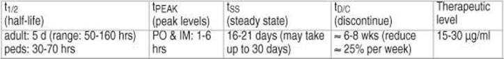
Pharmacokinetics: Unless otherwise specified, numbers are given for oral dosing form. t1/2 = half-life; tPEAK = time to peak serum level; tSS = time to steady state (approximately 5 x t1/2); tD/C = time to discontinue (recommended withdrawal period over which drug should be tapered); MDF = minimum dosing frequency. “Therapeutic level” is the average therapeutic range. |
phenytoin (PHT) (Dilantin®)DRUG INFO
INDICATIONS
GTC, S/C-P, occasionally in ABS.
PHARMACOKINETICS
Pharmacokinetics are complicated: at low concentrations, kinetics are 1st order (elimination proportional to concentration), metabolism saturates near the therapeutic level resulting in zero-order kinetics (elimination at a constant rate). ≈ 90% of total drug is protein bound. Oral bioavailability is ≈ 90% whereas IV bioavailability is ≈ 95%; this small difference may be significant when patients are near limits of therapeutic range (due to zero-order kinetics).

Renal failure: dosage adjustment not needed. However, serum protein binding may be altered in uremia which can obfuscate interpretation of serum phenytoin levels. Eq 17-1 may be used to convert serum PHT concentration in a uremic patient C (observed), to the expected PHT level in nonuremic patients C (nonuremic).
![]()
ORAL DOSE
Rx Adult: usual maintenance dose= 300-600 mg/d divided BID or TID (MDF = q d, for single daily dosing, either the phenytoin-sodium capsules or the extended release form should be used). Oral loadingdose: 300 mg PO q 4 hrs until 17 mg/kg are given. Peds: oral maintenance: 4-7 mg/kg/d (MDF = BID). SUPPLIED: (oral forms): 100 mg tablets of phenytoin-sodium (sodium-salt); 30 & 100 mg Kapseals® (extended release); 50 mg chewable Infatabs® (phenytoin-acid); oral suspension 125 mg/5-ml in 8 oz. (240 ml) bottles or individual 5 ml unit dose packs; pediatric suspension 30 mg/5-ml. Phenytek® 200 & 300 mg capsules.
Dosage changes
Because of zero-order kinetics, at near-therapeutic levels a small dosage change can cause large level changes. Although computer models are necessary for a high degree of accuracy, the dosing change guidelines in Table 17-9 or the nomogram in Figure 17-165 may be used as a quick approximation.
GI absorption of phenytoin suspension or capsules may be decreased by up to 70% when given with nasogastric feedings of Osmolyte® or Isocal®66, 67, and the sus-pension has been reported to have erratic absorption. Hold NG feeding for 2 hrs before and 1 hour after phenytoin dose.
Table 17-9 Guidelines for changing phenytoin dosage
|
Present level (mg/dl) |
Change to make |
|
< 6 |
100 mg/day |
|
6-8 |
50 mg/day |
|
> 8 |
25-30 mg/day |

Figure 17-1 Nomogram for adjusting phenytoin dose
PARENTERAL DOSE
Phenytoin is a negative inotrope and can cause hypotension.
Conventional phenytoin may be given slow IVP or by IV drip (see below). The IM route should NOT be used (unreliable absorption, crystallization and sterile abscesses may develop). IV must be given slowly to reduce risk of arrhythmias and hypotension, viz. Adult: < 50 mg/min, Peds: < 1-3 mg/kg/min. The only compatible solution is NS, inject at site nearest vein to avoid precipitation.
Rx loading. Adult: 18 mg/kg slow IV. Peds: 20 mg/kg slow IV.
Rx maintenance. Adult: 200-500 mg/d (MDF = q d). Most adults have therapeutic levels on 100 mg PO TID. Peds: 4-7 mg/kg/d (MDF = BID).
Drip loading method:
Requires cardiac monitoring, and BP check q 5 minutes.
Rx Add 500 mg PHT to 50 ml NS to yield 10 mg/ml, run at 2 ml/min (20 mg/min) long enough to give 18 mg/kg (for 70 kg patient: 1200 mg over 60 mins). For more rapid administration, up to 40 mg/min may be used, or use fosphenytoin (see below). Decrease rate if hypotension occurs.
Fosphenytoin sodium injection
Fosphenytoin sodium (FOS) injection (Cerebyx®) is a newer formulation for administering IV phenytoin, and is indicated for short term use (≤ 5 days) when the enteral route is not usable. It is completely converted in vivo to phenytoin by organ and blood phosphatases with a conversion half-life of 10 minutes. Product labeling is given in terms of phenytoin equivalents (PE). Safety in pediatric patients has not been established. SUPPLIED: 50 mg PE/ml in 2 & 10 ml vials (100 mg PE and 500 mg PE respectively).
Advantages of FOS (over conventional IV phenytoin):
1. less venous irritation (due to lower pH of 8.6-9 compared to 12 for phenytoin) resulting in less pain and IV extravasation
2. FOS is water soluble and therefore may be infused with dextrose or saline
3. tolerated by IM injection (IM route should not be used for status epilepticus)
4. does not come combined with propylene glycol (which can cause cardiac arrhythmias and/or hypotension itself)
5. the maximum administration rate is 3 x as fast (i.e. 150 mg PE/min)
SIDE EFFECTS OF PHENYTOIN
May interfere with cognitive function. May produce SLE-like syndrome, hepatic granulomas, megaloblastic anemia, cerebellar degeneration (chronic doses), hirsutism, gingival hypertrophy, hemorrhage in newborn if mother on PHT, toxic epidermal necrolysis (Stevens-Johnson variant). PHT antagonizes vitamin D → osteomalacia and rickets. Most hypersensitivity reactions occur within 2 months of initiating therapy66. In cases of maculopapular erythematous rash, the drug may be stopped and the patient may be re-challenged; often the rash will not recur the second time. Teratogenic (fetal hydantoin syndrome68).
Signs of phenytoin toxicity may develop at concentrations above 20 μg/ml (toxicity is more common at levels > 30 μg/ml) and include nystagmus (may also occur at therapeutic levels), diplopia, ataxia, asterixis, slurred speech, confusion, and CNS depression.
Drug-drug interactions: fluoxetine (Prozac®) results in elevated phenytoin levels (ave: 161% above baseline)69. Phenytoin may impair the efficacy of: corticosteroids, warfarin, digoxin, doxycycline, estrogens, furosemide, oral contraceptives, quinidine, rifampin, theophylline, vitamin D.
carbamazepine (CBZ) (Tegretol®)DRUG INFO
INDICATIONS
Partial seizures with or without secondary generalization. Trigeminal neuralgia. An IV form for use in e.g. status epilepticus is in development.
DOSE
Rx oral route. Adult range: 600-2000 mg/d. Peds: 20-30 mg/kg/d. MDF = BID.
Before starting, check: CBC & platelet count (consider reticulocyte count) & serum Fe. Package insert says “recheck at frequent intervals, perhaps q week x 3 mos, then q month x 3 yrs.”
Do not start CBZ (or discontinue it if patient already on CBZ) if: WBC < 4K, RBC < 3 x 10⁶, Hct < 32%, platelets < 100K, reticulocytes < 0.3%, Fe > 150 μg%.
Start low and increment slowly: 200 mg PO q d x 1 wk, BID x 1 wk, TID x 1 wk. As an inpatient, dosage changes may be made every 3 days, monitoring for signs of side effects. As an outpatient, changes should be made only ≈ weekly, with levels after each change. Carbatrol® (extended release CBZ) is usually dosed BID.
SUPPLIED: oral form. Scored tabs 200 mg. Chewable scored tabs 100 mg. Suspension 100 mg/5-ml. IV form: not available in the U.S. at the time of this writing. Carbatrol® (extended release CBZ): 200 & 300 mg tablets.
Caveats with oral forms: oral absorption is erratic, and smaller more frequent doses are preferred70. Oral suspension is absorbed more readily, and also ✖ should not be administered simultaneously with other liquid medicinal agents as it may result in the precipitation of a rubbery, orange mass. ✖ May aggravate hyponatremia by SIADH-like effect.
PHARMACOKINETICS

CBZ induces hepatic enzymes that result in increased metabolism of itself (autoinduction) as well as other drugs over a period of ≈ 3-4 weeks.
SIDE EFFECTS
✖ Drug-drug interaction: caution, propoxyphene (Darvon®), cimetidine, erythromycin and isoniazid may cause dramatic elevation of CBZ levels due to inhibition of hepatic cytochrome oxidase that degrades CBZ71. Side effects include:
1. drowsiness and GI upset: minimized by slow dose escalation
2. relative leukopenia in many: usually does not require discontinuing drug
3. transient diplopia
4. ataxia
5. less effect on cognitive function than PHT
6. hematological toxicity: rare. May be serious → agranulocytosis & aplastic anemia
7. Stevens-Johnson syndrome
8. SIADH
9. hepatitis (occasionally fatal) reported
oxcarbazepine (Trileptal®)DRUG INFO
Very similar efficacy profile to carbamazepine with the following differences:
1. there is no autoinduction (C-P450 is not involved in metabolism) and therefore minimal drug-drug interactions
2. no blood testing is required since:
A. there is no liver toxicity
B. there is no hematologic toxicity
C. there is no need to check drug levels
3. dosing is BID
4. kinetics are linear
5. more expensive
DOSE
Rx: starting dose for pain control is 150 mg PO BID, for seizures it is 300 mg PO BID. Maximum dose 2400 mg/day total. SUPPLIED: 150, 300 & 600 mg scored tablets. 300 mg/5 ml oral suspension.
valproateDRUG INFO
Available as valproic acid (Depakene®) and divalproex sodium (Depakote®).
INDICATIONS
Effective in primary GTC. Also useful in ABS with GTC, juvenile myoclonic epilepsy, and partial seizures (not FDA approved for latter). Also FDA approved for migraine prophylaxis. Note: severe GI upset and short half life make valproic acid much less useful than Depakote® (divalproex sodium).
DOSE
Adult range: 600-3000 mg/d. Peds range: 15-60 mg/kg/d. MDF = q d.
Rx Start at 15 mg/kg/d, increment at 1 wk intervals by 5-10 mg/kg/d. Max recommended adult dose: 60 mg/kg/d. If daily dose > 250 mg is required, it should be divided. SUPPLIED: Oral: capsules 250 mg. Syrup 250 mg/5-ml. Depakote® (enteric coated) tabs: 125, 250, & 500 mg; sprinkle capsules125 mg. IV: Depacon® for I.V. injection 500 mg/5 ml vial.
PHARMACOKINETICS

Valproic acid (VA) is 90% protein bound. ASA displaces VA from serum proteins.
SIDE EFFECTS
Serious side effects are rare. Pancreatitis has been reported. Fatal liver failure has occurred especially if age < 2 yrs and in combination with other AEDs. Teratogenic (see Contraindications below). Drowsiness (temporary), minimal cognitive deficits, N/V (minimized with Depakote), liver dysfunction, hyperammonemia (even without liver dysfunction), weight gain, mild hair loss, tremor (dose related; similar to benign familial tremor; if severe and valproic acid is absolutely necessary, the tremor may be treated with beta blockers). May interfere with platelet function, caution with surgery on these patients.
CONTRAINDICATIONS
✖ Pregnancy: causes neural tube defects (NTD) in ≈ 1-2% of patients72. Since a correlation between peak VA levels and the risk of NTDs has been found, if VA must be used, some experts recommend changing from BID to TID dosing. ✖ Patients ≤ 2 yrs age (risk of hepatotoxicity).
phenobarbitalDRUG INFO
INDICATIONS
Used as alternative in GTC and partial (not DOC). Had been DOC for febrile seizures, dubious benefit47. About as effective as PHT, but very sedating. Also used for status epilepticus (see page 406).
DOSE
Same dose PO, IV, or IM. MDF = q d73, 74. Start slowly to minimize sedation.
Rx Adult loading: 20 mg/kg slow IV (administer at rate < 100 mg/min). Maintenance: 30-250 mg/d (usually divided BID-TID). Peds loading: 15-20 mg/kg. Maintenance: 2-6 mg/kg/d (usually divided BID). SUPPLIED: tabs 15 mg, 30 mg, 60 mg, 100 mg; elixir 20 mg/5-ml.
PHARMACOKINETICS
Phenobarbital is a potent inducer of hepatic enzymes that metabolize other AEDs.
SIDE EFFECTS
Cognitive impairment (may be subtle and may outlast administration of the drug by at least several months47), thus avoid in peds; sedation; paradoxical hyperactivity (especially in peds); may cause hemorrhage in newborn if mother is on phenobarbital.
primidone (Mysoline®)DRUG INFO
INDICATIONS
Same as phenobarbital (not DOC). NB: when used in combination therapy, low doses (50-125 mg/day) may add significant seizure control to the primary AED with few side effects.
DOSE
Rx Adult: 250-1500 mg/d. Peds: 15-30 mg/kg/d; MDF = BID.
Start at 125 mg/d x 1 wk, and inc. slowly to avoid sedation. SUPPLIED: (oral only): scored tabs 50 mg, 250 mg; suspension 250 mg/5-ml.
PHARMACOKINETICS
Metabolites include phenylethylmalonamide (PEMA) and phenobarbital. Therefore always check phenobarbital level at same time as primidone level.

SIDE EFFECTS
Same as phenobarbital, plus: loss of libido, rare macrocytic anemia.
ethosuximide (Zarontin®)DRUG INFO
INDICATIONS
DOC in ABS.
DOSE
Rx Adult: 500-1500 mg/d. Peds: 10-40 mg/kg/d; MDF = q d. SUPPLIED: oral only; capsules 250 mg; syrup 250 mg/5-ml.
PHARMACOKINETICS

SIDE EFFECTS
N/V; lethargy; hiccups; H/A; rarely: eosinophilia, leukopenia, erythema multiforme, Stevens-Johnson syndrome, SLE-like syndrome. Toxic levels → psychotic behavior.
methsuximide (Celontin®)DRUG INFO
INDICATIONS
Indicated for absence seizures refractory to other drugs.
DOSE
Rx optimum dose must be determined by trial. Start with 300 mg PO q d, increase by 300 mg PRN at weekly intervals up to 1200 mg/d. SUPPLIED: 150 & 300 mg capsules.
felbamate (Felbatol®)DRUG INFO
✖ CAUTION: Due to an unacceptably high rate of aplastic anemia and hepatic failure, felbamate (FBM) should not be used except in those circumstances where the benefit clearly outweighs the risk; then, hematologic consultation is recommended by the manufacturer. See Side effects below (also for drug-drug interactions).
FBM is efficacious for monotherapy and adjunctive therapy for partial seizures (complex and secondary generalization), and reduces the frequency of atonic and GTC seizures in Lennox-Gastaut syndrome.
PHARMACOKINETICS

DOSE
Rx: CAUTION see above. Felbamate is not to be used as a first-line drug. Patient or guardian should sign informed consent release. Start with 1200 mg/d divided BID, TID, or QID, and decrease other AEDS by one third. Increase felbamate biweekly in 600 mg increments to usual dose of 1600-3600 mg/d (max: 45 mg/kg/d). Slow down increments and/or reduce other AEDs further if side effects become severe. Administer at upper end of range when used as monotherapy. SUPPLIED: (oral only) 400 & 600 mg scored tablets; sus-pension 600 mg/5-ml.
Table 17-10 Effect of felbamate on other AED levels
|
AED |
Change in level |
Recommended dosing change |
|
phenytoin |
↑ 30-50% |
↓ 20-33% |
|
carbamazepine |
↓ 30% total |
↓ 20-33% |
|
valproic acid |
↑ 25-100% |
↓ 33% |
SIDE EFFECTS
Felbamate has been associated with aplastic anemia (usually discovered after 5-30 wks of therapy) in ≈ 2-5 cases per million persons per yr, and hepatic failure (some fatal, necessitating baseline and serial LFTs every 1-2 wks). Other side effects: insomnia, anorexia, N/V, H/A. Felbamate is a potent metabolic inhibitor, thus it is necessary to reduce the dose of phenytoin, valproate or carbamazepine when used with felbamate75 (see Table 17-10) (general rule: drop dose by one third).
levetiracetam (Keppra®)DRUG INFO
No identified drug-drug interactions. Less than 10% protein bound. Linear pharmakokinetics, no level monitoring needed.
INDICATIONS
Adjunctive therapy for partial onset Sz with secondary generalization in patients 4 years of age and older. Myoclonic seizures (juvenile myoclonic epilepsy). Generalized tonic-clonic.
DOSE
Rx start with 500 mg PO BID. Increment by 1000 mg/d q 2 weeks PRN to a maximum of 3000 mg/d. Keppra XR: the same dose of levetiracetam can be converted to Keppra XR for q d dosing.
IV: 500-1500 mg diluted in 100 ml of diluent (LR, D5W, normal saline) infused over 15 minutes BID.
SUPPLIED: 250, 500, 750 & 1000 mg scored film-coated tabs; 100 mg/ml oral solution. Keppra XR (extended release) 500 mg.
IV: 1 vial (5 ml) contains 500 mg.
SIDE EFFECTS
PO or IV: somnolence and fatigue in 15%. Dizziness in 9%. Asthenia 15% and infection 13% (nasopharyngitis and influenza may or may not have been related).
Keppra XR: somnolence 8%, irritability 6%.
clonazepam (Klonopin®)DRUG INFO
A benzodiazepine derivative.
INDICATIONS
✖ Not a recommended drug for seizures (see below).
Used for myoclonic, atonic, and absence seizures (in absence, less effective than valproate or ethosuximide, and tolerance may develop).
NB: clonazepam usually works very well for several months, and then tends to become less effective, leaving only the sedating effects. Also, many cases have been reported of patients having seizures during withdrawal, including status epilepticus (even in patients with no history of status). Thus, may need to taper this drug over 3-6 months.
DOSE
Rx Adult: start at 1.5 mg/d DIV TID, increase by 0.5-1 mg q 3 d, usual dosage range is 1-12 mg/d (max 20 mg/d); MDF = q d. Peds: start at 0.01-0.03 mg/kg/d DIV BID or TID, increase by 0.25-0.5 mg/kg/d q 3 d; usual dosage range is 0.01-0.02 mg/kg/d; MDF = q d. SUPPLIED: oral only; scored tabs: 0.5 mg, 1 mg, 2 mg.
PHARMACOKINETICS

SIDE EFFECTS
Ataxia; drowsiness; behavior changes.
zonisamide (Zonegran®)DRUG INFO
INDICATIONS
Adjunctive therapy for partial Sz in adults.
acetazolamide (Diamox®)DRUG INFO
The anti-epileptic effect may be either due to direct inhibition of CNS carbonic anhydrase (also reduces CSF production) or due to the slight CNS acidosis that results.
INDICATIONS
Centricephalic epilepsies (absence, nonfocal seizures). Best results are in absence seizures; however benefit has also been observed in GTC, myoclonic jerk.
SIDE EFFECTS
Do not use in first trimester of pregnancy (may be teratogenic). The diuretic effect causes renal loss of HCO3 which may lead to an acidotic state with long-term therapy. A sulfonamide, therefore any typical reaction to this class may occur (anaphylaxis, fever, rash, Stevens-Johnson syndrome, toxic epidermal necrolysis…). Paresthesias: medication should be discontinued.
DOSE
Rx Adult: 8-30 mg/kg/d in divided doses (max 1 gm/d, higher doses do not improve control). When given with another AED, the suggested starting dose is 250 mg once daily, and this is gradually increased. SUPPLIED: tablets 125, 250 mg. Diamox sequels® are sustained release 500 mg capsules. Sterile cryodessicated powder is also available in 500 mg vials for parenteral (IV) use.
Although developed to be a GABA agonist, it does not interact at any known GABA
gabapentin (Neurontin®)DRUG INFO
receptor. Efficacious for primary generalized seizures and partial seizures (with or without secondary generalization). Ineffective for absence seizures. Very low incidence of known side-effects. No known drug interactions (probably because it is renally excreted). Also used for central pain
DOSE
Rx Adult: 300 mg PO x 1 day 1; 300 mg BID day 2; 300 mg TID day 3; then increase rapidly up to usual doses of ≈ 800-1800 mg per day. Doses of 1800-3600 may be needed in intractable patients. ✖Dosage must be reduced in patients with renal insufficiency or on dialysis (see Eq 3-1, page 46 to estimate). SUPPLIED: 100, 300, 400, 600, 800 mg capsules. 50 mg/ml suspension.
PHARMACOKINETICS
Gabapentin is not metabolized, and 93% is excreted unchanged renally with plasma clearance directly proportional to creatinine clearance76. Does not affect hepatic microsomal enzymes, and does not affect metabolism of other AEDs. Antacids decrease bioavailablilty by ≈ 20%, therefore give gabapentin > 2 hrs after the antacid77.

SIDE EFFECTS
Somnolence, dizziness, ataxia, fatigue, nystagmus; all reduce after 2-3 weeks of drug therapy. Increased appetite. Not known to be teratogenic.
lamotrigine (Lamictal®)DRUG INFO
Anticonvulsant effect may be due to presynaptic inhibition of glutamate release76.
Efficacious as adjunctive therapy for partial seizures (with or without secondary generalization) and Lennox-Gastaut syndrome. Preliminary data suggest it may also be useful as an adjunct for refractory generalized seizures, or as monotherapy for newly diagnosed partial or generalized seizures78. Also FDA approved for bipolar disorder.
SIDE EFFECTS
Somnolence, dizziness, diplopia. ✖ Serious rashes requiring hospitalization and discontinuation of therapy have been reported (rash usually begins 2 weeks after initiating therapy and may be severe and potentially life-threatening, including Stevens-Johnson syndrome (more of a concern with simultaneous use of valproate), and rarely, toxic epidermal necrolysis (TEN)). Incidence of significant epidermal reaction may be decreased by a slow ramping-up of dosage. May increase seizure frequency in severe myoclonic epilepsy of infancy79. Metabolism of lamotrigine is affected by other AEDs.
DOSE
Rx Adult: In adults receiving enzyme-inducing AEDs (PHT, CBZ, or phenobarbital), start with 50 mg PO q d x 2 wks, then 50 mg BID x 2 wks, then ↑ by 100 mg/d q week until the usual maintenance dose of 200-700 mg/d (divided into 2 doses) is reached. For patients on valproic acid (VA) alone, the maintenance dose was 100-200 mg/d (divided into 2 doses), and VA levels drop by ≈ 25% within a few weeks of starting lamotrigine. For patients on both enzyme-inducing AEDs and VA, the starting dose is 25 mg PO qod x 2 wks, then 25 mg qd x 2 wks, then ↑ by 25-50 mg/d q 1-2 wks up to a maintenance of 100-150 mg/d (divided into 2 doses). Instruct patients that rash, fever or lymphadenopathy may herald a serious reaction and that a physician should be contacted immediately. Peds: not indicated for use in patients < 16 yrs old due to higher incidence of potentially life-threatening rash in the pediatric population76. SUPPLIED: 25, 100, 150 & 200 mg tablets. 2, 5 & 25 mg chewable dispersible tablets.
PHARMACOKINETICS78

vigabatrinDRUG INFO
INDICATIONS
Effective in treating partial seizures. Less so for generalized seizures.
DOSE
Rx Adult: 1500-3000 mg/d.
topiramate (Topamax®)DRUG INFO
May block voltage-sensitive sodium channels and enhance GABA activity at GABAA receptors and attenuate some glutamate receptors76.
INDICATIONS80
As an oral adjunct to other drugs in treating refractory partial onset seizures.
DOSE
Rx Adult: start with 50 mg/d and increase slowly up to 200-400 mg/d81, with no significant benefit noted at dosages > 600 mg/d82. SUPPLIED: 25, 100, & 200 mg tabs.
PHARMACOKINETICS
30% is metabolized in the liver, the rest is excreted unchanged in the urine.
|
t1/2 (half-life) |
tSS (steady state) |
Therapeutic level |
|
19-25 hrs |
5-7 days |
not established |
SIDE EFFECTS
May increase phenytoin concentration by up to 25%. Levels of topiramate are reduced by other AEDs (phenytoin, carbamazepine, valproic acid and possibly others).
Cognitive impairment (word finding difficulty, problems with concentration…), weight loss, dizziness, ataxia, diplopia, paresthesias, nervousness and confusion have been troublesome. ≈ 1.5% incidence of renal stones which usually pass spontaneously76
Oligohidrosis (reduced sweating) and hyperthermia, primarily in children in association with elevated environmental temperatures and/or vigorous physical activity.
tiagabine (Gabitril®)DRUG INFO
A GABA uptake inhibitor, with cognitive problems of a similar frequency to that with topiramate83.
Rx Adult: start with 4 mg/d, increase weekly by 4-8 mg to a maximum of 32-56 mg (divided BID to QID). SUPPLIED: 4, 12, 16 & 20 mg tablets.
lacosamide (Vimpat®)DRUG INFO
Enhances slow inactivation of voltage gated sodium channels, affecting only neurons that are depolarized of active over a prolonged period (as in a seizure).
INDICATIONS
Partial onset seizures. Painful diabetic neuropathy.
DOSE
Rx Adult: 200-400 mg. SUPPLIED: 50, 100, 150 & 200 mg tabs. 10 mg/ml IV solution.
17.4.3.1. Withdrawal of antiepileptic drugs
Most seizure recurrences develop during the first 6 months after AED withdrawal84.
INDICATIONS FOR AED WITHDRAWAL
There is no agreement on how long a patient should be seizure-free before withdrawal of anticonvulsants, nor is there agreement on the prognostic value of EEGs and on the best time period over which to withdraw AEDs.
The following is based on a study of 92 patients with idiopathic epilepsy, who had been free of seizures for two years85. Generalization, e.g. to posttraumatic seizures, may not be appropriate. Taper was by 1 “unit” q 2 weeks (where a unit is defined as 200 mg for CBZ or valproic acid, or 100 mg for PHT). Follow-up: mean = 26 mos (range: 6-62).

31 patients (34%) relapsed, with the average time to relapse being 8 mos (range: 1-36). Using actuarial methods, the risk for recurrence is 5.9%/month for 3 months, then 2.7%/month for 3 months, then 0.5%/month for 3 months. Factors found to affect the likelihood of relapse include:
1. seizure type: 37% relapse rate for generalized seizures; 16% for complex or simple partial; 54% for complex partial with secondary generalization
2. number of seizures before control attained: those with ≥ 100 seizures before control had statistically significant higher relapse rate
3. the number of drugs that had to be tried before single drug therapy successfully controlled seizures: 29% if 1st drug worked, 40% if a change to a 2nd drug was needed, and 80% if a change to a 3rd drug was required
4. EEG class (see Table 17-11): class 4 had worst prognosis for relapse. Epileptiform discharges on EEG serves to discourage AED withdrawal86
In a larger randomized study87, the most important factors identified to predict freedom from recurrent seizures were:
1. longer seizure-free period
2. use of only one AED (vs. multiple AEDs)
3. seizures other than tonic-clonic seizures
WITHDRAWAL TIMES
The recommended withdrawal times in Table 17-12 should be used only as guide-lines.
Table 17-12 Recommended AED withdrawal times
|
AED |
Recommended withdrawal period |
|
phenytoin, valproic acid, carbamazepine |
2-4 weeks |
|
phenobarbital |
6-8 weeks (25% per week) |
|
clonazepam |
3-6 months (see CAUTION on page 415) |
17.4.3.2. Pregnancy and antiepileptic drugs
Women of childbearing potential with epilepsy should undergo counseling regarding pregnancy88.
BIRTH CONTROL
AEDs that induce liver microsomal cytochrome P450 enzymes (see Table 17-13) increase the failure rate of oral contraceptives up to fourfold90. Patients desiring to use BCPs should employ barrier contraceptive measures until ovulation is consistently suppressed, and they should watch for breakthrough bleeding which may indicate a need for a change in the hormone dosage84. Non-oral hormonal contraceptives (e.g. levonorgestrel implant (Nor-plant®)) circumvents first pass liver degradation but should combined with a barrier method because of declining effectiveness with time.
Table 17-13 Effect of AEDs on liver cytochrome P450*
|
Inducers |
Noninducers |
|
carbamazepine phenobarbital phenytoin felbamate primidone |
valproic acid benzodiazepines gabapentin lamotrigine |
* references84, 89
COMPLICATIONS DURING PREGNANCY
Women with epilepsy have more complications with pregnancy than mothers without epilepsy, but > 90% of pregnancies have a favorable outcome84.
There is an increase in the number of gravid seizures in ≈ 17% (reported range: 17-30%) of epileptic women, which may be due to noncompliance or to changes of free drug levels of AEDs during pregnancy (see Table 17-14). Isolated seizures can occasionally be deleterious, but usually cause no problem. Status epilepticus poses serious risk to mother and fetus during pregnancy and should be treated aggressively.
There is also a slightly increased risk of toxemia (HTN of pregnancy) and fetal loss.
Table 17-14 Changes in free AED levels during pregnancy91
|
Drug |
Change |
|
carbamazepine |
↓ 11% |
|
phenobarbital |
↓ 50% |
|
phenytoin |
↓ 31% |
|
valproic acid |
↑ 25% |
BIRTH DEFECTS
The incidence of fetal malformations in offspring of patients with a known seizure disorder is ≈ 4-5%, or approximately double that of the general population92. The degree to which this is due to the use of AEDs vs. genetic and environmental factors is unknown. All AEDs have the potential to cause deleterious effects on the infant. Polytherapy is associated with an increased risk over monotherapy in a more than additive manner.
Generally, the risk of seizures (with possible concomitant maternal and fetal hypoxia and acidosis) is felt to outweigh the teratogenic risk of most AEDs, but this must be evaluated on a case-by-case basis. Occasionally patients may be weaned off AEDs.
Specific drugs
Carbamazepine (CBZ) produced an increased incidence of “minor” malformations (but not of “major” malformations) in one study93 (this study may have had methodologic problems), and may increase the incidence of neural tube defects (NTD)94. In utero exposure to phenytoin may lead to the fetal hydantoin syndrome68, 95 and a child with an IQ lower by ≈ 10 points96. Phenobarbital produced the highest incidence of major malformations (9.1%) in one prospective study97 and was also associated with most of the increase in fetal death or anomalies in another study98. Valproate (VA) causes the highest incidence of NTD (1-2%72), which can be detected with amniocentesis and allow an abortion if desired. TID dosing may reduce the risk of NTD (see page 413). Benzodiazepines given shortly before delivery can produce the “floppy infant syndrome”99. Similar effects may occur with other sedating AEDs such as phenobarbital.
Drug recommendations
A general consensus is that for most women of childbearing potential who require AEDs, that monotherapy with the lowest dose of CBZ that is effective is the method of choice if the seizure disorder is responsive to it100. If ineffective, then monotherapy with valproic acid (with TID dosing) is currently the recommended second choice. Folate supplementation (after confirming normal B12 levels, see page 1187) should be used in all.
17.5. Seizure surgery
20% of patients continue to have seizures even with AEDs. Many of these patients may be candidates for surgical procedures to control their seizures6.
INDICATIONS
Seizure disorder must be severe, medically refractory with satisfactory trials of tolerable medication for at least ≈ 1 year, and disabling to the patient. Medically refractory is usually considered two attempts of high-dose monotherapy with two distinct AEDs, and one attempt at polytherapy.
The three general categories of patients suitable for seizure surgery have101:
1. partial seizures
A. temporal origin: the largest group of surgical candidates (especially mesial temporal epilepsy which is often medically refractory)
B. extratemporal origin
2. secondarily generalized seizures: e.g. Lennox-Gastaut
3. unilateral, multifocal epilepsy associated with infantile hemiplegia syndrome
EVALUATION
All patients should undergo imaging study to rule out neoplasm, AVM, etc. Noninvasive techniques allow localization in the majority of cases.
NONINVASIVE TECHNIQUES
MRI
The imaging modality of choice. Extremely good for detecting hippocampal asymmetry of mesial temporal sclerosis that may produce complex partial seizures (CPS)102.
CAT SCAN
A seizure focus may enhance with IV contrast shortly following a seizure. Subtle enhancement may be present on the side of the focus on interictal CT scan103.
VIDEO-EEG MONITORING
Most centers perform pre-operative long-term inpatient video-EEG monitoring to correlate the clinically disabling seizure with appropriate electrical abnormalities and possibly to identify the seizure focus.
PET SCAN (POSITRON EMISSION TOMOGRAPHY)
Interictal PET scan using fluorine-18 deoxyglucose (18FDG) shows hypometabolism lateralized to the side of temporal lobe focus in 70% of patients with medically refractory CPS (does not show actual siteof origin). Useful when MRI and EEG cannot localize.
SPECT SCAN (SINGLE PHOTON EMISSION TOMOGRAPHY)
Used to demonstrate increased blood flow during a seizure to help localize site of onset. [99m] Technetium (Tc) hexamethyl-propylene-amine-oxime (HMPAO) is usually administered immediately after onset of seizure, and the scan may be obtained within several hours104.
MILDLY INVASIVE TECHNIQUES
WADA TEST105
AKA intracarotid amytal test. Localizes dominant hemisphere (side of language function) and assesses ability of hemisphere without lesion to maintain memory when isolated. Usually reserved for candidates for large resections106.
Start with angiogram to assess cross flow and to R/O persistent trigeminal artery (see page 107). Significant cross-flow is a relative contraindication to anesthetizing the side of dominant supply (patient goes to sleep).
Wada test may be grossly inaccurate with high flow AVM. Also, portions of hippocampus may be supplied by posterior circulation (not anesthetized by ICA injection).
EEG monitoring is usually performed during the test when it is being done for seizure surgery. Patient will show delta waves during deepest level of anesthesia.
Technique
• instruct patient as to what is expected
• catheterize ICA: usually start on side of lesion
• have patient hold contralateral arm in air, and instruct them to hold it there
• inject 100-125 mg sodium amobarbital (Amytal®) rapidly into internal carotid artery (effect starts almost instantaneously, begins to subside after ≈ 8 minutes (may subside in ≈ 2 minutes with AVM where flow rates are high))
• determine adequacy of injection by assessing motor function in elevated arm (should be ≈ flaccid)
• assess language skills by showing patient pictures of objects and ask them to name each one out loud and remember each one
• assess memory function by asking patient to name as many of the pictures as they can ≈ 15 minutes after test: if they have difficulty, ask them to pick out pictures from a group that contains additional ones not shown to patient
• repeat procedure on other side (use lower Amytal doses with each subsequent injection)
SURGICAL TECHNIQUES
EEG OBTAINED WITH INVASIVE ELECTRODES
Risk of infection with depth electrodes106: 2-10%.
Surface strip electrodes may be placed through a burr hole.
Depth electrodes may be placed stereotactically. Temporal depth electrodes may be helpful for CPS, usually to determine the laterality of the mesiotemporal source of seizure. Frontal depth electrodes are also sometimes used. 2-3% risk of intracerebral hemorrhage106.
Subdural grid electrodes are placed with a craniotomy. These sometimes may allow sufficient mapping to permit surgery under general anesthesia without need for intraoperative mapping under local anesthesia (helpful in children or in the mentally retarded).
SURGICAL CONSIDERATIONS
Three basic types of procedure: resections, disconnections and stimulationA:
1. resections
A. resection of epileptic focus: higher chance of completely controlling seizures. Performed in noneloquent brain. Seizures must have focal onset (resection not encouraged if multifocal onset). Includes:
1. anterior temporal lobectomy: see below
2. amygdalo-hippocampectomy
3. neocortical resections: especially with neuronal migration abnormalities
B. resection of lesion in secondary epilepsy (e.g. tumor, AVM, cavernous malformation107…). In most cases the seizure focus is in or near the lesion, but some structural lesions are not responsible for the seizures. For seizure foci within the temporal lobe, seizure control is better when lesionectomy is accompanied by amygdalo-hippocampectomy108
2. disconnections: used when eloquent brain is involved, or to separate the electrical activity of the two cerebral hemispheres
A. section of corpus callosum (callosotomy): when drop attacks are the most disabling seizure type or for multiple bilateral foci (see below)
B. hemispherectomy: for unilateral seizures with widespread hemispheric lesions and profound contralateral neurologic deficit. If any cortex is left, must make sure it is functionally deafferented (disconnected)
1. anatomic hemispherectomy
2. functional hemispherectomy: preservation of the basal ganglia isolates the abnormal side with ≈ 80% seizure control rate (similar to anatomic hemispherectomy, but with lower complication rate).
C. multiple subpial transection109: for partial seizure originating in eloquent cortical areas. The cortex is transected at 5 mm intervals, thus interrupting the horizontal spread of the seizure while sparing the vertically oriented functional fibers
3. stimulation
A. vagal nerve stimulation: see page 424
B. deep brain stimulation (DBS)
1. centromedian nucleus of the thalamus110: better for generalized tonic-clonic seizures
2. bilateral anterior nucleus of the thalamus (report of 5 patients111, and the recent “SANTE” trial112): for partial seizures
3. hippocampus110: for partial seizures
A. all of the listed procedures are for refractory seizures. For the definition of refractory, see page 420
ANESTHETIC CONSIDERATIONS
If intraoperative electrocorticography is to be performed:
• under local anesthesia: the only anesthetic agents that may be used are narcotics (usually fentanyl) and droperidol
• under general anesthesia: avoid benzodiazepines and barbiturates
INTRAOPERATIVE ELECTROCORTICOGRAPHY (ECOG)
May be performed with surface matrix that includes superior temporal gyrus and inferior frontal gyrus. Depth electrodes in the amygdala (3 cm from temporal tip) and hippocampus (5 cm from temporal tip) may also be used.
Methohexital (Brevitol®) may be given to try to provoke a seizure: observe for ↓ fast activity in suspected focus.
INTRAOPERATIVE CORTICAL MAPPING
For techniques of cortical mapping, see page 150.
CORPUS CALLOSOTOMY
Partial or total section may be most effective for generalized major motor seizures. Of little benefit for simple or complex seizures. Benefit has been supported for:
1. frequent episodes of atonic seizures (“drop attacks”) where loss of postural tone → falls and injuries113 (70% reduction with callosotomy)
2. possibly for generalized seizure disorder with unilateral hemisphere damage (e.g. infantile hemiplegia syndrome); hemicortical resection may be better for this type, whereas callosotomy may promote partial seizures.
Note: a “functional hemispherectomy” is recommended over “anatomically complete” hemispherectomy to reduce morbidity and mortality101
3. some patients with generalized seizures without identifiable, resectable focus
Division of the anterior two thirds of the corpus callosum (CC) (minimizes the risk of disconnection syndrome, see below) may be advantageous over complete callosotomy (controversial). Some advocate sectioning the CC with intraoperative EEG until the typical bisynchronous discharges that are usually seen become asynchronous114. No need to section anterior commissure. Can usually be performed via a bifrontal craniotomy utilizing a bicoronal skin incision.
May produce post-op ↓ verbalization or akinetic mutism that usually resolves in weeks. ✖ Contraindication: major behavioral and/or language deficits may occur even with partial division in patients with speech and dominant handedness located in opposite hemispheres (“crossed dominance”). Thus, Wada test is recommended in all left handed patients.
MRI sagittal cuts are superb for assessing extent of division of the CC115.
DISCONNECTION SYNDROME
In a patient with a dominant left hemisphere, consists of left tactile anomia, left sided dyspraxia (may resemble hemiparesis), pseudohemianopsia, right sided anomia for smell, impaired spatial synthesis of right hand resulting in difficulty copying complex figures, decreased spontaneity of speech, incontinence.
More common with larger surgical sections of the CC. Risk is less if the anterior commissure is spared. Patients usually adapt after 2-3 months, with final function normal for most daily activities (deficits may show up on neuropsychological testing).
TEMPORAL LOBECTOMY
80% of patients with medically intractable seizures with demonstrable focus have foci in anterior temporal lobe. Most patients have neuronal loss and gliosis of mesial temporal structures. Thus, a standard resection of temporal tip (often with amygdalo-hippocampectomy) may be performed.
Limits of resection (without significant neurologic deficit)
Note that these values are generally considered safe, however, variations occur from patient to patient and only intraoperative mapping can reliably determine the location of language centers116. Some centers spare the superior temporal gyrus117. The following measurements are made along the middle temporal gyrus
• dominant temporal lobe: up to 4-5 cm may be removed. Over-resection may injure speech centers, which cannot be reliably localized visually
• non-dominant temporal lobe: 6-7 cm may be resected. Slight over-resection may → partial contralateral upper quadrant homonymous hemianopsia; resection of 8-9 cm → complete quadrantanopsia
Alternatively, intraoperative electrocorticography may be used to guide resection of electrically abnormal areas.
Resection should be performed in subpial plane to prevent injury to MCA branches.
AMYGDALO-HIPPOCAMPECTOMY
The amygdala lies in the roof of the anterior temporal horn of the lateral ventricle.
Two basic approaches:
1. transcortical: image guidance is very helpful
A. Niemeyer approach118: 2-3 cm longitudinal cortical incision through the middle temporal gyrus centered at a point ≈ 4 cm posterior to the temporal tip
B. approach through the anterior superior temporal gyrus
2. transsylvian: approach advocated by Yasargil. More restrictive and greater risk of injury to M1 portion of MCA within sylvian fissure
Complications: vascular injury is the most significant risk.
RISKS OF SEIZURE SURGERY
Major risks are related to119:
1. removal of essential areas of cortex
2. injury to medullary core underlying cortical resection (projection fibers, association fibers, and/or commissural fibers): the most common deficit after temporal lobectomy is a contralateral (homonymous) superior quadrantanopsia (so-called “pie-in-the-sky” defect, due to an injury to Meyer’s loop wherein the fibers for the superior visual field of the optic radiation take a slight rostral “detour” towards the temporal tip)
3. injury to vessels in area of resection → ischemic damage to areas supplied: especially sylvian branches during temporal lobectomy or ACA branches with corpus callosotomy
4. injury to nearby cranial nerves: especially third nerve during hippocampectomy where it lies medial to tentorium
PERIOPERATIVE MANAGEMENT FOR SEIZURE SURGERY
Management during evaluation:
During period when AEDs are being tapered, patient should be observed at all times (for patients not in ICU, a 24 hour-a-day sitter is required).
PRE-OP ORDERS (EPILEPTIC SURGERY)
1. taper anticonvulsants, completely D/C 1 day before surgery
2. 10 mg Decadron® PO hs before surgery, repeat PO or IV on AM of surgery
3. if seizures develop: phenobarbital 130 mg IV (@ < 100 mg/min)
POST-OP ORDERS (EPILEPTIC SURGERY)
1. for seizures in the immediate post-op period (“honeymoon seizures;”), not necessary to treat only one brief generalized seizure, otherwise load appropriately with phenytoin or phenobarbital;
2. continue 4 mg dexamethasone (Decadron®) PO q 6 hrs x 1 wk, then taper over next week (essential to maintain for full week)
3. anticonvulsants are continued x 1-2 years even if no post-op seizures occur
4. before discharge:
A. neuropsychiatric evaluation
B. serum anticonvulsant level
C. EEG
OUTCOME WITH RESECTION OF SEIZURE FOCUS
The greatest effect of seizure surgery is reduction of seizure frequency117, however, any surgical procedure may fail to have a beneficial effect.
Seizure control is usually assessed at 1,3 & 6 most post op, and then annually. A post-op MRI is usually obtained at 3 most post-op to assess extent of surgical resection. Most patients take anti-epileptic drugs (AEDs) for 2 years post-op, and then may be discontinued in those free of seizures.
Recurrent seizures: although late seizures may occur, 90% of seizures that recur do so within 2 years.
2 years post-op in patients maintained on AEDs: 50% are seizure-free, and 80% have over 50% reduction of seizure frequency.
For temporal lobectomies in the dominant hemisphere without intraoperative monitoring, there is a 6% risk of mild dysphasia. Significant memory deficits occur in ≈ 2%.
VAGAL NERVE STIMULATION (VNS)
Electrodes wrapped around the vagus nerve in the neck are connected to an implanted programmable generator to stimulate the nerve to reduce seizure frequency.
As is also true with many AEDs, the mechanism of action is not well understood.
Indications: Although it has been used (off label) for treatment resistant depression and other psychiatric conditions, the FDA approved indication is for adjunctive therapy for patients > 12 years old with partial onset seizures refractory to medical treatment.
Outcome: In a 12 year retrospective review of VNS in 12 patients120, mean seizure frequency decreased by 26% at 1 year, by 30% at 5 years, and by 52% after 12 years.
17.6. References
1. Commission on Classification and Terminology of the International League Against Epilepsy: Proposal for revised clinical and electroencephalographic classification of epileptic seizures. Epilepsia 22: 489-501, 1981.
2. Commission on Classification and Terminology of the International League Against Epilepsy: Guidelines for epidemiologic studies on epilepsy. Epilepsia 30: 389-99, 1989.
3. Mosewich R K, So E L: A clinical approach to the classification of seizures and epileptic syndromes. Mayo Clin Proc 71: 405-14, 1996.
4. French J A, Williamson P D, Thadani V M, et al.: Characteristics of medial temporal lobe epilepsy. I. Results of history and physical examination. Ann Neurol 34: 774-80, 1993.
5. Williamson P D, French J A, Thadani V M, et al.: Characteristics of medial temporal lobe epilepsy. II. Interictal and ictal scalp electroencephalography, neuropsychological testing, neuroimaging, surgical results, and pathology. Ann Neurol 34: 781-7, 1993.
6. Engel J J: Surgery for seizures. N Engl J Med 334: 647-52, 1996.
7. Grunewald R A, Panayiotopoulos C P: Juvenile myoclonic epilepsy: A review. Arch Neurol 50: 594-8, 1993.
8. Hauser W A, Annegers J F, Kurland L T: Incidence of epilepsy and unprovoked seizures in Rochester, Minnesota, 1935-1984. Epilepsia 34: 453-68, 1993.
9. Hauser W A, Anderson V E, Loewenson R B, et al.: Seizure recurrence after a first unprovoked seizure. New Engl J Med 307: 522-8, 1982.
10. Reith J, Jorgensen H S, Nakayama H, et al.: Seizures in acute stroke: Predictors and prognostic significance. The Copenhagen stroke study. Stroke 28: 1585-89, 1997.
11. Landfish N, Gieron-Korthals M, Weibley R E, et al.: New onset childhood seizures: Emergency department experience. J Fla Med Assoc 79: 697-700, 1992.
12. Hauser W A, Rich S S, Jacobs M P, et al.: Patterns of seizure occurrence and recurrence risks in patients with newly diagnosed epilepsy. Epilepsia 24: 516-7, 1983 (abstract).
13. van Donselaar C, Schimsheimer R-J, Geerts A T, et al.: Value of the electroencephalogram in adult patients with untreated idiopathic first seizures. Arch Neurol 49: 231-7, 1992.
14. Young B, Rapp R P, Norton J A, et al.: Failure of prophylactically administered phenytoin to prevent late posttraumatic seizures. J Neurosurg 58: 236-41, 1983.
15. Annegers J F, Grabow J D, Groover R V, et al.: Seizures after head trauma: A population study. Neurology 30: 683-9, 1980.
16. Bullock R, Chesnut R M, Clifton G, et al.: Guidelines for the management of severe head injury, The Brain Trauma Foundation (New York), The American Association of Neurological Surgeons (Park Ridge, Illinois), and The Joint Section of Neurotrauma and Critical Care, 1995.
17. McQueen J K, Blackwood D H R, Harris P, et al.: Low risk of late posttraumatic seizures following severe head injury. J Neurol Neurosurg Psychiatry 46: 899-904, 1983.
18. Young B, Rapp R P, Norton J A, et al.: Failure of prophylactically administered phenytoin to prevent early posttraumatic seizures. J Neurosurg 58: 231-5, 1983.
19. Hahn Y S, Fuchs S, Flannery A M, et al.: Factors influencing posttraumatic seizures in children. Neurosurgery 22: 864-7, 1988.
20. Weiss G H, Salazar A M, Vance S C, et al.: Predicting posttraumatic epilepsy in penetrating head injury. Arch Neurol 43: 771-3, 1986.
21. Temkin N R, Dikmen S S, Winn H R: Posttraumatic seizures. Neurosurg Clin North Amer 2: 425-35, 1991.
22. Temkin N R, Dikmen S S, Wilensky A J, et al.: A randomized, double-blind study of phenytoin for the prevention of post-traumatic seizures. N Engl J Med 323: 497-502, 1990.
23. Dikmen S S, Temkin N R, Miller B, et al.: Neurobehavioral effects of phenytoin prophylaxis of post-traumatic seizures. JAMA 265: 1271-7, 1991.
24. Brain Trauma Foundation: Antiseizure prophylaxis. J Neurotrauma 24: S83-6, 2007.
25. Haltiner A M, Newell D W, Temkin N R, et al.: Side effects and mortality associated with use of phenytoin for early posttraumatic seizure prophylaxis. J Neurosurg 91: 588-92, 1999.
26. Yablon S A: Posttraumatic seizures. Arch Phys Med Rehabil 74: 983-1001, 1993.
27. North J B, Penhall R K, Hanieh A, et al.: Phenytoin and postoperative epilepsy: A double-blind study. J Neurosurg 58: 672-7, 1983.
28. Charness M E, Simon R P, Greenberg D A: Ethanol and the nervous system. N Engl J Med 321: 442-54, 1989.
29. Lechtenberg R, Worner T M: Seizure risk with recurrent alcohol detoxification. Arch Neurol 47: 535-8, 1990.
30. Chabolla D R, Krahn L E, So E L, et al.: Psychogenic nonepileptic seizures. Mayo Clin Proc 71: 493-500, 1996.
31. Gates J R, Ramani V, Whalen S, et al.: Ictal characteristics of pseudoseizures. Arch Neurol 42: 1183-7, 1985.
32. Lempert T, Bauer M, Schmidt D: Syncope: A video-metric analysis of 56 episodes of transient cerebral hypoxia. Ann Neurol 36 (2): 233-7, 1994.
33. King D W, Gallagher B B, Marvin A J, et al.: Pseudoseizures: Diagnostic evaluation. Neurology 32: 18-23, 1982.
34. Henrichs T F, Tucker D M, Farha J, et al.: MMPI indices in the identification of patients evidencing pseudoseizures. Epilepsia 29: 184-7, 1988.
35. Wyllie E, Luders H, MacMillan J P, et al.: Serum prolactin levels after epileptic seizures. Neurology 34: 1601-4, 1984.
36. Dana-Haeri J, Trimble M R, Oxley J: Prolactin and gonadotropin changes following generalized and partial seizures. J Neurol Neurosurg Psychiatry 46: 331-5, 1983.
37. Prichard P B, Wannamaker B B, Sagel J, et al.: Serum prolactin and cortisol levels in evaluation of pseudoepileptic seizures. Ann Neurol 18: 87-9, 1985.
38. Laxer K D, Mullooly J P, Howell B: Prolactin changes after seizures classified by EEG monitoring. Neurology 35: 31-5, 1985.
39. Abbott R J, Browning M C K, Davidson D L W: Serum prolactin and cortisol concentrations after grand mal seizures. J Neurol Neurosurg Psychiatry 43: 163-7, 1980.
40. Jackel R A, Malkowicz D, Trivedi R, et al.: Reduction of prolactin response with repetitive seizures. Epilepsia 28: 588, 1987.
41. Tomson T, Lindbom U, Nilsson B Y, et al.: Serum prolactin during status epilepticus. J Neurol Neurosurg Psychiatry 52: 1435-7, 1989.
42. Sperling M R, Pritchard P B, Engel J, et al.: Prolactin in partial epilepsy: An indicator of limbic seizures. Ann Neurol 20: 716-22, 1986.
43. Meierkord H, Shorvon S, Lightman S, et al.: Comparison of the effects of frontal and temporal lobe partial seizures on prolactin levels. Arch Neurol 49: 225-30, 1992.
44. Dana-Haeri J, Trimble M R: Prolactin and gonadotropin changes following partial seizures in epileptic patients with and without psychopathology. Biol Psychiatry 19: 329-36, 1984.
45. Herzog A G: Prolactin: Quo vadis? Arch Neurol 49: 223-4, 1992 (editorial).
46. Verity C M, Golding J: Risk of epilepsy after febrile convulsions: A national cohort study. BMJ 303: 1373-6, 1991.
47. Farwell J R, Lee Y J, Hirtz D G, et al.: Phenobarbital for febrile seizures - effects on intelligence and on seizure recurrence. N Engl J Med 322: 364-9, 1990.
48. Rosman N P, Colton T, Labazzo J, et al.: A controlled trial of diazepam administered during febrile ilnesses to prevent recurrence of febrile seizures. N Engl J Med 329: 79-84, 1993.
49. Costello D J, Cole A J: Treatment of acute seizures and status epilepticus. J Intensive Care Med 22 (6): J Intensive Care Med: 319-47, 2007.
50. Abend N S, Dlugos D J: Treatment of refractory status epilepticus: Literature review and a proposed protocol. Pediatr Neurol 38 (6): Pediatr Neurol: 377-90, 2008.
51. Hauser W A: Status epilepticus: Epidemiologic considerations. Neurology 40: 9-13, 1990.
52. Phillips S A, Shanahan R J: Etiology and mortality of status epilepticus in children. Arch Neurol 46: 74-6, 1989.
53. Delorenzo R J, Pellock J M, Towne A R, et al.: Epidemiology of status epilepticus. J Clin Neurophysiol 12: 312-25, 1995.
54. Fountain N B, Lothman E W: Pathophysiology of status epilepticus. J Clin Neurophysiol 12: 326-42, 1995.
55. Scott R C, Besag F M, Neville B G: Buccal midazolam and rectal diazepam for treatment of prolonged seizures in childhood and adolescence: A randomised trial. Lancet 353 (9153): Lancet: 623-6, 1999.
56. Treiman D M, Meyers P D, Walton N Y, et al.: A comparison of four treatments for generalized convulsive status epilepticus. Veterans Affairs Status Epilepticus Cooperative Study Group. N Engl J Med339 (12): N Engl J Med: 792-8, 1998.
57. Mayer S A, Claassen J, Lokin J, et al.: Refractory status epilepticus: Frequency, risk factors, and impact on outcome. Arch Neurol 59 (2): Arch Neurol: 205-10, 2002.
58. Giang D, McBride M: Lorazepam versus diazepam for the treatment of status epilepticus. Pediatr Neurol 4: 358-61, 1988.
59. Appleton R, Sweeney A, Choonara I, et al.: Lorazepam versus diazepam in the acute treatment of epileptic seizures and status epilepticus. Dev Med Child Neurol 37: 682-8, 1995.
60. Crawford T O, Mitchell W G, Snodgrass S R: Lorazepam in childhood status epilepticus and serial seizures: Effectiveness and tachyphylaxis. Neurology 37: 190-5, 1987.
61. Brodie M J, Dichter M A: Antiepileptic drugs. N Engl J Med 334: 168-75, 1996.
62. Mattson R H, Cramer J A, Collins J F, et al.: Comparison of carbamezepine, phenobarbital, phenytoin, and primidone in partial and secondarily generalized tonic-clonic seizures. N Engl J Med 313: 145-51, 1985.
63. Mattson R H, Cramer J A, Collins J F, et al.: A comparison of valproate with carbamezepine for the treatment of complex partial seizures and secondarily generalized tonic-clonic seizures in adults. N Engl J Med 327: 765-71, 1992.
64. Drugs for epilepsy. Med Letter 28: 91-3, 1986.
65. Rambeck B, Boenigk H E, Dunlop A, et al.: Predicting phenytoin dose - a revised nomogram. Ther Drug Monit 1: 325-33, 1979.
66. Saklad J J, Graves R H, Sharp W P: Interaction of oral phenytoin with enteral feedings. J Parent Ent Nutr 10: 322-3, 1986.
67. Worden J P, Wood C A, Workman C H: Phenytoin and nasogastric feedings. Neurology 34: 132, 1984 (letter).
68. Buehler B A, Delimont D, van Waes M, et al.: Prenatal prediction of risk of the fetal hydantoin syndrome. N Engl J Med 322: 1567-72, 1990.
69. Public Health Service: Fluoxetine-phenytoin interaction. FDA Medical Bulletin 24: 3-4, 1994.
70. Winkler S R, Luer M S: Antiepileptic drug review: Part 1. Surg Neurol 49: 449-52, 1998.
71. Oles K S, Waqar M, Penry J K: Catastrophic neurologic signs due to drug interaction: Tegretol and darvon. Surg Neurol 32: 144-51, 1989.
72. Oakeshott P, Hunt G M: Valproate and spina bifida. Br Med J 298: 1300-1, 1989.
73. Wroblewski B A, Garvin W H: Once-daily administration of phenobarbital in adults: Clinical efficacy and benefit. Arch Neurol 42: 699-700, 1985.
74. Davis A G, Mutchie K D, Thompson J A, et al.: Once-daily dosing with phenobarbital in children with seizure disorders. Pediatrics 68: 824-7, 1981.
75. Felbamate. Med Letter 35: 107-9, 1993.
76. Winkler S R, Luer M S: Antiepileptic drug review: Part 2. Surg Neurol 49: 566-8, 1998.
77. Gabapentin - a new anticonvulsant. Med Letter 36: 39-40, 1994.
78. Lamotrigine for epilepsy. Med Letter 37: 21-3, 1995.
79. Guerrini R, Dravet, C, Genton P, et al.: Lamotrigine and seizure aggravation in severe myoclonic epilepsy. Epilepsia 39: 508-12, 1998.
80. Topiramate for epilepsy. Med Letter 39: 51-2, 1997.
81. Faught E, Wilder B J, Ramsay R E, et al.: Topira-mate placebo-controlled dose-ranging trial in refractory partial epilepsy using 200-, 400-, and 600-mg daily dosages. Neurology 46: 1684-90, 1996.
82. Privitera M, Fincham R, Penry J, et al.: Topiramate placebo-controlled dose-ranging trial in refractory partial epilepsy using 600-, 800-, and 1,000-mg daily dosages. Neurology 46: 1678-83, 1996.
83. Tiagabine for epilepsy. Med Letter 40: 45-6, 1998.
84. Shuster E A: Epilepsy in women. Mayo Clin Proc 71: 991-9, 1996.
85. Callaghan N, Garrett A, Goggin T: Withdrawal of anticonvulsant drugs in patients free of seizures for two years. N Engl J Med 318: 942-6, 1988.
86. Anderson T, Braathen G, Persson A, et al.: A comparison between one and three years of treatment in uncomplicated childhood epilepsy: A prospective study. II. The EEG as predictor of outcome after withdrawal of treatment. Epilepsia 38: 225-32, 1997.
87. Medical Research Council Antiepileptic Drug Withdrawal Study Group: Randomized study of antiepileptic drug withdrawal in patients in remission. Lancet 337 (8751): 1175-80, 1991.
88. Delgado-Escueta A, Janz D: Consensus guidelines: Preconception counseling, management, and care of the pregnant woman with epilepsy. Neurology 42: 149-60, 1992.
89. Perucca E, Hedges A, Makki K A, et al.: A comparative study of the relative enzyme inducing properties of anticonvulsant drugs in epileptic patients. Br J Clin Pharmacol 18: 401-10, 1984.
90. Mattson R H, Cramer J A, Darney P D, et al.: Use of oral contraceptives by women with epilepsy. JAMA 256: 238-40, 1986.
91. Yerby M S, Freil P N, McCormick K: Antiepileptic drug disposition during pregnancy. Neurology 42: 12-6, 1992.
92. Dias M S, Sekhar L N: Intracranial hemorrhage from aneurysms and arteriovenous malformations during pregnancy and the puerperium. Neurosurgery 27: 855-66, 1990.
93. Jones K L, Lacro R V, Johnson K A, et al.: Patterns of malformations in the children of women treated with carbamazepine during pregnancy. N Engl J Med 310: 1661-6, 1989.
94. Rosa F W: Spina bifida in infants of women treated with carbamazepine during pregnancy. N Engl J Med 324: 674-7, 1991.
95. Hanson J W, Smith D W: The fetal hydantoin syndrome. J Pediatr 87: 285-90, 1975.
96. Scolnik D, Nulman I, Rovet J, et al.: Neurodevelopment of children exposed in utero to phenytoin and carbamazepine monotherapy. JAMA 271: 767-70, 1994.
97. Nakane Y, Okuma T, Takahashi R, et al.: Multi-institutional study of the teratogenicity and fetal toxicity of antiepileptic drugs: A report of a collaberative study group in Japan. Epilepsia 21: 663-80, 1980.
98. Waters C H, Belai Y, Gott P S, et al.: Outcomes of pregnancy associated with antiepileptic drugs. Arch Neurol 51: 250-3, 1994.
99. Kanto J H: Use of benzodiazepines during pregnancy, labor, and lactation, with particular reference to pharmacokinetic considerations. Drugs 23: 354-80, 1982.
100. Saunders M: Epilepsy in women of childbearing age: If anticonvulsants cannot be avoided, use carbamazepine. Br Med J 199: 581, 1989.
101. National Institutes of Health Consensus Development Conference: Surgery for epilepsy. JAMA 264: 729-33, 1990.
102. Barkovich A J, Rowley H A, Anderman F: MR in partial epilepsy: Value of high-resolution volumetric techniques. AJNR 16: 339-43, 1995.
103. Oakley J, Ojemann G A, Ojemann L M, et al.: Identifying epileptic foci on contrast-enhanced cat scans. Arch Neurol 36: 669-71, 1979.
104. Harvey A S, Hopkins I J, Bowe J M, et al.: Frontal lobe epilepsy: Clinical seizure characteristics and localization with ictal [99m]Tc-HMPAO SPECT. Neurology 43: 1966-80, 1993.
105. Wada J, Rasmussen T: Intracranial injection of amytal for the lateralization of cerebral speech dominance. J Neurosurg 17: 266-82, 1960.
106. Queenan J V, Germano I M: Advances in the neurosurgical management of adult epilepsy. Contemp
107. Cohen D S, Zubay G P, Goodman R R: Seizure outcome after lesionectomy for cavernous malformations. J Neurosurg 83: 237-42, 1995.
108. Jooma R, Yeh H-S, Privitera M D, et al.: Lesionectomy versus electrophysiologically guided resection for temporal lobe tumors manifesting with complex partial seizures. J Neurosurg 83: 231-6, 1995.
109. Morrell F, Whisler W W, Bleck T P: Multiple subpial transection: A new approach to the surgical treatment of focal epilepsy. J Neurosurg 70: 231-9, 1989.
110. Velasco M, Velasco F, Velasco A L: Centromedianthalamic and hippocampal electrical stimulation for the control of intractable epileptic seizures. J Clin Neurophysiol 18 (6): J Clin Neurophysiol: 495-513, 2001.
111. Kerrigan J F, Litt B, Fisher R S, et al.: Electrical stimulation of the anterior nucleus of the thalamus for the treatment of intractable epilepsy. Epilepsia 45 (4): Epilepsia: 346-54, 2004.
112. Awan N R, Lozano A, Hamani C: Deep brain stimulation: Current and future perspectives. Neurosurgical Focus 27 (1): Neurosurgical Focus: E2, 2009.
113. Gates J R, Leppik I E, Yap J, et al.: Corpus callosotomy: Clinical and electroencephalographic effects. Epilepsia 25: 308-16, 1984.
114. Marino R, Ragazzo P C: In Epilepsy and the corpus callosum, Reeves A G, (ed.). Plenum Press, New York, 1985: pp 281-302.
115. Bogen J E, Schultz D H, Vogel P J: Completeness of callosotomy shown by MRI in the long term. Arch Neurol 45: 1203-5, 1988.
116. Ojemann G A: In Surgical treatment of the epilepsies, Engel J, (ed.). Raven Press, New York, 1987: pp 635-9.
117. Ojemann G A: Surgical therapy for medically intractable epilepsy. J Neurosurg 66: 489-99, 1987.
118. Niemeyer P: The transventricular amygdala-hippocampectomy in temporal lobe epilepsy. In Temporal lobe epilepsy, Baldwin M and Bailey P, (eds.). Charles C Thomas, Springfield, 1958: pp 461-82.
119. Crandall P H: Cortical resections. In Surgical treatment of the epilepsies, Engel J, (ed.). Raven Press, New York, 1987: pp 377-404.
120. Uthman B M, Reichl A M, Dean J C, et al.: Effectiveness of vagus nerve stimulation in epilepsy patients: A 12-year observation. Neurology 63 (6): Neurology: 1124-6, 2004.
121. Sondergaard Khinchi M, Nielsen K A, Dahl M, et al.: Lamotrigine therapeutic thresholds. Seizure 17 (5): 391-5, 2008.