Glenn Dranoff
INTRODUCTION
Significant progress towards elucidating the requirements for effective antitumor immunity has invigorated efforts to develop cancer immunotherapies. Novel genetic, biochemical, and bioinformatics technologies have uncovered a large number of tumor-associated gene products that evoke immune recognition. A critical function for endogenous host reactions in modulating spontaneous tumor formation and progression has been revealed through studies of mice rendered immune-deficient by gene-targeting techniques in embryonic stem cells. Specific types of human antitumor immune responses have been linked to improved patient outcomes in diverse cancers. The importance of dendritic cell activation in priming potent antitumor reactions and the key role of immune regulatory networks in attenuating antitumor immunity have been delineated.
Together, these powerful insights provide a rich scientific foundation for crafting therapeutic strategies aimed at augmenting antitumor immunity. Whereas the passive transfer of antitumor antibodies and T cells already has proven efficacious for some hematologic and solid malignancies, the active immunization of cancer patients has become a dynamic investigative enterprise. Multiple cancer vaccination schemes have achieved impressive antitumor effects in preclinical models, and the major requirements for tumor rejection have been clarified. Many of these approaches have been translated into early stage clinical evaluation, and preliminary evidence of immunologic activity, safety, and tumor destruction has been obtained in several phase I studies. A small number of immunization strategies have progressed further to definitive efficacy testing in ongoing randomized phase III trials. Indeed, cancer vaccines have entered the mainstream of clinical investigation in oncology. In this review, I discuss recent progress in cancer immunology and highlight the ways in which a deeper understanding of the antitumor response has improved the prospects for realizing therapeutic and prophylactic cancer vaccines.
ENDOGENOUS ANTITUMOR RESPONSES
Tumors emerge from and are sculpted by a microenvironment composed of stroma, vascular elements, and immune cells.1 Cross-talk among these populations modulates tumor growth, survival, invasion, and metastasis. Cytokines produced in response to transformation play a key role in disease pathogenesis, in part through triggering immune reactions aimed at minimizing cellular stress and tissue damage.2
Infiltrating immune cells recognize cancers through two major pathways. Innate immune effectors, which include granulocytes, mast cells, macrophages, dendritic cells, and natural killer (NK) cells, detect tumor cells directly. NK cells and phagocytes express NKG2D molecules that function as receptors for stress-related genes, such as MICA and MICB, which are induced as a consequence of cellular transformation.3 NK cells further scan for the loss of MHC class I molecules on the surface of tumor cells.4 Dendritic cells employ a variety of scavenger receptors to accomplish the phagocytosis of dying tumor cells.5
The adaptive immune system, composed of CD4+ and CD8+ T cells and B cells, recognizes cancer initially through an indirect pathway termed cross-priming. In this scheme, dendritic cells process tumor cell debris and migrate to regional lymph nodes to stimulate CD4+ and CD8+ T cells6; these specific lymphocytes react with major histocompatibility complex (MHC)–restricted tumor peptides derived from mutated proteins, aberrantly expressed gene products, and normal differentiation antigens.7 Primed T cells acquire the capacity to detect tumor cells directly in an MHC-restricted fashion. CD4+ T cells also contribute to B-cell production of antibodies directed against amplified or mutated tumor-associated gene products.8
The outcomes of cancer immune recognition have been explored through the use of immune-deficient mice generated through gene-targeting technologies. Mice with defective interferon-γ(IFN-γ) function manifested more tumors and a shorter latency (time to develop cancer) in response to chemical carcinogens compared to wild type controls.9 Similarly, mice lacking the adaptive immune response showed an increased susceptibility to methylcholanthrene exposure.10Mice doubly deficient in IFN-γ and adaptive immunity succumbed to spontaneous adenocarcinomas of the colon, breast, and lung,10 whereas mice lacking IFN-γ and granulocyte-macrophage colony-stimulating factor (GM-CSF) developed diverse hematological and solid neoplasms in a background of chronic infection and inflammation.11
Together, these studies reveal an important role for host responses in attenuating tumor growth in experimental models. Consistent with these findings, dense intratumoral lymphocyte infiltrates in early-stage neoplasms are strongly correlated with reduced frequencies of metastasis and improved patient survival in multiple cancer types.12, 13 Moreover, lymphocyte infiltrates predict for subsequent responsiveness to standard oncologic therapy, particularly in advanced ovarian carcinoma.14 Viewed in this light, the development of clinically evident cancers indicates a failure of host immunity. Some of the mechanisms responsible for loss of immune control include tumor-derived immunosuppressive factors, regulatory pathways that maintain immune tolerance to self-antigens, inefficient cross-priming, and tumor genomic instability, which might promote the emergence of escape variants.15
In contrast to this protective role, however, tumor cells can in some cases exploit host responses to promote disease progression. In this context, unresolved inflammation elicits cell turnover in an effort to restore tissue homeostasis, which together with carcinogen-induced or phagocyte-induced DNA damage can eventually culminate in transformation.16 Studies of immune-deficient mice underscore the ways in which tumor cells subvert immune reactions. Mice deficient in tumor necrosis factor-α and interleukin-6 (IL-6) were protected from the development of skin tumors and lymphomas by chemical carcinogens, establishing critical roles for these cytokines in promoting tumor formation.17, 18 Similarly, mice lacking macrophage colony-stimulating factor, a key growth and differentiation factor for monocyte/macrophages, showed impaired breast cancer invasion and metastasis.19 Innate immune cells were required for the development of squamous cell carcinomas in mice transgenic for human papilloma virus, in part through the production of matrix metalloproteinase-9, an important cofactor for angiogenesis.20 Moreover, tumor cells use the expression of chemokine receptors to subvert the normal signals for cell migration, thus fostering metastasis.21 Consistent with the idea of immune subversion, the attenuation of chronic inflammation with cyclooxygenase-2 inhibitors suppresses tumor formation in multiple murine models and in patients with diverse malignancies.22
Collectively, these studies of endogenous host reactions to tumors illuminate a dual role for immunity in cancer initiation and progression. The mechanisms that dictate whether a host will develop a beneficial or permissive response remain to be established but are likely to include the mixture of cytokines produced in the tumor microenvironment. While this dual role needs to be considered in designing immunotherapy, altering the cytokine balance, as discussed later, can prove therapeutic by promoting the generation of protective antitumor reactions.
CANCER VACCINE MODELS
Experiments with murine transplantable tumors have provided important insights into the mechanisms underlying immune-mediated tumor destruction. In syngeneic models of chemically-induced neoplasms, tumor cell death, accomplished by irradiation or surgical excision, can serve as a vaccine that engenders protective immunity against subsequent wild-type tumor challenge.23 Tumor elimination involves T-cell and B-cell responses directed against mutated cancer-associated gene products.24 Rejection of this class of neoplasms is highly specific, reflecting the random targeting of distinct genes by chemical carcinogens.25The autochthonous host can similarly mount an effective response against these relatively immunogenic tumors.26 In contrast, vaccination against spontaneous neoplasms is significantly more stringent and therefore not easily achieved by surgical excision or vaccination with irradiated tumor cells. Nonetheless, exposing tumor cells to mutagens before immunization can evoke protection against subsequent wild-type tumor challenge; in this scenario, mutated gene products trigger initial immune recognition, which subsequently spreads to other gene products expressed by wild-type tumor cells.27 Thus, once a threshold for effective immune priming is overcome, the activated effectors are capable of destroying even poorly immunogenic tumors.
The efficient priming of antitumor immunity depends on proper stimulation of professional antigen-presenting cells, particularly dendritic cells. An improved understanding of dendritic cell development and maturation has catalyzed a large number of studies exploring the use of these cells for cancer vaccination.28In one approach, dendritic cells are expanded ex vivo from hematopoietic progenitors (by culture in GM-CSF and other cytokines), loaded with tumor antigens, and then transplanted into tumor-bearing hosts to enhance cancer immunity. Whole tumor cell lysates, tumor cell fusions, and defined tumor antigens (in the form of RNA, DNA, peptides, or proteins) have all augmented tumor rejection in various models.29 In a second scheme, dendritic cell function is modulated in vivo, resulting in improved tumor antigen presentation. The inoculation of naked DNA encoding defined tumor antigens promotes dendritic cell activation in situ, primarily through engagement of toll-like receptor-9 by unmethylated CpG oligonucleotides.30 Tumor-derived heat shock proteins also trigger dendritic cell maturation while chaperoning partially digested tumor moieties into dendritic cell antigen presentation pathways.31 Tumor antigen–expressing recombinant viral vectors derived from adenovirus, herpes simplex virus, and pox viruses present an additional approach to stimulate dendritic cell function in vivo.32
Manipulating the cytokine milieu can further regulate dendritic cell activities. The systemic administration of Flt3 ligand induces a marked increase in dendritic cell numbers in many tissues and thereby improves tumor antigen presentation.33 Altering the mixture of cytokines present in the tumor microenvironment similarly shifts the outcome of the host response. Forni and colleagues were the first to show that the peritumoral injection of specific cytokines, such as IL-2, provoked tumor rejection through the recruitment and activation of neutrophils, eosinophils, macrophages, NK cells, dendritic cells, and lymphocytes.34 The application of gene transfer techniques for the stable modification of tumor cells significantly advanced this line of inquiry by enabling a comparison of the relative abilities of multiple cytokines to stimulate tumor rejection. Of a large number of gene products examined, IL-12 and GM-CSF proved to be the most potent in multiple tumor models.35 IL-12 induced antitumor effects involving Th1 responses (characterized by robust IFN-γ production), increased lymphocyte cytotoxicity, and angiogenesis inhibition.36 GM-CSF enhanced the local activation of dendritic cells, macrophages, granulocytes, and NKT cells, resulting in a coordinated humoral and cellular response that mediated tumor destruction.37
Immunotherapy has also been explored for transgenic murine systems in which tumor formation is driven by the expression of defined oncogenes. These models recapitulate the multiple stages of tumor progression and provide a more physiologic context to examine spontaneous and elicited antitumor host reactions. The strains that have been productively analyzed include Min mice, which harbor a mutation in the APC tumor suppressor gene that results in gastrointestinal carcinomas38; TRAMP mice, which express the SV40 early T and t genes in the prostate epithelium and thereby develop metastatic prostate adenocarcinomas39; and BALB-neu T mice, which express a mutated rat HER2/neu oncogene in the breast epithelium and thus generate multiple breast carcinomas.40 Remarkably, when dendritic cell or cytokine-secreting tumor cell vaccines are administered to animals manifesting precursor stages of disease (i.e., atypical hyperplasia or dysplasia), progression to invasive carcinoma can be abrogated.41, 42 Vaccine-induced antitumor humoral and cellular responses inhibit tumor progression through a coordinated attack on transformed cells, vasculature, and stroma. These provocative findings serve as a strong foundation for initiating clinical trials of prophylactic vaccination in patients harboring inherited mutations that confer a high risk of cancer development.43
CANCER ANTIGEN–BASED VACCINES
The investigations in murine tumor models indicate that potent immunotherapy involves efficient tumor antigen presentation by activated dendritic cells and a coordinated effector response that includes antibodies and multiple innate and adaptive cell types. The translation of these principles into clinical trials of immunotherapy has been supported by the development of several informative techniques for immunologic monitoring. When defined cancer antigens are used for vaccination, these methods allow for detailed analysis of elicited B-cell and T-cell responses. Antibody titers and isotypes can be readily measured with ELISAs that use recombinant protein produced in bacteria or insect cells.8 Bioinformatic algorithms facilitate the identification of cancer antigen–derived, MHC-restricted peptide epitopes that are the targets of CD4+ and CD8+ tumor-specific T cells. These peptides can be incorporated into fluorochrome-labeled, soluble MHC tetramers for the detection and quantification of tumor antigen–specific T cells.44 Tetramer binding cells can be further characterized phenotypically for activation status and tissue-trafficking patterns. Functional analysis of tumor-specific T cells can be performed with ELISPOT (cytokine production) and cytotoxicity (killing) assays. The immunohistochemistry of resected lesions can identify types of tumor-infiltrating leukocytes and determine tumor antigen expression.
The advantages afforded by these techniques have promoted the undertaking of a large number of clinical trials based on defined cancer antigens.45 Because of space limitations, a comprehensive review of all these efforts is not possible; rather, selected approaches will be highlighted to illustrate the breadth of investigation. A key issue for all studies using specific antigens is target selection.7 Unfortunately, the criteria for choosing the optimal target currently remain unclear; paradoxically, definition of the “best” antigens might need to await the validation of a particular target and vaccination strategy in large patient studies that employ standard clinical endpoints (prolonged survival and/or regression). In view of this limited information, several classes of antigens have entered clinical testing. One group, exemplified by the idiotype of B-cell malignancies, encompasses gene products with expression limited to neoplastic cells.46 These targets might be most advantageous for vaccination, since they should be subjected to less stringent tolerance mechanisms that delete self-reactive cells. A second group includes genes with high-level expression in tumors but restricted expression in normal tissues (typically testes and placenta); intensively studied examples are MAGE-3 and NY-ESO-1.47 These genes likely manifest intermediate susceptibilty to tolerance pathways. A third class of targets are nonmutated differentiation antigens that are expressed in both tumors and their normal tissue counterparts; members of this large group include Her2/neu and the melanosomal pigmentation proteins MART-1, gp100, and tyrosinase.48 These gene products should stimulate the strongest tolerizing mechanisms yet confer the greatest risk for autoimmunity.
As multiple strategies to enhance tumor antigen presentation have been successful in experimental models, a wide array of vaccination strategies are being tested clinically. Whereas immunization with MAGE-3–derived peptides alone can evoke some antitumor effects,49 most peptide-based vaccinations incorporate adjuvants to improve antigen presentation. A number of cytokines, including GM-CSF, Flt3 ligand, IL-12, and IL-2, augment immunity to various peptides in melanoma, breast, and ovarian cancer patients.50, 51, 52 Although the relative immunostimulatory activities of these molecules remain to be clarified in humans, preliminary evidence suggests that GM-CSF might be more effective than Flt3 ligand as an adjuvant.53
Several trials of ex vivo expanded dendritic cells pulsed with peptides similarly demonstrated the consistent induction of T-cell responses and occasional tumor regressions.28 However, key issues regarding the optimal source of hematopoietic progenitors for dendritic cell expansion (peripheral blood mononuclear cells, bone marrow, purified CD34+ cells, with or without prior systemic treatment with Flt3 ligand), the best method for inducing dendritic cell maturation (i.e., tumor necrosis factor, poly I-C, CD40 ligation) and the optimal dose and route of administration remain to be clarified. Moreover, the relative immunogenicity of peptide-pulsed dendritic cells versus peptides plus cytokine needs to be defined. An excellent study that directly compared vaccination with a mixture of four peptides derived from gp100 and tyrosinase, either in an emulsion with GM-CSF and Montanide ISA-51 (a saponin-type adjuvant) or with monocyte-derived dendritic cells, demonstrated a stronger overall immune response in the GM-CSF arm.54
Whereas peptide vaccines can target specific MHC molecules, whole gene product immunizations should be broadly applicable to a population with diverse MHC haplotypes. Idiotype vaccinations, either with GM-CSF as an adjuvant or with pulsed dendritic cells, elicit cellular and humoral responses associated with tumor regressions in patients with non-Hodgkin's lymphoma55, 56; these encouraging findings have motivated current phase III trials. Immunization with NY-ESO-1 protein admixed with ISCOMATRIX, a novel saponin-type adjuvant, consistently resulted in a broad response consisting of high titer antibodies, delayed-type hypersensitivity reactions, and specific CD4+ and CD8+ T cells.57 Vaccination with dendritic cells pulsed with a fusion protein composed of prostatic acid phosphatase and GM-CSF triggered immune reactivity in advanced prostate cancer patients.58 Similarly, recombinant viruses expressing full-length tumor antigens manifest biologic activity in humans. Prime-boost regimens with vaccinia and fowl pox vectors (which elicit non-cross-reactive responses) encoding prostate-specific antigen enhance immunity and may delay tumor progression in relapsed prostate cancer patients.59
Glycolipid antigens can also be effectively incorporated into vaccines. The inoculation of a mucin-related O-linked glycopeptide, α-N-acetylgalatosamine-O-serine/threonine emulsified in the saponin QS-21, stimulated high-titer antibodies in prostate cancer patients.60 Moreover, the injection of dendritic cells pulsed with α-galactosylceramide, a glycolipid that binds to the MHC class I–related gene CD1d, triggered invariant NKT-cell activation and subsequent adaptive immune responses in patients with diverse malignanices.61
CELL-BASED CANCER VACCINES
While defined cancer antigen–based vaccinations augment tumor immunity and mediate tumor destruction, they might be currently limited by incomplete knowledge of the most immunogenic targets. The evolution of antigen-loss variants under immune selection further suggests that a diversified response encompassing multiple targets might evoke a more durable antitumor effect. These considerations underlie the application of whole tumor cells as vaccines. In this approach, if antigens shared among multiple tumors are sufficiently immunogenic, then standardized cancer cell vaccines might prove broadly efficacious. Alternatively, if tumor-specific, mutated gene products (generated as a consequence of cancer genomic instability) confer greater immunostimulation, then patient-specific vaccines might be preferable, despite the increased manufacturing complexity.
Several standardized cellular vaccine strategies are under active investigation. Allogeneic melanoma cell lines admixed with BCG appear to prolong survival in patients with fully resected stage IV melanoma, although the results of ongoing randomized Phase III trials are required to resolve this issue definitively.62Vaccination with allogeneic pancreatic cancer cells engineered to secrete GM-CSF elicited surprisingly long survival in a minority of patients with advanced disease, and larger studies to define efficacy are underway.63 Immunization with GM-CSF–secreting, allogeneic prostate carcinoma cells also might be associated with improved survival in patients with metastatic disease, and phase III studies to clarify this finding are in progress.64 Lysates of allogeneic melanoma cell lines pulsed onto ex vivo expanded dendritic cells similarly evoked significant antitumor immunity.65
Multiple autologous tumor cell vaccines are in various stages of clinical development. Melanoma cells that have been coupled to haptens, small chemical moieties that provoke hypersensitivity reactions, might prolong survival in resected stage III patients, particularly when delayed-type hypersensitivity responses to unmodified tumor cells are elicited.66 Vaccination with autologous solid tumor lysates pulsed onto dendritic cells and fusions of autologous breast carcinomas or renal cell carcinomas with dendritic cells augmented tumor immunity and accomplished tumor destruction in some patients with advanced disease.67, 68 The pulsing of dendritic cells with RNA derived from autologous renal cell carcinomas stimulated cellular responses to a diverse array of target antigens and was associated with unexpectedly prolonged survival.69 Immunization with heat shock proteins isolated from autologous melanomas provoked strong cellular responses and some tumor regressions, and phase III trials of this approach have been initiated.70 The use of adenoviral vectors to engineer autologous GM-CSF–secreting melanoma cell vaccines consistently stimulated immunity in stage IV patients and might be associated with prolonged survival.71The same immunization strategy in stage IV non–small cell lung carcinoma patients mediated some durable complete responses, and large phase II studies to validate these findings are underway.72, 73
IMMUNE REGULATORY NETWORKS
Many cancer antigen–based and whole cell–based vaccines augment tumor immunity in patients with advanced tumors, but most subjects eventually succumb to progressive disease. An important mechanism that restrains tumor immunity involves regulatory circuits that normally function to maintain tolerance to self-antigens. A critical role for cytotoxic T lymphocyte–associated antigen-4 (CTLA-4) has emerged in this context.74 Upon T-cell activation, surface expression of CTLA-4 is up-regulated, whereupon engagement by B7-1 or B7-2 results in cell cycle arrest and diminished cytokine production.75 Furthermore, regulatory T cells, a distinct population devoted to modulating effector T-cell activities, constitutively express CTLA-4.76 The importance of this pathway for immune homeostasis was underscored by the development of a lethal lymphoproliferative disease in young CTLA-4–deficient mice.77 Nonetheless, the transient inhibition of CTLA-4 function with blocking antibodies improved the efficacy of tumor vaccines in murine models, albeit with a loss of tolerance to some normal antigens.78
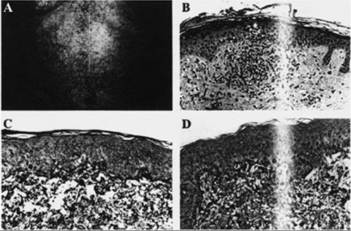
To obtain an initial assessment of the biologic activity of antagonizing CTLA-4 function in humans, we administered a single infusion of the humanized CTLA-4–blocking monoclonal antibody MDX-CTLA4 (3 mg/kg) to nine previously vaccinated metastatic melanoma or ovarian carcinoma patients.79 Tolerance was partially compromised by therapy, as manifested by low titers of autoantibodies in four patients and rashes due to CD4+ and CD8+ T cells directed against normal melanocytes in five patients (Fig. 34-1). Nonetheless, MDX-CTLA4 provoked extensive tumor necrosis with lymphocyte and granulocyte infiltrates in three of three metastatic melanoma patients and a reduction or stabilization of CA-125 levels in two of two metastatic ovarian carcinoma patients previously vaccinated with irradiated, autologous GM-CSF–secreting tumor cells. In contrast, MDX-CTLA4 did not elicit tumor necrosis in four of four metastatic melanoma patients previously immunized with defined melanosomal antigens. Pathologic examination of the responding metastases revealed CD4+ and CD8+ T cells, CD20+ B cells producing immunoglobulin, and granulocytes leading to tumor destruction (Fig. 34-2). Moreover, a striking circumferential lymphoid infiltrate was also detected in occluded tumor blood vessels, resulting in extensive ischemic necrosis. Since these pathologic features were similar to those previously observed in patients responding to GM-CSF–secreting tumor vaccines, they raise the possibility that MDX-CTLA4 may amplify a long-lived memory response.
|
|
|
Figure 34.1 MDX-CTLA4 compromises tolerance to normal melanocytes. A: Reticular erythematous rash. B: Perivascular lymphocyte infiltrate extending into epidermis with interface dermatitis. C: CD4+ T cells apposed to dying melanocytes. D: CD8+ T cells apposed to dying melanocytes. (Reprinted with permission from Hodi FS, Mihm MC, Soiffer RJ et al. Biologic activity of cytotoxic T lymphocyte–associated antigen 4 antibody blockade in previously vaccinated metastatic melanoma and ovarian carcinoma patients. Proc Natl Acad Sci USA 2003;100:4712–4717.) |
Additional studies examined serial infusions of MDX-CTLA4 together with melanosomal antigen–derived peptide vaccines.80 Of 14 patients tested, 3 achieved objective tumor responses, while 6 developed serious autoimmune disorders, including enterocolitis, hepatitis, dermatitis, and hypophysitis. This work highlights the potential for CTLA-4 antibody blockade to elicit serious autoimmune toxicities, thereby underscoring the need to determine optimal dosages and schedules for selective tumor destruction.
|
|
|
Figure 34.2 MDX-CTLA4 provokes extensive tumor necrosis. A, B: Tumor necrosis with granulocytes and lymphocytes. C: CD4+ T cells. D: CD8+ T cells. E: CD20+B cells. F: Vasculopathy with perivascular and intramural lymphoid infiltrates associated with luminal thrombosis. (Reprinted with permission from Hodi FS, Mihm MC, Soiffer RJ et al. Biologic activity of cytotoxic T lymphocyte–associated antigen 4 antibody blockade in previously vaccinated metastatic melanoma and ovarian carcinoma patients. Proc Natl Acad Sci USA 2003;100: 4712–4717.) |
CONCLUSION
An improved understanding of the mechanisms underlying antitumor immunity provides a strong foundation for undertaking detailed clinical studies of cancer vaccination. The development of new technologies to measure antitumor immune responses with precision substantially elevates the scientific rigor that can be applied to investigations in human cancer immunology. Indeed, several promising strategies to improve dendritic cell–mediated tumor antigen presentation have been validated in patients with diverse cancers. Moreover, initial trials of CTLA-4 antibody blockade have illuminated the therapeutic potential of interrupting tolerance networks that otherwise attenuate tumor immunity, albeit with a risk for autoimmunity. Determining the proper balance of immune stimulation and inhibition of negative regulation poses a significant clinical challenge, but one that the maturing field of tumor immunology most welcomes.
REFERENCES
1. Hanahan D, Weinberg RA. The hallmarks of cancer. Cell 2000;100: 57–70.
2. Dranoff G. Cytokines in cancer pathogenesis and cancer therapy. Nat Rev Cancer 2004;4:11–22.
3. Diefenbach A, Raulet D. The innate immune response to tumors and its role in the induction of T-cell immunity. Immunol Rev 2002;188:9–21.
4. Karre K. NK cells, MHC class I molecules and the missing self. Scan J Immunol 2002;55:221–228.
5. Albert M, Pearce S, Francisco L, et al. Immature dendritic cells phagocytose apoptotic cells via avb5 and CD36, and cross-present antigens to cytotoxic T lymphocytes. J Exp Med 1998;188: 1359–1368.
6. Banchereau J, Steinman R. Dendritic cells and the control of immunity. Nature 1998;392:245–252.
7. Gilboa E. The makings of a tumor rejection antigen. Immunity 1999;11:263–270.
8. Old L, Chen Y-T. New paths in human cancer serology. J Exp Med 1998;187:1163–1167.
9. Kaplan D, Shankaran V, Dighe A, et al. Demonstration of an interferon γ–dependent tumor surveillance system in immunocompetent mice. Proc Natl Acad Sci USA 1998;95:7556–7561.
10. Shankaran V, Ikeda H, Bruce AT, et al. IFN-γ and lymphocytes prevent primary tumour development and shape tumour immunogenicity. Nature 2001;410:1107–1111.
11. Enzler T, Gillessen S, Manis JP, et al. Deficiencies of GM-CSF and interferon-gamma link inflammation and cancer. J Exp Med 2003;197:1213–1219.
12. Clark W, Elder D, Guerry D, et al. Model predicting survival in stage I melanoma based on tumor progression. J Natl Cancer Inst 1989;81:1893–1904.
13. Naito Y, Saito K, Shiiba K, et al. CD8+ T cells infiltrated within cancer cell nests as a prognostic factor in human colorectal cancer. Cancer Res 1998;58:3491–3494.
14. Zhang L, Conejo-Garcia J, Katsaros D, et al. Intratumoral T cells, recurrence, and survival in epithelial ovarian cancer. N Engl J Med 2003;348:203–213.
15. Marincola FM, Jaffee EM, Hicklin DJ, et al. Escape of human solid tumors from T-cell recognition: molecular mechanisms and functional significance. Adv Immunol 2000;74:181–273.
16. Ames BN, Gold LS, Willett WC. The causes and prevention of cancer. Proc Natl Acad Sci USA 1995;92:5258–5265.
17. Moore RJ, Owens DM, Stamp G, et al. Mice deficient in tumor necrosis factor-a are resistant to skin carcinogenesis. Nature Med 1999;5:828–831.
18. Hilbert DM, Kopf M, Mock BA, et al. Interleukin 6 is essential for in vivo development of B lineage neoplasms. J Exp Med 1995;182:243–248.
19. Lin E, Nguyen A, Russell R, et al. Colony-stimulating factor 1 promotes progression of mammary tumors to malignancy. J Exp Med 2001;193:727–739.
20. Coussens LM, Tinkle CL, Hanahan D, et al. MMP-9 supplied by bone marrow–derived cells contributes to skin carcinogenesis. Cell 2000;103:481–490.
21. Muller A, Homey B, Soto H, et al. Involvement of chemokine receptors in breast cancer metastasis. Nature 2001;410:50–56.
22. Wang D, DuBois RN. Cyclooxygenase 2–derived prostaglandin E2 regulates the angiogenic switch. Proc Natl Acad Sci USA 2004; 101:415–416.
23. Prehn RT, Main JM. Immunity to methylcholanthrene-induced sarcomas. J Natl Cancer Inst 1957;18:769–778.
24. Ikeda H, Ohta N, Furukawa K, et al. Mutated mitogen–activated protein kinase: a tumor rejection antigen of mouse sarcoma. Proc Natl Acad Sci USA 1997;94:6375–6379.
25. Basombrio MA. Search for common antigenicity among twenty-five sarcomas induced by methylcholanthrene. Cancer Res 1970;30:2458–2462.
26. Klein G, Sjogren HO, Klein E, et al. Demonstration of resistance against methylcholanthrene-induced sarcomas in the primary autochtonous host. Cancer Res 1960;20:1561–1572.
27. Van Pel A, Boon T. Protection against a non-immunogenic mouse leukemia by an immunogenic variant obtained by mutagenesis. Proc Natl Acad Sci USA 1982;79:4718–4722.
28. Cerundolo V, Hermans IF, Salio M. Dendritic cells: a journey from laboratory to clinic. Nat Immunol 2004;5:7–10.
29. Young JW, Inaba K. Dendritic cells as adjuvants for class I major histocompatibility complex–restricted antitumor immunity. J Exp Med 1996;183:7–11.
30. Krieg AM. CpG motifs: the active ingredient in bacterial extracts? Nat Med 2003;9:831–835.
31. Suto R, Srivastava PK. A mechanism for the specific immunogenicity of heat shock protein–chaperoned peptides. Science 1995;269:1585–1588.
32. Pardoll DM. Spinning molecular immunology into successful immunotherapy. Nat Rev Immunol 2002;2:227–238.
33. Lynch D, Andreasen A, Maraskovsky E, et al. Flt3 ligand induces tumor regression and antitumor immune responses in vivo. Nature Med 1997;3:625–631.
34. Forni G, Fujiwara H, Martino F, et al. Helper strategy in tumor immunology: expansion of helper lymphocytes and utilization of helper lymphokines for experimental and clinical immunotherapy. Cancer Metast Rev 1988;7:289–309.
35. Mach N, Dranoff G. Cytokine-secreting tumor cell vaccines. Curr Opin Immunol 2000;12:571–575.
36. Cavallo F, Signorelli P, Giovarelli M, et al. Antitumor efficacy of adenocarcinoma cells engineered to produce interleukin 12 (IL-12) or other cytokines compared with exogenous IL-12. J Natl Cancer Inst 1997;89:1049–1058.
37. Dranoff G. GM-CSF–based cancer vaccines. Immunol Rev 2002; 188:147–154.
38. Haigis KM, Hoff PD, White A, et al. Tumor regionality in the mouse intestine reflects the mechanism of loss of Apc function. Proc Natl Acad Sci USA 2004.
39. Greenberg NM, Demayo F, Finegold MJ, et al. Prostate cancer in a transgenic mouse. Proc Natl Acad Sci USA 1995;92:3439–3443.
40. Nanni P, Nicoletti G, De Giovanni C, et al. Combined allogeneic tumor cell vaccination and systemic interleukin 12 prevents mammary carcinogenesis in HER-2/neu transgenic mice. J Exp Med 2001;194:1195–1205.
41. Iinuma T, Homma S, Noda T, et al. Prevention of gastrointestinal tumors based on adenomatous polyposis coli gene mutation by dendritic cell vaccine. J Clin Invest 2004;113:1307–1317.
42. De Giovanni C, Nicoletti G, Landuzzi L, et al. Immunoprevention of HER-2/neu transgenic mammary carcinoma through an interleukin 12–engineered allogeneic cell vaccine. Cancer Res 2004;64: 4001–4009.
43. Finn OJ, Forni G. Prophylactic cancer vaccines. Curr Opin Immunol 2002;14:172–177.
44. Altman J, Moss P, Goulder P, et al. Direct visualization and phenotypic analysis of virus-specific T lymphocytes in HIV-infected individuals. Science 1996;274:94–96.
45. Finn OJ. Cancer vaccines: between the idea and the reality. Nat Rev Immunol 2003;3:630–641.
46. Kwak LW, Campbell MJ, Czerwinski DK, et al. Induction of immune responses in patients with B-cell lymphoma against the surface-immunoglobulin idiotype expressed by their tumors. N Engl J Med 1992;327:1209–1215.
47. Boon T, van der Bruggen P. Human tumor antigens recognized by T lymphocytes. J Exp Med 1996;183:725–729.
48. Rosenberg SA. Progress in human tumour immunology and immunotherapy. Nature 2001;411:380–384.
49. Marchand M, van Baren N, Weynants P, et al. Tumor regressions observed in patients with metastatic melanoma treated with an antigenic peptide encoded by gene MAGE-3 and presented by HLA-A1. Int J Cancer 1999;80:219–230.
50. Disis ML, Schiffman K, Guthrie K, et al. Effect of dose on immune response in patients vaccinated with an her-2/neu intracellular domain protein–based vaccine. J Clin Oncol 2004;22: 1916–1925.
51. Peterson AC, Harlin H, Gajewski TF. Immunization with Melan-A peptide–pulsed peripheral blood mononuclear cells plus recombinant human interleukin-12 induces clinical activity and T-cell responses in advanced melanoma. J Clin Oncol 2003;21: 2342–2348.
52. Rosenberg S, Yang J, Schwartzentruber D, et al. Immunologic and therapeutic evaluation of a synthetic peptide vaccine for the treatment of patients with metastatic melanoma. Nature Med 1998;4: 321–327.
53. Disis ML, Rinn K, Knutson KL, et al. Flt3 ligand as a vaccine adjuvant in association with HER-2/neu peptide–based vaccines in patients with HER-2/neu–overexpressing cancers. Blood 2002;99: 2845–2850.
54. Slingluff CL Jr, Petroni GR, Yamshchikov GV, et al. Clinical and immunologic results of a randomized phase II trial of vaccination using four melanoma peptides either administered in granulocyte-macrophage colony-stimulating factor in adjuvant or pulsed on dendritic cells. J Clin Oncol 2003;21:4016–4026.
55. Bendandi M, Gocke C, Kobrin C, et al. Complete molecular remissions induced by patient-specific vaccination plus granulocyte-monocyte colony-stimulating factor against lymphoma. Nature Med 1999;5:1171–1177.
56. Timmerman JM, Czerwinski DK, Davis TA, et al. Idiotype-pulsed dendritic cell vaccination for B-cell lymphoma: clinical and immune responses in 35 patients. Blood 2002;99:1517–1526.
57. Davis ID, Chen W, Jackson H, et al. Recombinant NY-ESO-1 protein with ISCOMATRIX adjuvant induces broad integrated antibody and CD4(+) and CD8(+) T cell responses in humans. Proc Natl Acad Sci USA 2004;101:10697–10702.
58. Small EJ, Fratesi P, Reese DM, et al. Immunotherapy of hormone-refractory prostate cancer with antigen-loaded dendritic cells. J Clin Oncol 2000;18:3894–3903.
59. Kaufman HL, Wang W, Manola J, et al. Phase II randomized study of vaccine treatment of advanced prostate cancer (E7897): a trial of the Eastern Cooperative Oncology Group. J Clin Oncol 2004;22: 2122–2132.
60. Slovin SF, Ragupathi G, Musselli C, et al. Fully synthetic carbohydrate-based vaccines in biochemically relapsed prostate cancer: clinical trial results with alpha-N-acetylgalactosamine-O-serine/threonine conjugate vaccine. J Clin Oncol 2003;21:4292–4298.
61. Nieda M, Okai M, Tazbirkova A, et al. Therapeutic activation of Valpha24+Vbeta11+ NKT cells in human subjects results in highly coordinated secondary activation of acquired and innate immunity. Blood 2004;103:383–389.
62. Hsueh EC, Essner R, Foshag LJ, et al. Prolonged survival after complete resection of disseminated melanoma and active immunotherapy with a therapeutic cancer vaccine. J Clin Oncol 2002;20:4549–4554.
63. Jaffee E, Hruban R, Biedrzycki B, et al. Novel allogeneic granulocyte-macrophage colony-stimulating factor–secreting tumor vaccine for pancreatic cancer: a phase I trial of safety and immune activation. J Clin Oncol 2001;19:145–156.
64. Simons J, Mikhak B, Chang JF, et al. Induction of immunity to prostate cancer antigens: results of a clinical trial of vaccination with irradiated autologous prostate tumor cells engineered to secrete granulocyte-macrophage colony-stimulating factor using ex vivo gene transfer. Cancer Res 1999;59:5160–5168.
65. Nestle F, Alijagic S, Gilliet M, et al. Vaccination of melanoma patients with peptide- or tumor lysate-pulsed dendritic cells. Nature Med 1998;4:328–332.
66. Berd D, Sato T, Maguire HC Jr, et al. Immunopharmacologic analysis of an autologous, hapten-modified human melanoma vaccine. J Clin Oncol 2004;22:403–415.
67. Stift A, Friedl J, Dubsky P, et al. Dendritic cell–based vaccination in solid cancer. J Clin Oncol 2003;21:135–142.
68. Avigan D, Vasir B, Gong J, et al. Fusion cell vaccination of patients with metastatic breast and renal cancer induces immunological and clinical responses. Clin Cancer Res 2004;10:4699–4708.
69. Su Z, Dannull J, Heiser A, et al. Immunological and clinical responses in metastatic renal cancer patients vaccinated with tumor RNA–transfected dendritic cells. Cancer Res 2003;63:2127–2133.
70. Belli F, Testori A, Rivoltini L, et al. Vaccination of metastatic melanoma patients with autologous tumor-derived heat shock protein gp96–peptide complexes: clinical and immunologic findings. J Clin Oncol 2002;20:4169–4180.
71. Soiffer R, Hodi FS, Haluska F, et al. Vaccination with irradiated, autologous melanoma cells engineered to secrete granulocyte-macrophage colony-stimulating factor by adenoviral-mediated gene transfer augments antitumor immunity in patients with metastatic melanoma. J Clin Oncol 2003;21:3343–3350.
72. Salgia R, Lynch T, Skarin A, et al. Vaccination with irradiated autologous tumor cells engineered to secrete granulocyte-macrophage colony-stimulating factor augments antitumor immunity in some patients with metastatic non-small-cell lung carcinoma. J Clin Oncol 2003;21:624–630.
73. Nemunaitis J, Sterman D, Jablons D, et al. Granulocyte-macrophage colony-stimulating factor gene-modified autologous tumor vaccines in non-small-cell lung cancer. J Natl Cancer Inst 2004;96:326–331.
74. Chambers CA, Kuhns MS, Egen JG, et al. CTLA-4–mediated inhibition in regulation of T cell responses: mechanisms and manipulation in tumor immunotherapy. Annu Rev Immunol 2001;19: 565–594.
75. Salomon B, Bluestone JA. Complexities of CD28/B7: CTLA-4 costimulatory pathways in autoimmunity and transplantation. Annu Rev Immunol 2001;19:225–252.
76. Shevach EM, McHugh RS, Piccirillo CA, et al. Control of T-cell activation by CD4+ CD25+ suppressor T cells. Immunol Rev 2001;182:58–67.
77. Tivol EA, Borriello F, Schweitzer AN, et al. Loss of CTLA-4 leads to massive lymphoproliferation and fatal multiorgan tissue destruction, revealing a critical negative regulatory role of CTLA-4. Immunity 1995;3:541–547.
78. van Elsas A, Hurwitz A, Allison J. Combination immunotherapy of B16 melanoma using anti-cytotoxic T lymphocyte–associated antigen 4 (CTLA-4) and granulocyte/macrophage colony-stimulating factor (GM-CSF)–producing vaccines induces rejection of subcutaneous and metastatic tumors accompanied by autoimmune depigmentation. J Exp Med 1999;190: 355–366.
79. Hodi FS, Mihm MC, Soiffer RJ, et al. Biologic activity of cytotoxic T lymphocyte–associated antigen 4 antibody blockade in previously vaccinated metastatic melanoma and ovarian carcinoma patients. Proc Natl Acad Sci USA 2003;100:4712–4717.
80. Phan GQ, Yang JC, Sherry RM, et al. Cancer regression and autoimmunity induced by cytotoxic T lymphocyte–associated antigen 4 blockade in patients with metastatic melanoma. Proc Natl Acad Sci USA 2003;100:8372–8377.
FOOTNOTE
Work on this chapter was supported by NIH grants CA74886, CA66996, and by the Leukemia and Lymphoma Society.