
Khanh T. Do, Shivaani Kummar, James H. Doroshow, and Yves Pommier
CLASSIFICATION, BIOCHEMICAL, AND BIOLOGIC FUNCTIONS OF TOPOISOMERASES
Nucleic acids (DNA and RNA) being long polymers, topoisomerases fulfill the need for cellular DNA to be densely packaged in the cell nucleus, transcribed, replicated, and evenly distributed between daughter cells following replication without tangles. Topoisomerases are ubiquitous and essential for all organisms as they prevent and resolve DNA and RNA entanglements and resolve DNA supercoiling during transcription and replication. This chapter first summarizes the basic elements necessary to understand the mechanism of action of topoisomerases and their inhibitors. More detailed information can be found in recent reviews1–7 and two recent books.8,9 The second part of the chapter summarizes the use of topoisomerase inhibitors as anticancer drugs.
Classification of Topoisomerases
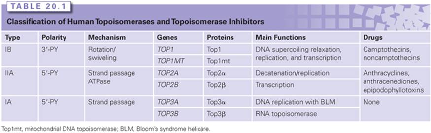
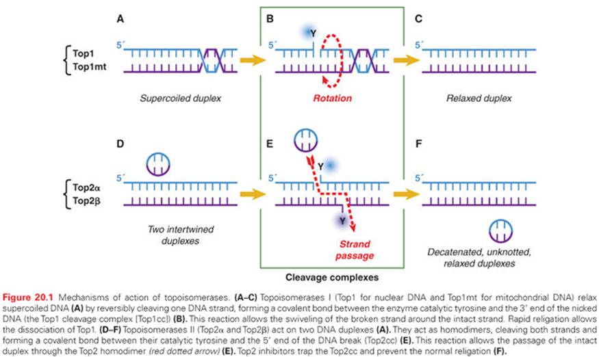
Human cells contain six topoisomerase genes (Table 20.1), which have been numbered historically. The commonly used abbreviations are Top1 for topoisomerases I (Top1mt being the mitochondrial topoisomerase whose gene is encoded in the cell nucleus),10 Top2 for topoisomerases II, and Top3 for topoisomerases III. Top1 was the first eukaryotic topoisomerase discovered by Champoux and Dulbecco.11Topoisomerases solve DNA topologic problems by cutting the DNA backbone and religating without the assistance of any additional ligase. Top1 and Top3 act by cleaving/religating a single strand of the DNA duplex, whereas Top2 enzymes cleave and religate both strands, making a four–base pair reversible staggered cut (Fig. 20.1). It is convenient to remember that odd-numbered topoisomerases (Top1 and Top3) cleave and religate one strand, whereas the even numbered topoisomerases (Top2s) cleave and religate both strands.
Biochemical Characteristics and Cleavage Complexes of the Different Topoisomerases
The DNA cutting/relegation mechanism is common to all topoisomerases and utilizes an enzyme catalytic tyrosine residue acting as a nucleophile and becoming covalently attached to the end of the broken DNA. These catalytic intermediates are referred to as cleavage complexes (see Fig. 20.1B, E). The reverse religation reaction is carried out by the attack of the ribose hydroxyl ends toward the tyrosyl-DNA bond.
Top1 (and Top1mt) attaches to the 3′-end of the break, whereas the other topoisomerases (Top2 and Top3) have opposite polarity and covalently attach to the 5′-end of the breaks (see Table 20.1 [second column] and Fig. 20.1B, E). Topoisomerases have distinct biochemical requirements. Top1 and Top1mt are the simplest, nicking/closing, and relaxing DNA as monomers in the absence of cofactor, and even at ice temperature. Top2 enzymes, on the other hand, are the most complex topoisomerases working as dimers, requiring ATP binding and hydrolysis, and a divalent metal (Mg2+) for catalysis. Top3 enzymes also require Mg2+ for catalysis but function as monomers without ATP requirement. Notably, the DNA substrates differ for Top3 enzymes. Whereas both Top1 and Top2 process double-stranded DNA, the Top3 substrates need to be single-stranded nucleic acids (DNA for Top3α and DNA or RNA for Top3β).10,12,13
Differential Topoisomerization Mechanisms: Swiveling Versus Strand Passage, DNA Versus RNA Topoisomerases
Topoisomerases use two main mechanisms to change nucleic topology. The first is by “untwisting” the DNA duplex. This mechanism is unique to Top1, which, by an enzyme-associated single-strand break, allows the broken strand to rotate around the intact strand (see Fig. 20.1B) until DNA supercoiling is dissipated. At this point, the stacking energy of adjacent DNA bases realigns the broken ends, and the 5′-hydroxyl end attacks the 3′-phosphotyrosyl end, thereby relegating the DNA. A remarkable feature of this Top1 untwisting mechanism is its extreme efficiency with a rotation speed around 6,000 rpm and relative independence from torque, thereby allowing full relaxation of DNA supercoiling.14
The second topologic mechanism is by “strand passage.” This mechanism allows the passage of a double- or a single-stranded DNA (or RNA) through the cleavage complexes. Top2α and Top2β both act by allowing the passage of an intact DNA duplex through the DNA double-strand break generated by the enzymes. After which, Top2 religates the broken duplex. Such reactions permit DNA decatenation, unknotting, and relaxation of supercoils.3 Top3 enzymes also act by strand passage but only pass one nucleic acid strand through the single-strand break generated by the enzymes. In the case of Top3α, the substrate is a single-stranded DNA segment (such as a double-Holliday junction), whereas in the case of Top3β, the substrate can be a single-stranded RNA segment, with Top3β acting as a RNA topoisomerase.13,15
TOPOISOMERASE INHIBITORS AS INTERFACIAL POISONS
Topoisomerase Inhibitors Act as Interfacial Inhibitors by Binding at the Topoisomerase–DNA Interface and Trapping Topoisomerase Cleavage Complexes
Relegation of the cleavage complexes is dependent on the structure of the ends of the broken DNA (i.e., the realignment of the broken ends). Binding the drugs at the enzyme–DNA interface misaligns the ends of the DNA and precludes relegation, resulting in the stabilization of the topoisomerase cleavage complexes (Top1cc and Top2cc). Crystal structures of drug-bound cleavage complexes have firmly established this mechanism for both Top1- and Top2-targeted drugs.16
It is critical to understand that the cytotoxic mechanism of topoisomerase inhibitors requires the drugs to trap the topoisomerase cleavage complexes rather than block catalytic activity. This sets apart topoisomerase inhibitors from classical enzyme inhibitors such as antifolates. Indeed, knocking out Top1 renders yeast cells totally immune to camptothecin,17,18 and reducing enzyme levels in cancer cells confers drug resistance. Conversely, in breast cancers, amplification of TOP2A, which is on the same locus as HER2, contributes to the efficacy of doxorubicin.19 Also, cellular mutations of Top1 and Top2 that renders cells insensitive to the trapping of topoisomerase cleavage complexes produce high resistance to Top1 or Top2 inhibitors. Based on this trapping of cleavage complexes mechanism, we refer to topoisomerase inhibitors as topoisomerase cleavage complex-targeted drugs.
Top1cc-Targeted Drugs (Camptothecin and Noncamptothecin Derivatives) Kill Cancer Cells by Replication Collisions
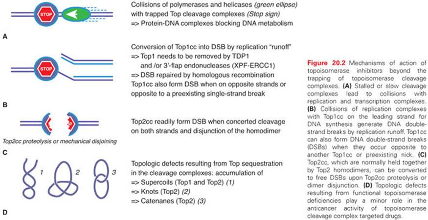
Top1cc are cytotoxic by their conversion into DNA damage by replication and transcription fork collisions. This explains why cytotoxicity is directly related to drug exposure and why arresting DNA replication protects cells from camptothecin.20,21 The collisions arise from the fact that the drugs, by slowing down the nicking/closing activity of Top1, uncouple the kinetics of Top1 with the polymerases and helicases, which lead polymerases to collide into Top1cc (Fig. 20.2A). Such collisions have two consequences. They generate double-strand breaks (replication and transcription runoff) and irreversible Top1–DNA adducts (see Fig. 20.2B). The replication double-strand breaks are repaired by homologous recombination, which explains the hypersensitivity of BRCA-deficient cancer cells to Top1cc-targeted drugs.22The Top1-covalent complexes can be removed by two pathways, the excision pathway centered around tyrosyl-DNA-phosphodiesterase 1 (TDP1)23 and the endonuclease pathway involving 3′-flap endonucleases such as XPF-ERCC1.24 It is also possible that drug-trapped Top1cc directly generate DNA double-strand breaks when they are within 10 base pairs on opposite strands of the DNA duplex or when they occur next to a preexisting single-strand break on the opposite strand. Finally, it is not excluded that topologic defects contribute to the cytotoxicity of Top1cc-targeted drugs (the accumulation of supercoils25 and the formation of alternative structures such as R-loops) (see Fig. 20.2D).26
Cytotoxic Mechanisms of Top2cc-Targeted Drugs (Intercalators and Demethyl Epipodophyllotoxins)
Contrary to camptothecins, Top2 inhibitors kill cancer cells without requiring DNA replication fork collisions. Indeed, even after a 30-minute exposure, doxorubicin and other Top2cc-targeted drugs can kill over 99% of the cells, which is in vast excess of the fraction of S-phase cells in tissue culture (generally less than 50%).27,28 The collision mechanism in the case of Top2cc-targeted drugs (see Fig. 20.2A) appears to involve transcription and proteolysis of both Top2 and RNA polymerase II.29 Such situation would then lead to DNA double-strand breaks by disruption of the Top2 dimer interface (see Fig. 20.2C). Alternatively, the Top2 homodimer interface could be disjoined by mechanical tension (see Fig. 20.2C). Yet, it is important to bear in mind that 90% of Top2cc trapped by etoposide are not concerted and, therefore, consist in single-strand breaks,3,30,31 which is different from doxorubicin, which traps both Top2 monomers and produces a majority of DNA double-strand breaks.32 Finally, it is not excluded that topologic defects resulting from Top2 sequestration by the drug-induced cleavage complexes could contribute to the cytotoxicity of Top2cc-targeted drugs (see Fig. 20.2D). Such topologic defects would include persistent DNA knots and catenanes, potentially leading to chromosome breaks during mitosis.
TOPOISOMERASE I INHIBITORS: CAMPTOTHECINS AND BEYOND
Camptothecin is an alkaloid identified in the 1960s by Wall and Wani33 in a screen of plant extracts for antineoplastic drugs. The two water-soluble derivatives of camptothecin containing the active lactone form are topotecan and irinotecan, which are approved by the U.S. Food and Drug Administration (FDA) for the treatment of several cancers. In addition, several Top1cc-targeting drugs are in clinical development, including camptothecin derivatives and formulations (including high–molecular-weight conjugates or liposomal formulations), as well as noncamptothecin compounds that exhibit greater potency or noncross resistance to irinotecan and topotecan in preclinical cancer models.31,34–36
Irinotecan
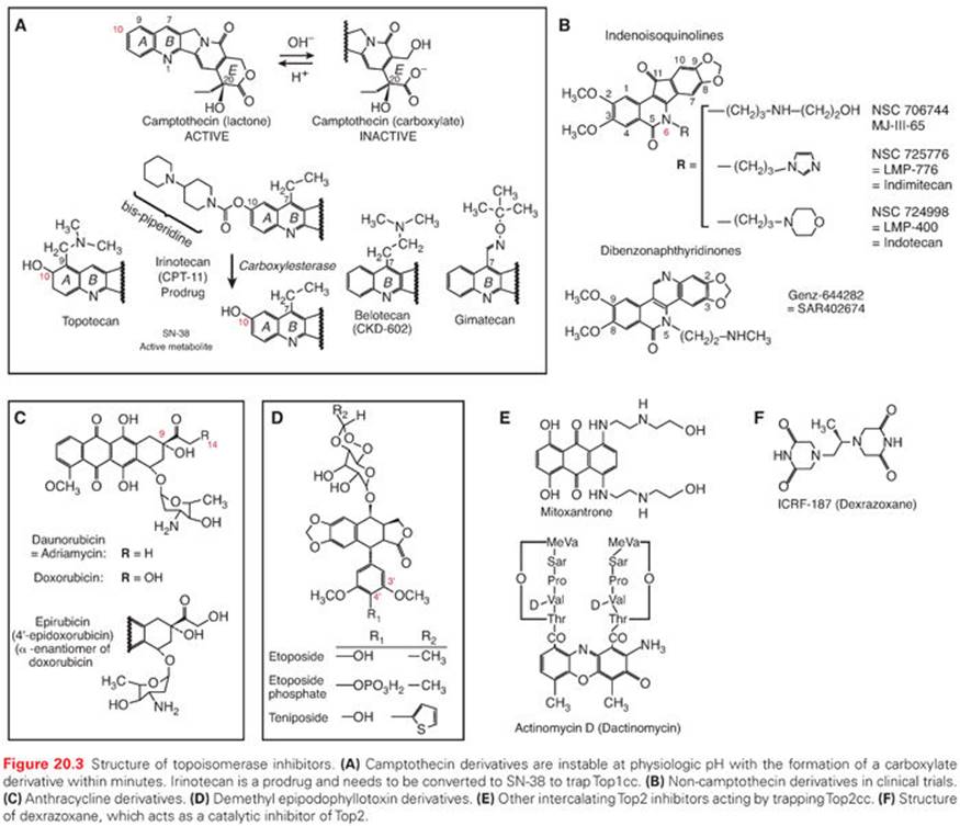
Irinotecan, a prodrug containing a bulky dipiperidine side chain at C-10 (Fig. 20.3), is cleaved by a carboxylesterase-converting enzyme in the liver and other tissues to generate the active metabolite, SN-38. Irinotecan is FDA approved for the treatment of colorectal cancer in the metastatic setting as first-line treatment in combination with 5-fluorouracil/leucovorin (5-FU/LV) and as a single agent in the second-line treatment of progressive colorectal cancer after 5-FU–based therapy (see Table 20.1).37,38 Newer therapeutic uses of irinotecan include a combination with oxaliplatin and 5-FU as first-line treatment in pancreatic cancer.39 Irinotecan is additionally used in combination with cisplatin or carboplatin in extensive-stage small-cell lung cancer40,41 as well as refractory esophageal and gastroesophageal junction (GEJ) cancers, gastric cancer, cervical cancer, anaplastic gliomas and glioblastomas, and non–small-cell lung cancer (Table 20.2). Irinotecan is usually administered intravenously at a dose of 125 mg/m2 for 4 weeks with a 2-week rest period in combination with bolus 5-FU/LV, 180 mg/m2 every 2 weeks in combination with an infusion of 5-FU/LV, or 350 mg/m2 every 3 weeks as a single agent.
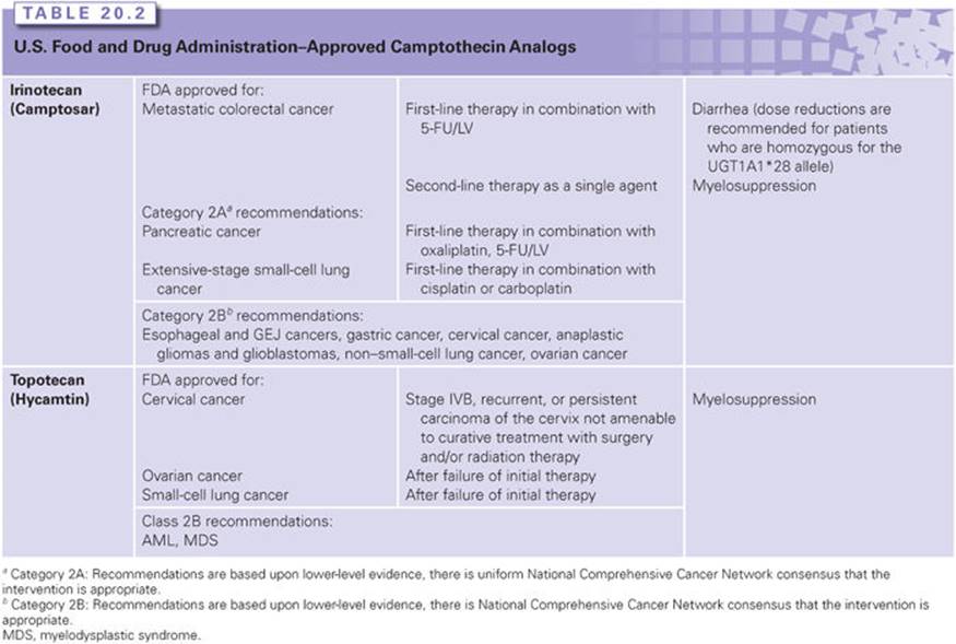
Diarrhea and myelosuppression are the most common toxicities associated with irinotecan administration. Two mechanisms explain irinotecan-induced diarrhea. Acute cholinergic effects resulting in abdominal cramping and diarrhea occur within 24 hours of drug administration are the result of acetylcholinesterase inhibition by the prodrug, and can be treated with the administration of atropine. Direct mucosal cytotoxicity with diarrhea is typically observed after 24 hours and can result in significant morbidity. Symptoms are managed with loperamide. Hepatic metabolism and biliary excretion accounts for >70% of the elimination of the administered dose, with renal excretion accounting for the remainder of the dose. SN-38 is glucuronidated in the liver by UGT1A1, and deficiencies in this pathway increase the risk of diarrhea and myelosuppression. Dose reductions are recommended for patients who are homozygous for the UGT1A1*28 allele, for which an FDA-approved test for detection of the UGT1A1*28 allele in patients is available.42,43 Additionally, dose reductions of irinotecan are recommended for patients with hepatic dysfunction, with bilirubin greater than 1.5 mg/mL.44
Topotecan
Topotecan contains a basic side chain at position C-9 that enhances its water solubility (see Fig. 20.3). Topotecan is approved for the treatment of ovarian cancer,45 small-cell lung cancer,46 and as a single agent and in combination with cisplatin for cervical cancer.47 Additionally, it is active in acute myeloid leukemia (AML) and myelodysplastic syndrome (see Table 20.2). Topotecan is administered intravenously as a single agent at a dose of 1.5 mg/m2 as a 30-minute infusion daily for 5 days, followed by a 2-week period of rest for the treatment of solid tumors or at a dose of 0.75 mg/m2 as a 30-minute infusion daily for 3 days in combination with cisplatin on day 1, every 3 weeks, for the treatment of cervical cancer.
Myelosuppression is the most common dose-limiting toxicity. Extensive prior radiation or previous bone marrow–suppressive chemotherapy increases the risk of topotecan-induced myelosuppression. Other toxicities include nausea, vomiting, diarrhea, fatigue, alopecia, and transient hepatic transaminitis.
Topotecan and its metabolites are primarily cleared by the kidneys, requiring dose reduction in patients with renal dysfunction. A 50% dose reduction is recommended for patients with moderate renal impairment (creatinine clearance 20 to 39 mL per minute). There are no formal guidelines for dose reductions in patients with hepatic dysfunction (defined as serum bilirubin >1.5 mg/dL to <10 mg/dL). Topotecan additionally penetrates the blood-brain barrier, achieving concentrations in cerebrospinal fluid that are approximately 30% that of plasma levels.48
Camptothecin Conjugates and Analogs
New formulations of camptothecin conjugates and analogs are currently in clinical development in an effort to improve the therapeutic index (Table 20.3). The development of camptothecin conjugates is based on the notion that the addition of a bulky conjugate would allow for a more consistent delivery system and extend the half-life of the molecule.
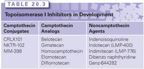
CRLX101, formerly IT-101, a covalent cyclodextrin-polyethylene glycol copolymer camptothecin conjugate, has plasma concentrations and area under the curve (AUC) that are approximately 100-fold higher than camptothecin, with a half-life in the range of 17 to 20 hours compared to 1.3 hours for camptothecin.49 It has demonstrated antitumor activity in preclinical studies in irinotecan-resistant tumors with complete tumor regression in human non–small-cell lung cancer, Ewing sarcoma, and lymphoma xenograft models.50 Preliminary data from Phase 1 studies indicate that CRLX101 is well tolerated at a dose of 15 mg/m2 administered in a biweekly administration schedule.51 It is currently being studied in Phase 2 studies as a single agent and in combination with chemotherapeutic agents in lung, renal cell cancer, and gynecologic malignancies.52–54
Etirinotecan pegol (NKTR-102), an irinotecan polymer conjugate, has a longer plasma circulation time with a lower maximum concentration of SN-38 compared with irinotecan. It was evaluated in a Phase 2 study in platinum-resistant refractory epithelial ovarian cancer at a dose of 145 mg/m2 administered on a schedule of every 21 days; a median progression-free survival of 5.3 months and median overall survival of 11.7 months was observed.55 Two schedules of administration, 145 mg/m2 administered every 14 days versus every 21 days, have been tested in a Phase 2 study of NKTR-102 in patients with previously treated metastatic breast cancer.56 Of the 70 patients evaluated in this study, 20 patients achieved an objective response (29%; 95% confidence interval [CI] 18.4 to 40.6). For both these studies, the most common adverse events on the 21-day administration schedule were dehydration and diarrhea. Etirinotecan pegol is currently being evaluated in several phase 2 studies in lung cancer, colorectal cancer, and high-grade gliomas,57–60 with evidence of clinical activity in refractory solid tumors. A Phase 3 trial (The BEACON Study) is underway evaluating NKTR-102 against the physicians’ choice in refractory breast cancer.61
As an alternative to macromolecular conjugates, attempts have also been made to alter the camptothecin pentacyclic ring structure with modifications of the A and B ring (see Fig. 20.3A) in an effort to improve solubility and enhance antitumor activity. Structure–activity relationship studies have shown that substitutions at the 7, 9, and 10 positions serve to enhance the antitumor activity of camptothecin.62Belotecan, a novel camptothecin analog, has a water-solubilizing group at the 7 position of the B ring of camptothecin (see Fig. 20.3A). Several Phase 2 studies have evaluated belotecan in combination with carboplatin in recurrent ovarian cancer63 and in combination with cisplatin in extensive-stage small-cell lung cancer,64 demonstrating activity in these cancers; however, these combinations were associated with prominent hematologic toxicities. Phase 2 studies evaluating belotecan as a single agent in patients with recurrent or progressive carcinoma of the uterine cervix failed to show activity.65 Gimatecan is a lipophilic oral camptothecin analog (see Fig. 20.3A). Pharmacokinetic studies demonstrate that gimatecan is primarily present in plasma as the lactone form (>85%), and has a long half-life of 77.1 +/− 29.6 hours, with an increase in maximum concentration (Cmax) and AUC of three- to six-fold after multiple dosing.66 Phase 2 studies show that gimatecan has demonstrated activity in previously treated ovarian cancer, with myelosuppression as the main toxicity.67
Newer development of analogs have attempted to modify the E-ring through introduction of an electron-withdrawing group at the α position in an effort to overcome the instability of the E-ring while maintaining the binding capability of the camptothecin analog to the Top1-DNA cleavage complex. Collectively called homocamptothecin analogs, two have been tested in clinical trials and include diflomotecan68 and elomotecan.69 The dose-limiting toxicity in the Phase I study of elomotecan was neutropenia. A five-member E-ring derivative has also been developed and has reached a Phase 1 clinical trial.70,71
Noncamptothecin Topoisomerase I Inhibitors
Noncamptothecin Top1 inhibitors are in clinical development, and include indenoisoquinolines and dibenzonaphthyridines (see Fig. 20.3B). Two indenoisoquinoline derivatives are currently in clinical development, indotecan (LMP400) and indimitecan (LMP776).72,73 Early in vitro studies show enhanced potency compared with camptothecins, and persistence of Top1 cleavage complexes.74 Genz-644282, a dibenzonaphthyridine derivative, demonstrated enhanced antitumor activity in preclinical studies75 and is currently being evaluated in Phase 1 clinical trials.76
TOPOISOMERASE II INHIBITORS: INTERCALATORS AND NONINTERCALATORS
Topoisomerase II inhibitors can be classified in two main classes: DNA intercalators, which encompass different chemical classes (Fig. 20.3C, E), and nonintercalators represented by the epipodophyllotoxin derivatives (see Fig. 20.3D). Although both act by trapping Top2 cleavage complexes (Top2cc), DNA intercalators exhibit a second effect as drug concentrations increase above low micromolar values: they block the formation of Top2cc by intercalating into DNA and destabilizing the binding of Top2 to DNA. This explains why Top2α and Top2β are trapped over a relatively narrow concentration range by anthracyclines, and why intercalators have additional effects besides trapping Top2cc, namely inhibition of a broad range of DNA processing enzymes including helicases, polymerase, and even nucleosome destabilization.
Doxorubicin
Doxorubicin and daunorubicin were the first anthracyclines discovered in the 1960s and remain among the most widely used anticancer agents over a broad spectrum of malignancies. Although doxorubicin only differs by one hydroxyl substitution on position 14 (see Fig. 20.3C), doxorubicin has a much broader anticancer activity than daunorubicin. Anthracyclines are natural products derived from Streptomyces peucetius variation caesius. They were found to target Top2 well after their clinical approval.77 Subsequent searches for less toxic drugs and formulations led to the approval of liposomal doxorubicin, idarubicin, and epirubicin.
Anthracyclines are flat, planar molecules that are relatively hydrophobic. The quinone structure of anthracyclines (see Fig. 20.3C) enhances the catalysis of oxidation-reduction reactions, thereby promoting the generation of oxygen free radicals, which may be involved in antitumor effects as well as the cardiotoxicity associated with these drugs.78,79 Anthracyclines are also substrates for P-glycoprotein and Mrp-1, and drug efflux is thought to be a major drug resistance determinant.80,81
Doxorubicin is available in a standard salt form and as a liposomal formulation. FDA-labeled indications for standard doxorubicin include acute lymphocytic leukemia (ALL), AML, chronic lymphoid leukemia, Hodgkin lymphoma, non-Hodgkin lymphoma, mantle cell lymphoma, multiple myeloma, mycosis fungoides, Kaposi sarcoma, breast cancer (adjuvant therapy and advanced), advanced prostate cancer, advanced gastric cancer, Ewing sarcoma, thyroid cancer, advanced nephroblastoma, advanced neuroblastoma, advanced non–small-cell lung cancer, advanced ovarian cancer, advanced transitional cell bladder cancer, cervical cancer, and Langerhans cell tumors. Doxorubicin has activity in other malignancies as well, including soft tissue sarcoma, osteosarcoma, carcinoid, and liver cancer (Table 20.4). Doxorubicin is typically administered at a recommended dose of 30 to 75 mg/m2 every 3 weeks intravenously.
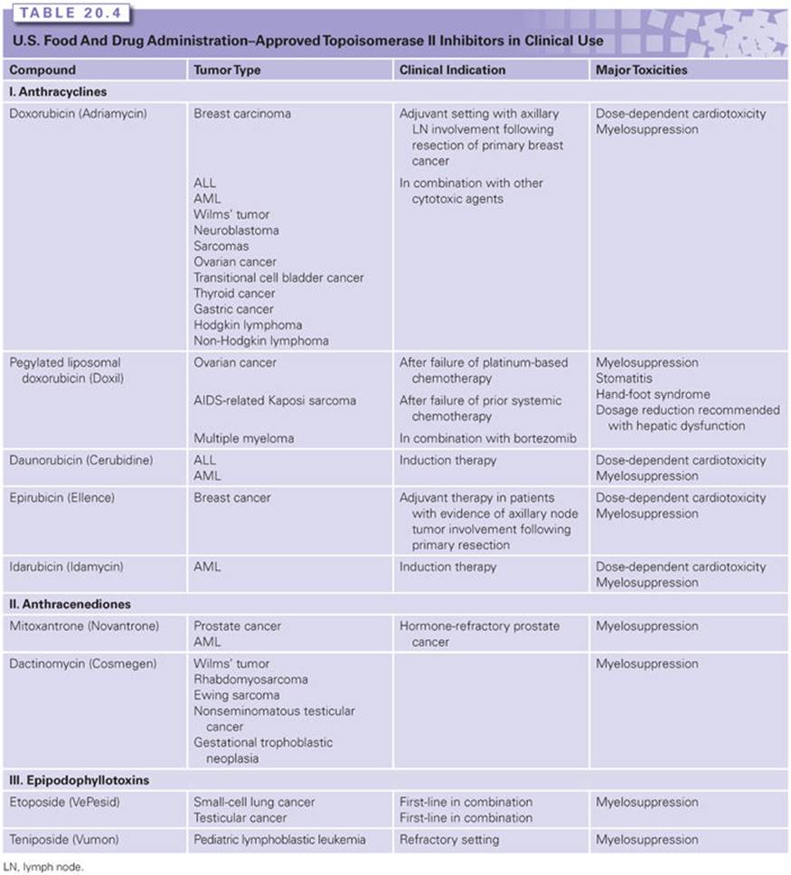
Major acute toxicities of doxorubicin include myelosuppression, mucositis, alopecia, nausea, and vomiting. Myelosuppression is the acute dose-limiting toxicity. Other toxicities, including diarrhea, nausea, vomiting, mucositis, and alopecia, are dose and schedule related. Prophylactic antiemetics are routinely given with bolus doses of doxorubicin, and longer infusions are associated with less nausea and less cardiotoxicity. Patients should also be warned to expect their urine to redden after drug administration. Doxorubicin is a potent vesicant, and extravasation can lead to severe necrosis of skin and local tissues, requiring surgical debridement and skin grafts. Infusions via a central venous catheter are recommended. Other toxicities of doxorubicin include radiation recall and the risk of developing secondary leukemia. Radiation recall is an inflammatory reaction at sites of previous radiation and can lead to pericarditis, pleural effusion, and skin rash. Secondary leukemias are thought to be a result of balanced translocations that result from Top2 poisoning by the anthracyclines, albeit to lesser degree than other Top2 poisons, such as the epipodophyllotoxins (see the following).82
Anthracyclines are cleared mainly by metabolism to less active forms and by biliary excretion. Less than 10% of the administered dose is cleared by the kidneys. Dose reductions should be made in patients with elevated plasma bilirubin. Doxorubicin should be dose reduced by 50% for plasma bilirubin concentrations ranging from 1.2 to 3.0 mg/dL, by 75% for values of 3.1 to 5.0 mg/dL, and withheld for values greater than 5 mg/dL.
Liposomal Doxorubicin
Doxorubicin is also available in a polyethylene glycol (PEG)ylated liposomal form, which allows for enhancement of drug delivery. Use of liposomal doxorubicin has been associated with less cardiotoxicity even at doses exceeding 500 mg/m2.83 Additionally, liposomal doxorubicin produces less nausea and vomiting and relatively mild myelosuppression compared to doxorubicin. Unique to the liposomal formulation is the risk of hand–foot syndrome and an acute infusion reaction manifested by flushing, dyspnea, edema, fever, chills, rash, bronchospasm, and hypertension. These infusion reactions are related to the rate of infusion; therefore, the recommended administration schedule is set at an initial rate of 1 mg per minute for the first 10 to 15 minutes. The rate may be slowly increased to complete infusion over 60 minutes if no reaction occurs. Typical dosing schedules include 50 mg/m2 intravenous infusion every 4 weeks for four courses in ovarian cancer, 20 mg/m2 intravenous infusion every 3 weeks in AIDS-related Kaposi sarcoma, and 30 mg/m2 intravenous infusion in combination with bortezomib to be given on days 1, 4, 8, and 11 every 3 weeks in multiple myeloma.
Daunorubicin
Despite its chemical similarity (see Fig. 20.3C), daunorubicin is considerably less active in solid tumors compared to doxorubicin. It is FDA approved for the treatment of ALL and AML. Daunorubicin is typically administered via intravenous push over 3 to 5 minutes at a dose of 30 to 45 mg/m2 per day on 3 consecutive days in combination chemotherapy. For induction therapy for pediatric acute lymphoblastic leukemia, daunorubicin is dosed at 25 mg/m2intravenously in combination with vincristine and prednisone. In children less than 2 years of age or in those who have a body surface area less than 0.5 m2, current recommendations are based on body mass index (1 mg/kg) rather than body surface area. A higher dose of daunorubicin at 60 mg/m2 per day to 90 mg/m2 per day intravenously for 3 consecutive days is currently recommended as part of the induction combination regimen for the treatment of acute myeloblastic leukemia. Daunorubicin has similar toxicities to doxorubicin, including myelosuppression, cardiac toxicity, nausea, vomiting, alopecia, and is also a vesicant. Daunorubicin is metabolized by the liver and undergoes substantial elimination by the kidneys, requiring dose reductions for both renal and hepatic dysfunction. A 50% dose reduction is recommended for either serum creatinine or bilirubin greater than 3 mg/dL, and a 25% reduction in dose for bilirubin concentrations ranging from 1.2 to 3.0 mg/dL.
Epirubicin
Epirubicin is an epimer of doxorubicin (see Fig. 20.3C) with increased lipophilicity. It is FDA approved for adjuvant therapy of breast cancer but is also used in combination for the treatment of a variety of malignancies. Epirubicin is administered intravenously at doses ranging from 60 to 120 mg/m2 every 3 to 4 weeks. Epirubicin has a similar toxicity profile to doxorubicin but is overall better tolerated.
In addition to being converted to an enol by an aldose reductase, epirubicin has a unique steric orientation of the C-4 hydroxyl group that allows it to serve as a substrate for conjugation reactions mediated by liver glucuronosyltransferases and sulfatases. As such, dose adjustments are recommended in the setting of hepatic dysfunction. For patients with serum bilirubin of 1.2 to 3 mg/dL or aspartate aminotransferase of 2 to 4 times the upper limit of normal, a 50% dose reduction is recommended. For patients with bilirubin greater than 3 mg/dL or aspartate aminotransferase greater than 4 times the upper limit of normal, a dose reduction of 75% is recommended. Due to limited data, no specific dose recommendations are currently available for patients with renal impairment, although current recommendations are for consideration of dose adjustments in patients with serum creatinine greater than 5 mg/dL.
Idarubicin
Idarubicin is a synthetic derivative of daunorubicin, but lacks the 4-methoxy group (see Fig. 20.3C). It is FDA approved as part of combination chemotherapy regimen for AML and is also active in ALL. It is given intravenously at a dose of 12 mg/m2 for 3 consecutive days, typically in combination with cytarabine. Idarubicin has similar toxicities as daunorubicin. Its primary active metabolite is idarubicinol, and elimination is mainly through the biliary system and, to a lesser extent, through renal excretion. A 50% dose reduction is recommended for serum bilirubin of 2.6 to 5 mg/dL and idarubicin should not be given if the bilirubin is greater than 5 mg/dL. Additionally, dose reductions in renal impairment are advised, but specific guidelines are not available.
Cardiac Toxicity of Anthracyclines
Anthracyclines are responsible for cardiac toxicities, and special considerations are necessary to minimize this severe side effect. Acute doxorubicin cardiotoxicity is reversible, and clinical signs include tachycardia, hypotension, electrocardiogram changes, and arrhythmias. It develops during or within days of anthracycline infusion, and its incidence can be significantly reduced by slowing doxorubicin infusion rates.
Chronic and delayed cardiotoxicity is more common and more severe because it is irreversible. Chronic cardiotoxicity with congestive heart failure peaks at 1 to 3 months but can occur even years after therapy. Myocardial damage has been shown to occur by several mechanisms. The classical mechanism is by the direct generation of reactive oxygen species (ROS) during the electron transfer from the semiquinone to quinone moieties of the anthracycline,84 which leads to myocardial damage. ROS can also be generated by mitochondrial damage resulting from drug-mediated inactivation of the oxidative phosphorylation chain because doxorubicin accumulates not only in chromatin, but also in mitochondria.78,79 A recent study has also related doxorubicin cardiotoxicity to the poisoning of Top2β cleavage complexes in myocardiocytes.85 Endomyocardial biopsy is characterized by a predominant finding of multifocal areas of patchy and interstitial fibrosis (stellate scars) and occasional vacuolated myocardial cells (Adria cells). Myocyte hypertrophy and degeneration, loss of cross-striations, and the absence of myocarditis are also characteristic of this diagnosis.86 The incidence of cardiomyopathy is related to both the cumulative dose and the schedule of administration, and predisposition to cardiac damage includes a previous history of heart disease, hypertension, radiation to the mediastinum, age greater than 65 years or younger than 4 years, prior use of anthracyclines or other cardiac toxins, and coadministration of other chemotherapy agents (e.g., paclitaxel, cyclophosphamide, or trastuzumab).87,88 Sequential administration of paclitaxel followed by doxorubicin in breast cancer patients is associated with cardiomyopathy at total doxorubicin doses above 340 to 380 mg/m2, whereas the reverse sequence of drug administration did not yield the same systemic toxicities at these doses.89 When doxorubicin is given in a low-dose weekly regimen (10 to 20 mg/m2 per week) or by slow continuous infusion over 96 hours, cumulative doses of more than 500 mg/m2 can be given. Doses of epirubicin less than 1,000 mg/m2 and daunorubicin less than 550 mg/m2 are considered safe. Additionally, liposomal doxorubicin is associated with less cardiac toxicity.
Cardiac function can be monitored during treatment with anthracyclines by electrocardiography, echocardiography, or radionuclide scans. Numerous studies have established the danger of embarking on anthracycline therapy in patients with underlying cardiac disease (e.g., a baseline left ventricular ejection fraction of less than 50%) and of continuing therapy after a documented decrease in the ejection fraction by more than 10% (if this decrease falls below the lower limit of normal). Because anthracycline-induced cardiotoxicity has been related to the generation of free radicals, efforts have been aimed at attenuating this effect through the targeting of redox response and reduction in oxidative stress. Dexrazoxane is a metal chelator that decreases the myocardial toxicity of doxorubicin in breast cancer patients. In two multicenter, double-blind studies, advanced breast cancer patients were randomized to chemotherapy with dexrazoxane or a placebo; dexrazoxane was shown to have a cardioprotective effect based on serial, noninvasive cardiac testing during the course of the trial and is approved for that use by the FDA.90Dexrazoxane chelates iron and copper, thereby interfering with the redox reactions that generate free radicals and damage myocardial lipids. Notably, dexrazoxane is also a Top2 catalytic inhibitor (see Fig. 20.3F), which potentially might minimize the therapeutic activity of anthracyclines by interfering with the trapping of Top2 cleavage complexes by anthracyclines.2,3,91 Other agents currently in use include β-blockers and statins. A recent meta-analysis of 12 randomized controlled trials and 2 observational studies involving the use of agents to prevent the cardiotoxicity associated with anthracyclines demonstrated relatively similar efficacy regardless of which prophylactic treatment was used.92
Anthracenediones
Mitoxantrone (see Fig. 20.3E) is currently the only clinically approved anthracenedione. Compared to anthracyclines, mitoxantrone is less cardiotoxic owing to a decreased ability to undergo oxidation-reduction reactions and form free radicals.
Mitoxantrone is FDA approved for the treatment of advanced hormone-refractory prostate cancer93 and AML.94 It is typically administered intravenously at a dose of 12 to 14 mg/m2 every 3 weeks in the treatment of prostate cancer, and at a dose of 12 mg/m2 in combination with cytosine arabinoside for 3 days in the treatment of AML.
Toxicities are generally less severe compared to doxorubicin and include myelosuppression, nausea, vomiting, alopecia, and mucositis. Cardiac toxicity can be seen at cumulative doses greater than 160 mg/m2.95 Mitoxantrone is rapidly cleared from the plasma and is highly concentrated in tissues. The majority of the drug is eliminated in the feces, with a small amount undergoing renal excretion. Dose adjustments for hepatic dysfunction are recommended, but formal guidelines are currently not available.
Dactinomycin
Dactinomycin was the first antibiotic shown to have antitumor activity96 and consists of a planar phenoxazone ring attached to two peptide side chains. This unique structure allows for tight intercalation into DNA between adjacent guanine–cytosine bases, leading to Top2 and Top1 poisoning and transcription inhibition.97 Dactinomycin was one of the first drugs shown to be transported by P-glycoprotein, and represents the major mechanism of resistance.98
Dactinomycin is FDA approved for Ewing sarcoma,99 gestational trophoblastic neoplasm,100 metastatic nonseminomatous testicular cancer,101 nephroblastoma,102 and rhabdomyosarcoma.103 Typically, it is administered intravenously at doses of 15 μg/kg for 5 days in combination with other chemotherapeutic agents for the treatment of nephroblastoma, rhabdomyosarcoma, and Ewing sarcoma; at does of 12 μg/kg intravenously as a single agent in the treatment of gestational trophoblastic neoplasias; and at doses of 1,000 μg/m2 intravenously on day 1 as part of a combination regimen with cyclophosphamide, bleomycin, vinblastine, and cisplatin in the treatment of metastatic nonseminomatous testicular cancer. Toxicities include myelosuppression, veno-occlusive disease of the liver, nausea, vomiting, alopecia, erythema, and acne. Additionally, similar to doxorubicin, dactinomycin can cause radiation recall and severe tissue necrosis in cases of extravasation. Dactinomycin is largely excreted unchanged in the feces and urine. Guidelines for dosing in patients with impaired renal or liver function are currently not available.
Epipodophyllotoxins
Epipodophyllotoxins are glycoside derivatives of podophyllotoxin, an antimicrotubule agent extracted from the mandrake plant. Two derivatives, demethylated on the pendant ring (see R1 in Fig. 20.3D), etoposide and teniposide were shown to primarily function as Top2 poisons rather than through antimicrotubule mechanisms.104,105 Epipodophyllotoxins poison Top2 through a mechanism distinct from that of anthracyclines and other DNA intercalators.106without intercalating into normal DNA in the absence of Top2. Therefore, they are “cleaner” Top2 inhibitors than the anthracyclines, anthracenediones, and dactinomycin. However, etoposide and teniposide trap Top2 cleavage complexes by base stacking in a ternary complex at the interface of the DNA and the Top2 homodimer. Mechanisms that have been implicated in resistance to etoposide include drug efflux, because epipodophyllotoxins are substrates for P-glycoprotein107; altered localization of Top2α; decreased cellular expression of Top2α108; and impaired phosphorylation of Top2.109
Etoposide
Etoposide (see Fig. 20.3D) is available in intravenous and oral forms. It is FDA approved for the treatment of small-cell lung cancer110 and refractory testicular cancer.111 It also has activity in hematologic malignancies and various solid tumors. The intravenous form is generally administered at doses of 35 to 50 mg/m2 for 4 to 5 days every 3 to 4 weeks in combination therapy for small-cell lung cancer, and 50 to 100 mg/m2 for 5 days every 3 to 4 weeks in combination therapy for refractory testicular cancer. The dose of oral etoposide is usually twice the intravenous dose. Oral bioavailability is highly variable due to dependence on intestinal P-glycoprotein.112
The dose-limiting toxicity for etoposide is myelosuppression, with white blood cell count nadirs typically occurring on days 10 to 14. Thrombocytopenia is less common than leukopenia. Additionally, mild to moderate nausea, vomiting, diarrhea, mucositis, and alopecia are associated with etoposide. Among topoisomerase inhibitors, epipodophyllotoxins have the greatest association with secondary malignancies, with etoposide having the highest risk, with an estimated 4% 6-year cumulative risk.113 The majority of etoposide is cleared unchanged by the kidneys, and a 25% dose reduction is recommended in patients with a creatinine clearance of 15 to 50 mL per minute. A 50% dose reduction is recommended in patients with a creatinine clearance less than 15 mL per minute. Because the unbound fraction of etoposide is dependent on albumin and bilirubin concentrations, dose adjustments for hepatic dysfunction are advised, but consensus guidelines are currently not available.
Teniposide
Teniposide contains a thiophene group in place of the methyl group on the glucose moiety of etoposide (Fig. 20.3D). Teniposide is FDA approved for refractory pediatric ALL.114,115 In pediatric ALL studies, doses ranged from 165 mg/m2 intravenously in combination with cytarabine to 250 mg/m2 intravenously weekly in combination with vincristine and prednisone. Similar to etoposide, the dose-limiting toxicity of teniposide is myelosuppression. Additional toxicities include mild-to-moderate nausea, vomiting, diarrhea, alopecia, and secondary leukemia. Teniposide is associated with greater frequency of hypersensitivity reactions compared to etoposide.
Teniposide is 99% bound to albumin and, as compared to etoposide, undergoes hepatic metabolism more extensively and renal clearance less extensively. No specific guidelines are currently available on dose adjustments for renal or hepatic dysfunction.
THERAPY-RELATED SECONDARY ACUTE LEUKEMIA
One of the major complications of Top2 inhibitor therapies, especially for etoposide and mitoxantrone, is acute secondary leukemia, which occurs in approximately 5% of patients. Therapy-related AMLs (t-AML) are characterized by their relatively rapid onset (they can occur only a few months after therapy) and the presence of recurrent balanced translocations involving the mixed lineage leukemia (MLL) locus on 11q23 and over 50 partner genes.116 The molecular mechanism is likely from the disjoining of two drug-trapped Top2 cleavage complexes on different chromosomes (see Fig. 20.2C) in relationship with transcription collisions and illegitimate relegation.117 Top2β, rather than Top2α, has been implicated in the generation of these disjoined cleavage complexes.117,118
FUTURE DIRECTIONS
Current challenges in the development of topoisomerase inhibitors lie in the inherent chemical instability of current and established agents. In addition to recent developments designed to enhance the stability with semisynthetic analogs and the development of novel delivery systems in an effort to achieve higher intratumoral concentrations, attention is also being focused on targeting other topoisomerase isoenzymes. Driving this trend has been the recent elucidation of the role of Top2β inhibition in the development of treatment-related cardiotoxicity and secondary AML.86,117,118 In addition to combination chemotherapy regimens already in use, attempts have also been made for the sequential inhibition of Top1 and Top2. Based on early preclinical models suggesting synergy with sequential inhibition of Top1 and Top2,120 phase 1 studies have evaluated the sequential administration of topotecan and etoposide in extensive-stage small-cell lung cancer and ovarian cancer, with significant myelosuppression as the dose-limiting toxicity.121,122 Future rational drug combinations include targeting DNA repair pathways in combination with Top1 inhibition, although further characterization is needed of the specific DNA repair and stress response pathways invoked in response to DNA damage as a result of Top1 inhibition. However, one such attempt of combining topotecan with veliparib, a small molecule inhibitor of poly (ADP-ribose) polymerase, was poorly tolerated due to significant myelosuppression, thus limiting the doses of topotecan that could be safely administered.123
Molecular characterization of tumors to better define patient selection and the development of pharmacodynamic biomarkers to monitor the response to treatment and to optimize the combination dose and schedules is needed for the further clinical development of topoisomerase inhibitors. Validated assays have been developed to evaluate topoisomerase 1 levels and levels of plosphorylated histone H2AX (gamma-H2AX) as a marker of DNA damage response to topoisomerase inhibition,124,125 and are being incorporated in current phase I studies of indenoisoquinolines.72,73
REFERENCES
1. Nitiss JL. DNA topoisomerase II and its growing repertoire of biological functions. Nat Rev Cancer 2009;9(5):327–337.
2. Nitiss JL. Targeting DNA topoisomerase II in cancer chemotherapy. Nat Rev Cancer 2009;9(5):338–350.
3. Pommier Y, Leo E, Zhang H, et al. DNA topoisomerases and their poisoning by anticancer and antibacterial drugs. Chem Biol 2010;17(5):421–433.
4. Fortune JM, Osheroff N. Topoisomerase II as a target for anticancer drugs: when enzymes stop being nice. Prog Nucleic Acid Res Mol Biol 2000;64:221–253.
5. Wang JC. A journey in the world of DNA rings and beyond. Annu Rev Biochem 2009;78:31–54.
6. Wang JC. Cellular roles of DNA topoisomerases: a molecular perspective. Nat Rev Mol Cell Biol 2002;3(6):430–440.
7. Champoux JJ. DNA topoisomerases: structure, function, and mechanism. Annu Rev Biochem 2001;70:369–413.
8. Wang JC. Untangling the Double Helix: DNA Entanglements and the Action of DNA Topoisomerases. Cold Spring Harbor, NY: Cold Spring Harbor Laboratory Press; 2009.
9. Pommier Y. DNA Topoisomerases and cancer. In: Teicher BA, ed. Cancer Discovery and Development. New York: Springer & Humana Press; 2012.
10. Zhang H, Barceló JM, Lee B, et al. Human mitochondrial topoisomerase I. Proc Natl Acad Sci U S A 2001;98(10):10608–10613.
11. Champoux JJ, Dulbecco R. An activity from mammalian cells that untwists superhelical DNA—a possible swivel for DNA replication (polyoma-ethidium bromide-mouse-embryo cells-dye binding assay). Proc Natl Acad Sci U S A 1972;69(1):143–146.
12. Chen SH, Wu CH, Plank JL, et al. Essential functions of C terminus of Drosophila Topoisomerase IIIα in double Holliday junction dissolution. J Biol Chem 2012;287(23):19346–19353.
13. Xu D, Shen W, Guo R, et al. Top3β is an RNA topoisomerase that works with fragile X syndrome protein to promote synapse formation. Nat Neurosci 2013;16(9):1238–1247.
14. Seol Y, Gentry AC, Osheroff N, et al. Chiral discrimination and writhe-dependent relaxation mechanism of human topoisomerase IIα. J Biol Chem 2013;288(19):13695–13703.
15. Stoll G, Pietiläinen OP, Linder B, et al. Deletion of TOP3b, a component of FMRP-containing mRNPs, contributes to neurodevelopmental disorders. Nat Neurosci 2013;16(9):1228–1237.
16. Pommier Y, Marchand C. Interfacial inhibitors: targeting macromolecular complexes. Nat Rev Drug Discov 2011;11(1):25–36.
17. Nitiss J, Wang JC. DNA topoisomerase-targeting antitumor drugs can be studied in yeast. Proc Natl Acad Sci U S A 1988;85(20):7501–7505.
18. Bjornsti MA, Benedetti P, Vigilanti GA, et al. Expression of human DNA topoisomerase I in yeast cells lacking yeast DNA topoisomerase I: restoration of sensitivity of the cells to the antitumor drug camptothecin. Cancer Res 1989;49(22):6318–6323.
19. Dressler LG, Berry DA, Broadwater G, et al. Comparison of HER2 status by fluorescence in situ hybridization and immunohistochemistry to predict benefit from dose escalation of adjuvant doxorubicin-based therapy in node-positive breast cancer patients. J Clin Oncol2005;23(19):4287–4297.
20. Holm C, Covey JM, Kerrigan D, et al. Differential requirement of DNA replication for the cytotoxicity of DNA topoisomerase I and II inhibitors in Chinese hamster DC3F cell. Cancer Res 1989;49(22):6365–6368.
21. Hsiang YH, Lihou MG, Liu LF. Arrest of DNA replication by drug-stabilized topoisomerase I-DNA cleavable complexes as a mechanism of cell killing by camptothecin. Cancer Res 1989;49(18):5077–5082.
22. Maede Y, Shimizu H, Fukushima T, et al. Differential and common DNA repair pathways for topoisomerase I- and II-targeted drug in a genetic DT40 repair screen panel. Mol Cancer Ther 2014;13(1):214–220.
23. Huang SN, Pommier Y, Marchand C. Tyrosyl-DNA Phosphodiesterase 1(Tdp1) inhibitors. Expert Opinion Ther Pat 2011;21(9):1285–1292.
24. Zhang YW, Regairaz M, Seiler JA, et al. Poly(ADP-ribose) polymerase and XPF-ERCC1 participate in distinct pathways for the repair of topoisomerase I-induced DNA damage in mammalian cells. Nucleic Acids Res 2011;39(9):3607–3620.
25. Koster DA, Palle K, Bot ES, et al. Antitumor drugs impede DNA uncoiling by topoisomerase I. Nature 2007;448(7150):213–217.
26. Sordet O, Redon CE, Guirouilh-Barbat J, et al. Ataxia telangiectasia mutated activation by transcription- and topoisomerase I-induced DNA double-strand breaks. EMBO Rep 2009;10(8):887–893.
27. Pommier Y, Zwelling LA, Mattern MR, et al. Effects of dimethyl sulfoxide and thiourea upon intercalator-induced DNA single-strand breaks in mouse leukemia (L1210) cells. Cancer Res 1983;43(12 Pt 1):5718–5724.
28. Long BH, Musial ST, Brattain MG. Comparison of cytotoxicity and DNA breakage activity of congeners of podophyllotoxin including VP16-213 and VM26: a quantitative structure-activity relationship. Biochemistry 1984;23(6):1183–1188.
29. Ban Y, Ho CW, Lin RK, et al. Activation of a novel ubiquitin-independent proteasome pathway when RNA polymerase II encounters a protein roadblock. Mol Cell Biol 2013;33(20):4008–4016.
30. Long BH, Musial ST, Brattain MG. Single- and double-strand DNA breakage and repair in human lung adenocarcinoma cells exposed to etoposide and teniposide. Cancer Res 1985;45(7):3106–3112.
31. Pommier Y. Drugging topoisomerases: lessons and challenges. ACS Chem Biol 2013;8(1):82–95.
32. Zwelling LA, Michaels S, Erickson LC, et al. Protein-associated deoxyribonucleic acid strand breaks in L1210 cells treated with the deoxyribonucleic acid intercalating agents 4’-(9-acridinylamino) methanesulfon-m-anisidide and adriamycin. Biochemistry 1981;20(23):6553–6563.
33. Wall ME, Wani MC. Camptothecin and taxol: discovery to clinic—thirteenth Bruce F. Cain Memorial Award Lecture. Cancer Res 1995;55:753–760.
34. Pommier Y. Topoisomerase I inhibitors: camptothecins and beyond. Nat Rev Cancer 2006;6(10):789–802.
35. Teicher BA. Next generation topoisomerase I inhibitors: rationale and biomarker strategies. Biochem Pharmacol 2008;75(6):1262–1271.
36. Pommier Y, Cushman M. The indenoisoquinoline noncamptothecin topoisomerase I inhibitors: update and perspectives. Mol Cancer Ther 2009;8(5):1008–1014.
37. Douillard JY, Cunningham D, Roth AD, et al. Irinotecan combined with fluorouracil compared with fluorouracil alone as first-line treatment for metastatic colorectal cancer: a multicentre randomised trial. Lancet 2000;355:1041–1047.
38. Saltz LB, Cox JV, Blanke C, et al. Irinotecan plus fluorouracil and leucovorin for metastatic colorectal cancer. Irinotecan Study Group. N Engl J Med 2000;343:905–914.
39. Conroy T, Desseigne F, Tchou M, et al. FOLFIRINOX versus gemcitabine for metastatic pancreatic cancer. N Engl J Med 2011;364:1817–1825.
40. Hanna N, Bunn PA Jr, Langer C, et al. Randomized phase III trial comparing irinotecan/cisplatin with etoposide/cisplatin in patients with previously untreated extensive-stage disease small-cell lung cancer. J Clin Oncol 2006;24:2038–2043.
41. Schmittel A, Fischer von Weikersthal L, Sebastian M, et al. A randomized phase II trial of irinotecan plus carboplatin versus carboplatin treatment in patients with extended disease small-cell lung cancer. Ann Oncol 2006;17:663–667.
42. Iyer L, King CD, Whitington PF, et al. Genetic predisposition to the metabolism of irinotecan (CPT-11). Role of uridine diphosphate glucuronosyltransferase isoform 1A1 in the glucuronidation of its active metabolite (SN-38) in human liver microsomes. J Clin Invest1998;101:847–854.
43. Innocenti F, Undevia SD, Iyer L, et al. Genetic variants in the UDP-glucuronosyltransferase 1A1 gene predict the risk of severe neutropenia of irinotecan. J Clin Oncol 2004;22:1382–1388.
44. Schaaf LJ, Hammond LA, Tipping SJ, et al. Phase 1 and pharmacokinetic study of intravenous irinotecan in refractory solid tumor patients with hepatic dysfunction. Clin Cancer Res 2006;12:3782–3791.
45. ten Bokkel Huinink W, Gore M, Carmichael J, et al. Topotecan versus paclitaxel for the treatment of recurrent epithelial ovarian cancer. J Clin Oncol 1997;15:2183–2193.
46. Ardizzoni A, Hansen H, Dombernowsky P, et al. Topotecan, a new active drug in the second-line treatment of small-cell lung cancer: a phase II study in patients with refractory and sensitive disease. The European Organization for Research and Treatment of Cancer Early Clinical Studies Group and New Drug Development Office, and the Lung Cancer Cooperative Group. J Clin Oncol 1997;15:2090–2096.
47. Long HJ 3rd, Bundy BN, Grendys EC Jr, et al. Randomized phase III trial of cisplatin with or without topotecan in carcinoma of the uterine cervix: a Gynecologic Oncology Group Study. J Clin Oncol 2005;23:4626–4633.
48. Baker SD, Heideman RL, Crom WR, et al. Cerebrospinal fluid pharmacokinetics and penetration of continuous infusion topotecan in children with central nervous system tumors. Cancer Chemother Pharmacol 1996;37:195–202.
49. Schluep T, Cheng J, Khin KT, et al. Pharmacokinetics and biodistribution of the camptothecin-polymer conjugate IT-101 in rats and tumor-bearing mice. Cancer Chemother Pharmacol 2006;57:654–662.
50. Young C, Schluep T, Hwang J, et al. CRLX101 (formerly IT-101)-A novel nanopharmaceutical of camptothecin in clinical development. Curr Bioact Compd 2011;7:8–14.
51. Weiss GJ, Chao J, Neidhart JD, et al. First-in-human phase 1/2a trial of CRLX101, a cyclodextrin-containing polymer-camptothecin nanopharmaceutical in patients with advanced solid tumor malignancies. Invest New Drugs 2013;31:986–1000.
52. University of Chicago. A randomized phase II study of IV Topotecan versus CRLX101 in the second line treatment of recurrent small cell lung cancer. ClinicalTrials.gov Identifier: NCT01803269.
53. Cerulean Pharma Inc. A randomized, phase 2, study to assess the safety and activity of CRLX101, a nanoparticle formulation of camptothecin, in patients with advanced non-small cell lung cancer who have failed one or two previous regimens of chemotherapy. ClinicalTrials Identifier: NCT01380769.
54. Massachusetts General Hospital. A Phase II, 2-stage Trial of CRLX101-202 in recurrent ovarian, tubal and peritoneal cancer. ClinicalTrials.gov Identifier: NCT01652079.
55. Vergote IB, Garcia A, Micha J et al. Randomized multicentre phase II trial comparing two schedules of etirinotecan pegol (NKTR-102) in women with recurrent platinum-resistant/refractory epithelial ovarian cancer. J Clin Oncol 2013;31(32):4060–4066.
56. Awada A, Garcia AA, Chan S et al. Two schedules of etirinotecan pegol (NKTR-102) in patients with previously treated metastatic breast cancer: a randomized phase 2 study. Lancet Oncol 2013;14(12):1216–1225.
57. Roswell Park Cancer Institute. A phase II study of single agent topoisomerase-I inhibitor polymer conjugate, Etirinotecan Pegol (NKTR-102), in patients with relapsed small cell lung cancer. ClinicalTrials Identifier: NCT01876446.
58. Abramson Cancer Center of the University of Pennsylvania. Phase 2 study of Etirinotecan Pegol (NKTR-102) in the treatment of patients with metastatic and recurrent non-small cell lung cancer (NSCLC) after failure of 2nd line treatment. ClinicalTrials Identifier: NCT01773109.
59. Nektar Therapeutics. A multicentre, open-label, randomized, phase 2 study to evaluate the efficacy and safety of NKTR-102 versus irinotecan in patients with second-line, irinotecan-naive, KRAS-mutant, metastatic colorectal cancer (mCRC). ClinicalTrials Identifier: NCT00856375.
60. Lawrence Recht. A phase II, single arm, open label study of NKTR-102 in bevacizumab-resistant high-grade glioma. ClinicalTrials Identifier: NCT01663012.
61. Nektar Therapeutics. The BEACON study (breast cancer outcomes with NKTR-102): a phase 3 open-label, randomized, multicenter study of NKTR-102 versus treatment of physician’s choice (TPC) in patients with locally recurrent or metastatic breast cancer previously treated with an anthracycline, a taxane and capecitabine. ClinicalTrials Identifier: NCT01492101.
62. Basili S, Moro S. Novel camptothecin derivatives as topoisomerase I inhibitors. Expert Opin Ther Pat 2009;19:555–574.
63. Choi CH, Lee YY, Song TJ, et al. Phase II study of belotecan, a camptothecin analogue, in combination with carboplatin for the treatment of recurrent ovarian cancer. Cancer 2011;117:2104–2111.
64. Rhee CK, Lee SH, Kim JS, et al. A multicentre phase II study of belotecan, a new camptothecin analogue, as a second-line therapy in patients with small cell lung cancer. Lung Cancer 2011;72(1):64–67.
65. Hwang JH, Lim MC, Seo SS, et al. Phase II study of belotecan (CKD 602) as a single agent in patients with recurrent or progressive carcinoma of the uterine cervix. Jpn J Clin Oncol 2011;41:624–629.
66. Frapolli R, Zucchetti M, Sessa C, et al. Clinical pharmacokinetics of the new oral camptothecin gimatecan: the inter-patient variability is related to alpha1-acid glycoprotein plasma levels. Eur J Cancer 2010;46:505–516.
67. Pecorelli S, Ray-Coquard I, Tredan O, et al. Phase II of oral gimatecan in patients with recurrent epithelial ovarian, fallopian tube or peritoneal cancer, previously treated with platinum and taxanes. Ann Oncol 2010;21:759–765.
68. Graham JS, Falk S, Samuel LM et al. A multi-centre dose escalation and pharmacokinetic study of diflomotecan in patients with advanced malignancy. Cancer Chemother Pharmacol 2009;63:945–952.
69. Trocóniz IF, Cendrós JM, Soto E, et al. Population pharmacokinetic/pharmacodynamics modeling of drug-induced adverse effects of a novel homocamptothecin analog, elomotecan (BN80927), in a Phase I dose finding study in patients with advanced solid tumors. Cancer Chemother Pharmacol 2012;70:239–250.
70. Takagi K, Dexheimer TS, Redon C, et al. Novel E-ring camptothecin keto analogues (S38809 and S39625) are stable, potent, and selective topoisomerase I inhibitors without being substrates of drug efflux transporters. Mol Cancer Ther 2007;6(12 Pt 1):3229–3238.
71. Lansiaux A, Léonce S, Kraus-Berthier L, et al. Novel stable camptothecin derivatives replacing the E-ring lactone by a ketone function are potent inhibitors of topoisomerase I and promising antitumor drugs. Mol Pharmacol 2007;72(2):311–319.
72. National Cancer Institute. A Phase I Study of Indenoisoquinolines LMP400 and LMP776 in Adults With Relapsed Solid Tumors and Lymphomas. ClinicalTrials Identifier: NCT01051635.
73. National Cancer Institute. A phase I trial of weekly Indenoisoquinolines LMP400 in adults with relapsed solid tumors and lymphomas. ClinicalTrials Identifier: NCT01794104.
74. Antony S, Agama KK, Miao ZH, et al. Novel indenoisoquinolines NSC 725776 and NSC 724998 produce persistent topoisomerase I cleavage complexes and overcome multidrug resistance. Cancer Res 2007;67:10397–10405.
75. Kurtzberg LS, Roth S, Krumbholz R, et al. Genz-644282, a novel non-camptothecin topoisomerase I inhibitor for cancer treatment. Clin Cancer Res 2011;17:2777–2787.
76. Genzyme, a Sanofi Company. Dose Escalation Study to Assess the Safety and Tolerability of Genz-644282 in Patients With Solid Tumors. ClinicalTrials Identifier: NCT00942799.
77. Capranico G, Zunino F, Kohn KW, et al. Sequence-selective topoisomerase II inhibition by anthracycline derivatives in SV40 DNA: relationship with DNA binding affinity and cytotoxicity. Biochemistry 1990;29(2):562–569.
78. Davies KJ, Doroshow JH. Redox cycling of anthracyclines by cardiac mitochondria. I. Anthracycline radical formation by NADH dehydrogenase. J Biol Chem 1986;261(7):3060–3067.
79. Doroshow JH, Davies KJ. Redox cycling of anthracyclines by cardiac mitochondria. II. Formation of superoxide anion, hydrogen peroxide, and hydroxyl radical. J Biol Chem 1986;261:3068–3074.
80. Schneider E, Cowan KH. Multiple drug resistance in cancer therapy. Med J Aust 1994;160(6):371–373.
81. Alvarez M, Paull K, Monks A, et al. Generation of a drug resistance profile by quantitation of mdr-1/P-glycoprotein in the cell lines of the National Cancer Institute Anticancer Drug Screen. J Clin Invest 1995;95(5):2205–2214.
82. Felix CA, Kolaris CP, Osheroff N. Topoisomerase II and the etiology of chromosomal translocations. DNA Repair 2006;5:1093–1108.
83. O’Brien ME, Wigler N, Inbar M, et al. Reduced cardiotoxicity and comparable efficacy in a phase III trial of pegylated liposomal doxorubicin HCl (CAELYX/Doxil) versus conventional doxorubicin for first-line treatment of metastatic breast cancer. Ann Oncol2004;15(3):440–449.
84. Doroshow JH. Effect of anthracycline antibiotics on oxygen radical formation in rat heart. Cancer Res 1983;43(2):460–472.
85. Zhang S, Liu X, Bawa-Khalfe T, et al. Identification of the molecular basis of doxorubicin-induced cardiotoxicity. Nat Med 2012;18:1639–1642.
86. Speyer J, Wasserheit C. Strategies for reduction of anthracycline cardiac toxicity. Sem Oncol 1998;25:525–537.
87. Chanan-Khan A, Srinivasan S, Czuczman MS. Prevention and management of cardiotoxicity from antineoplastic therapy. J Support Oncol 2004;2:251–256.
88. Von Hoff DD, Layard MW, Basa P, et al. Risk factors for doxorubicin-induced congestive heart failure. Ann Intern Med 1979;91:710–717.
89. Shan K, Lincoff AM, Young JB. Anthracycline-induced cardiotoxicity. Ann Intern Med 1996;125:47–58.
90. Swain SM, Whaley FS, Gerber MC, et al. Cardioprotection with dexrazoxane for doxorubicin-containing therapy in advanced breast cancer. J Clin Oncol 15(4):1318–1332.
91. Andoh T, Ishida R. Catalytic inhibitors of DNA topoisomerase II. Biochim Biophys Acta 1998;1400(1–3):155–171.
92. Kalam K, Marwick TH. Role of cardioprotective therapy for prevention of cardiotoxicity with chemotherapy: a systematic review and meta-analysis. Eur J Cancer 2013;49:2900–2909.
93. Tannock IF, Osoba D, Stockler MR, et al. Chemotherapy with mitoxantrone plus prednisone or prednisone alone for symptomatic hormone-resistant prostate cancer: a Canadian randomized trial with palliative end points. J Clin Oncol 1996;14:1756–1764.
94. Reece DE, Elmongy MB, Barnett MJ, et al. Chemotherapy with high-dose cytosine arabinoside and mitoxantrone for poor-prognosis myeloid leukemias. Cancer Invest 1993;11:509–516.
95. Shenkenberg TD, Von Hoff DD. Mitoxantrone: a new anticancer drug with significant clinical activity. Ann Intern Med 1986;105:67–81.
96. Hollstein U. Actinomycin. Chemistry and mechanism of action. Chem Rev 1974;74(6):625–652.
97. Wassermann K, Markovits J, Jaxel C, et al. Effects of morpholinyl doxorubicins, doxorubicin, and actinomycin D on mammalian DNA topoisomerases I and II. Mol Pharmacol 1990;38(1):38–45.
98. Biedler JL, Riehm H. Cellular resistance to actinomycin D in Chinese hamster cells in vitro: cross-resistance, radioautographic, and cytogenetic studies. Cancer Res 1970;30:1174–1184.
99. Jaffe N, Paed D, Traggis D, et al. Improved outlook for Ewing’s sarcoma with combination chemotherapy (vincristine, actinomycin D and cyclophosphamide) and radiation therapy. Cancer 1976;38(5):1925–1930.
100. Turan T, Karacay O, Tulunay G, et al. Results of EMA/CO (etoposide, methotrexate, actinomycin D, cyclophosphamide, vincristine) chemotherapy in gestational trophoblastic neoplasia. Int J Gynecol Cancer 2006;16(3):1432–1438.
101. Early KS, Albert DJ. Single agent chemotherapy (actinomycin D) in the treatment of metastatic testicular carcinoma. South Med J 1976;69(8):1017–1021.
102. Fernbach DJ, Martyn DT. Role of dactinomycin in the improved survival of children with Wilm’s tumor. JAMA 1966;195(1222):1005–1009.
103. Maurer HM, Moon T, Donaldson M, et al. The intergroup rhabdomyosarcoma study: a preliminary report. Cancer 1977;40(5):2015–2026.
104. Chen GL, Yang L, Rowe TC, et al. Nonintercalative antitumor drugs interfere with the breakage-reunion reaction of mammalian DNA topoisomerase. J Biol Chem 1984;259(21):13560–13566.
105. Long BH, Musial ST, Brattain MG. Comparison of cytotoxicity and DNA breakage activity of cogeners of podophyllotoxin including VP16-213 and VM26: a quantitative structure-activity relationship. Biochemistry 1984;23(6):1183–1188.
106. Ross W, Rowe T, Glisson B, et al. Role of topoisomerase II in mediating epipodophyllotoxin-induced DNA cleavage. Cancer Res 1984;44:5857–5860.
107. Meresse P, Dechaux E, Monneret C, et al. Etoposide: discovery and medicinal chemistry. Curr Med Chem 2004;11:2443–2466.
108. Valkov NI, Gump JL, Engel R, et al. Cell density-dependent VP-16 sensitivity of leukaemic cells is accompanied by the translocation of topoisomerase IIalpha from the nucleus to the cytoplasm. Br J Haematol 2000;108:331–345.
109. Takano H, Kohno K, Ono M, et al. Increased phosphorylation of DNA topoisomerase II in etoposide-resistant mutants of human cancer KB cells. Cancer Res 1991;51:3951–3957.
110. Sundstrøm S, Bremnes RM, Kaasa S, et al. Cisplatin and etoposide regimen is superior to cyclophosphamide, epirubicin, and vincristine regimen in small-cell lung cancer: results from a randomized phase III trial with 5 years’ follow-up. J Clin Oncol 2002;20:4665–4672.
111. Nichols CR, Catalano PJ, Crawford ED, et al. Randomized comparison of cisplatin and etoposide and either bleomycin or ifosfamide in treatment of advanced disseminated germ cell tumors: an Eastern Cooperative Oncology Group, Southwest Oncology Group, and Cancer and Leukemia Group B Study. J Clin Oncol 1998;16:1287–1293.
112. Leu BL, Huang JD. Inhibition of intestinal P-glycoprotein and effects on etoposide absorption. Cancer Chemother Pharmacol 1995;35:432–436.
113. Smith MA, Rubinstein L, Anderson JR, et al. Secondary leukemia or myelodysplastic syndrome after treatment with epipodophyllotoxins. J Clin Oncol 1999;17:569–577.
114. Maluf PT, Odone Filho V, Cristofani LM, et al. Teniposide plus cytarabine as intensification therapy and in continuation therapy for advanced nonlymphoblastic lymphomas of childhood. J Clin Oncol 1994;12:1963–1968.
115. Rivera G, Bowman WP, Murphy SB, et al. VM-26 with prednisone and vincristine for treatment of refractory acute lymphocytic leukemia. Med Pediatr Oncol 1982;10:439–446.
116. Lovett BD, Lo Nigro L, Rappaport EF, et al. Near-precise interchromosomal recombination and functional DNA topoisomerase II cleavage sites at MLL and AF-4 genomic breakpoints in treatment-related acute lymphoblastic leukemia with t(4;11) translocation. Proc Natl Acad Sci U S A 2001;98(17):9802–9807.
117. Cowell IG, Sondka Z, Smith K, et al. Model for MLL translocations in therapy-related leukemia involving topoisomerase IIb-mediated DNA strand breaks and gene proximity. Proc Natl Acad Sci U S A 2012;109(23):8989–8994.
118. Azarova AM, Lyu YL, Lin CP, et al. Roles of DNA topoisomerases II isozymes in chemotherapy and secondary malignancies. Proc Natl Acad Sci U S A 2007;104:11014–11019.
119. Changela A, DiGate RJ, Mondragón A. Structural studies of E. Coli topoisomerase III-DNA complexes reveal a novel type IA topoisomerase-DNA conformational intermediate. J Mol Biol 2007;368:105–118.
120. Bertrand R, O’Connor PM, Kerrigan D, et al. Sequential administration of camptothecin and etoposide circumvents the antagonistic cytotoxicity of simultaneous drug administration in slowly growing human colon carcinoma HT-29 cells. Eur J Cancer 1992;28A(4–5):743–748.
121. Miller AA, Al Omari A, Murry DJ, et al. Phase I and pharmacologic study of sequential topotecan-carboplatin-etoposide in patients with extensive stage small cell lung cancer. Lung Cancer 2006;54:379–385.
122. Rose PG, Markham M, Bell JG, et al. Sequential prolonged oral topotecan and prolonged oral etoposide as second-line therapy in ovarian or peritoneal carcinoma: a phase I Gynecologic Oncology Study Group study. Gynecol Oncol 2006;102:236–239.
123. Kummar S, Chen A, Ji J, et al. Phase I study of PARP inhibitor ABT-888 in combination with topotecan in adults with refractory solid tumors and lymphomas. Cancer Res 2011;71(17):5626–5634.
124. Pfister TD, Hollingshead M, Kinders RJ, et al. Development and validation of an immunoassay for quantification of topoisomerase I in solid tumor tissues. PLoS One 2012;7:e50494.
125. Kinders RJ, Hollingshead M, Lawrence S, et al. Development of a validated immunofluorescence assay for gH2AX as a pharmacodynamics marker of topoisomerase I inhibitor activity. Clin Cancer Res 2010;16:5447–5457.