
Cindy H. Chau and William Douglas Figg, Sr.
INTRODUCTION
Blood vessels are indispensable for tumor growth and metastasis, and the formation of a new network of blood vessels from the existing vasculature, termed angiogenesis, is one of the essential hallmarks of cancer development.1Indeed, it was over 70 years ago that the existence of tumor-derived factors responsible for promoting new vessel growth was postulated,2 and that tumor growth is essentially dependent on vascular induction and the development of a neovascular supply.3 By the late 1960s, Dr. Judah Folkman and colleagues4 had begun the search for a tumor angiogenesis factor. In the 1971 landmark report, Folkman5 proposed that inhibition of angiogenesis by means of holding tumors in a nonvascularized dormant state would be an effective strategy to treat human cancer, and hence laid the groundwork for the concept behind the development of antiangiogenesis agents. This fostered the search for angiogenic factors, regulators of angiogenesis, and antiangiogenic molecules over the next few decades and shed light on angiogenesis as an important therapeutic target for the treatment of cancer and other diseases.
A decade has passed since the regulatory approval of the first antiangiogenic drug bevacizumab, and while initial results were regarded as highly promising, clinical evidence indicated that antiangiogenic therapy also had limitations. Successful development and clinical translation of this novel class of agents depends on the complete understanding of the biology of angiogenesis and the regulatory proteins that govern this angiogenic process, topics that have been covered in greater detail in another section of this textbook. This chapter will briefly review the mechanisms underlying tumor angiogenesis followed by an in-depth discussion of antiangiogenic therapy, the modes of action of angiogenesis inhibitors, and the successes and challenges of this treatment modality.
UNDERSTANDING THE ANGIOGENIC PROCESS
Angiogenic Switch and Regulatory Proteins
Tumor development and progression depend on angiogenesis. Recruitment of new blood vessels to the tumor site is required for the delivery of nutrients and oxygen to the cancerous growths and for the removal of waste products.6Cancer cells promote angiogenesis at an early stage of tumorigenesis, beginning with the release of molecules that send signals to the surrounding normal host tissue and stimulate the migration of microvascular endothelial cells (EC) in the direction of the angiogenic stimulus. These angiogenic factors not only mediate EC migration, but also EC proliferation and microvessel formation in tumors undergoing the switch to the angiogenic phenotype.7Experimental evidence for this angiogenic switch was observed when hyperplastic islets in transgenic mice (RIP-Tag model) switch from small (<1 mm), white microscopic dormant tumors to red, rapidly growing tumors.7 Dormant tumors have been discovered during autopsies of individuals who died of causes other than cancer.8 These autopsy studies suggest that the vast majority of microscopic in situ cancers never switch to the angiogenic phenotype during a normal lifetime. Such incipient tumors are usually not neovascularized and can remain harmless to the host for long periods of time as microscopic lesions that are in a state of dormancy.9,10 These nonangiogenic tumors cannot expand beyond the initial microscopic size and cannot become clinically detectable, lethal tumors until they have switched to the angiogenic phenotype11–13 through neovascularization and/or blood vessel cooption.14 Depending on the tumor type and the environment, this switch can occur at different stages of the tumor progression pathway and ultimately depends on a net balance of positive and negative regulators. Thus, the angiogenic phenotype may result from the production of growth factors by tumor cells and/or the downregulation of negative modulators.
Changes in this angiogenic balance affecting the levels of activator and inhibitor molecules dictate whether an EC will be in a quiescent or an angiogenic state. Normally, the inhibitors predominate, thereby blocking growth. Once the balance shifts in favor of the angiogenic state, proangiogenic factors prompts the activation, growth, and division of vascular ECs, resulting in the formation of new blood vessels. Activated ECs produce and release matrix metalloproteinases (MMP) into the surrounding tissue to break down the extracellular matrix to allow the ECs to migrate and organize themselves into hollow tubes that eventually evolve into a mature network of blood vessels. Proangiogenic factors or positive regulators of angiogenesis include vascular endothelial growth factor (VEGF), basic fibroblast growth factor (PlGF), platelet-derived growth factor (PDGF), placental growth factor, transforming growth factor-β, pleiotrophins, and others.15 Activation of the hypoxia-inducible factor 1 (HIF-1) via tumor-associated hypoxic conditions is also involved in the upregulation of several angiogenic factors.16 The angiogenic switch also involves the downregulation of angiogenesis suppressor proteins, which include endostatin, angiostatin, thrombospondin, and others.17,18 Most notably, however, is the link between many oncogenes and angiogenesis and the significant role oncogenes play in driving the angiogenic switch.19,20 These proangiogenic oncogenes not only induce the expression of stimulators, but may also downregulate inhibitors of angiogenesis.21
Endogenous Inhibitors of Angiogenesis
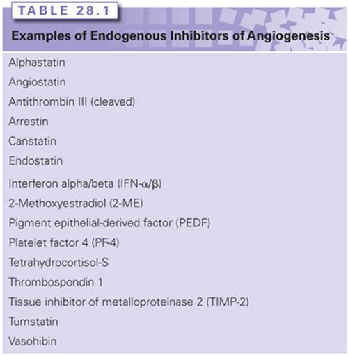
The infrequency of microscopic in situ tumors that actually undergo the angiogenic switch (<1%) suggests that naturally occurring endogenous inhibitors exist in the body to defend against the angiogenic switch in pathologic conditions and to limit physiologic angiogenesis.9 These circulating endogenous inhibitors could also prevent microscopic metastases from growing into visible tumors. Early studies by Langer et al.22,23 demonstrated the possible existence of such inhibitors through the extraction of a functional inhibitor from cartilage, a tissue that is poorly vascularized. Since then, dozens of endogenous angiogenesis inhibitors have been identified, some of which are listed in Table 28.1.17,18,24 Many of the endogenous inhibitors of angiogenesis that have been discovered to date are proteolytically cleaved fragments of larger proteins that are members of either the clotting/coagulation system or members of the extracellular matrix family of glycoproteins. Endostatin is the most well-studied endogenous angiogenesis inhibitor.25,26 Other potent endogenous angiogenesis inhibitors include thrombospondin-127 and tumstatin.28 The discovery of vasohibin, an endogenous inhibitor that is selectively induced in ECs by proangiogenic stimulatory growth factors such as VEGF, demonstrated the existence of an intrinsic and EC-specific feedback inhibitor control mechanism,29,30 whereas most endogenous inhibitors of angiogenesis are extrinsic to ECs. More recently, a second endothelium-produced negative regulator of angiogenesis has been discovered, the Dll4-Notch signaling system.31,32Both intrinsic factors have since been shown to control tumor angiogenesis by an autoregulatory or negative-feedback mechanism. The Dll4-Notch axis has emerged as a critical regulator of tumor angiogenesis, and inhibitors of this pathway (e.g., demcizumab, the anti-Dll4 monoclonal antibody) are currently being investigated in early phase trials of solid tumors.33
Perhaps the most compelling genetic evidence that endogenous inhibitors suppress pathologic angiogenesis was observed in studies using mice deficient in tumstatin, endostatin, or thrombospondin 1 (TSP-1).34 These experiments demonstrate that normal physiologic levels of the inhibitors can retard the tumor growth and that their absence leads to enhanced angiogenesis and increased tumor growth by two- to threefold, strongly suggesting that endogenous inhibitors of angiogenesis can act as endothelium-specific tumor suppressors. The connection between a tumor suppressor protein and angiogenesis is best illustrated by the classic tumor suppressor p53. p53 inhibits angiogenesis by increasing the expression of TSP-135 by repressing VEGF36 and basic fibroblast growth factor–binding protein,37 and by degrading HIF-1,38 which blocks the downstream induction of VEGF expression. New evidence suggests that p53 also indirectly downregulates VEGF expression via the retinoblastoma pathway in a p21-dependent manner during sustained hypoxia.39 Furthermore, p53-mediated inhibition of angiogenesis may also occur in part via the antiangiogenic activity of endostatin and tumstatin.40 This landmark finding clearly demonstrates that p53 not only controls cell proliferation, but can also repress tumor angiogenesis through enzymatic mobilization of these endogenous angiogenesis inhibitor proteins to prevent ECs from being recruited into the dormant, microscopic tumors, thereby preventing the switch to the angiogenic phenotype.41 The discovery that these endogenous angiogenesis inhibitors can suppress the growth of primary tumors raises the possibility that such inhibitors might also be able to slow tumor metastasis. Indeed, the inhibition of angiogenesis by angiostatin significantly reduced the rate of metastatic spread.
DRUG DEVELOPMENT OF ANGIOGENESIS INHIBITORS
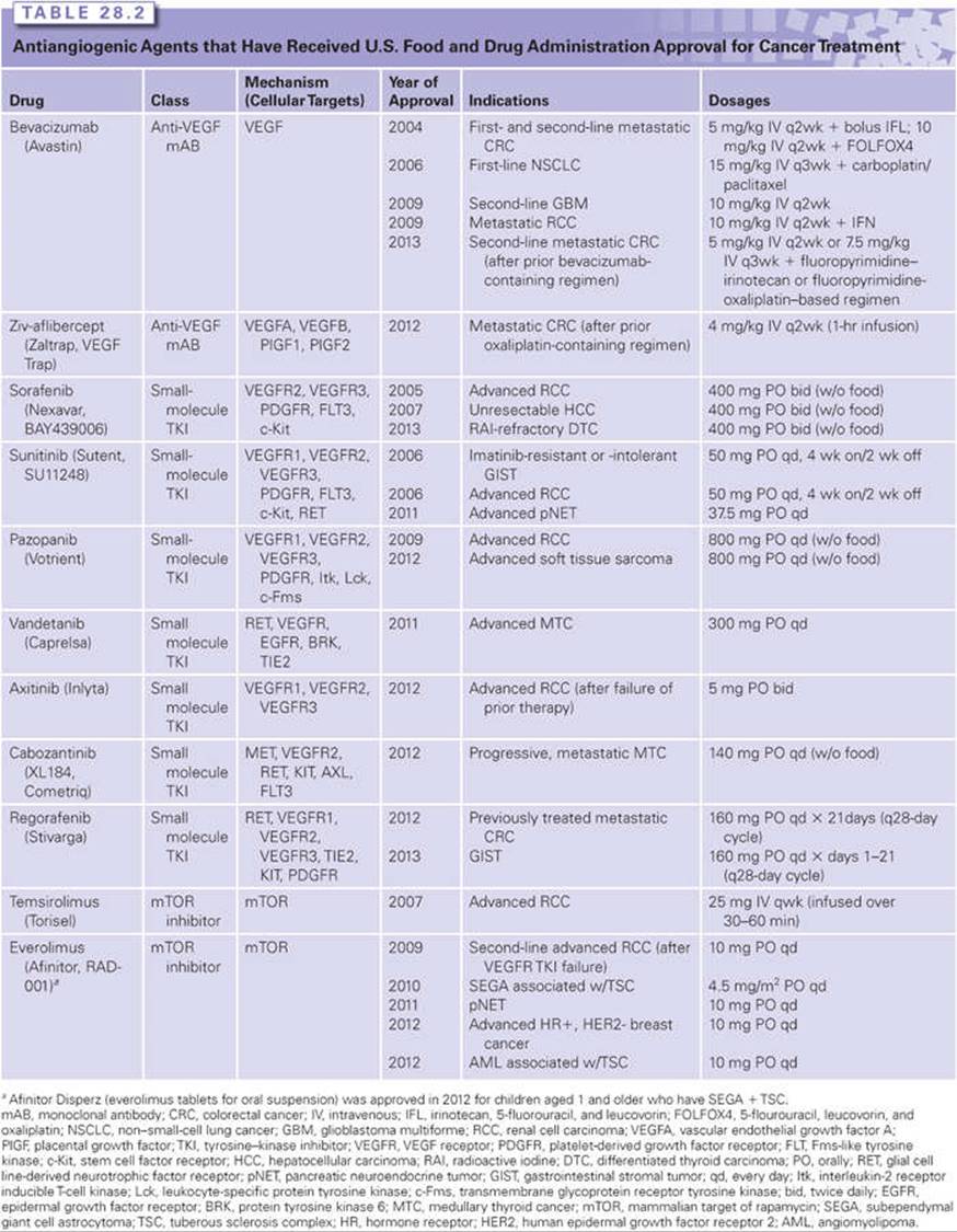
The first angiogenesis inhibitor was reported in 1980 and involved the low-dose administration of interferon α (IFN-α).42–44 Over the next decade, several compounds were discovered to have potent antiangiogenic activity, including protamine and platelet factor 4,45 trahydrocortisol,46 and the fumagillin analog TNP-470.47 The proof of concept that targeting angiogenesis is an effective strategy for treating cancer came with the approval of the first angiogenesis inhibitor, bevacizumab, by the U.S. Food and Drug Administration (FDA). Since then, several antiangiogenic agents have received FDA approval for cancer treatment (Table 28.2), and three additional agents (pegaptanib, ranibizumab, and aflibercept) are approved for the treatment of wet age-related macular degeneration.
Rationale for Antiangiogenic Therapy
Antiangiogenic therapy stems from the fundamental concept that tumor growth, invasion, and metastasis are angiogenesis dependent; thus, blocking blood vessel recruitment to starve primary and metastatic tumors is a rational approach. The microvascular EC recruited by a tumor has become an important second target in cancer therapy. Unlike the cancer cell (the primary target of cytotoxic chemotherapy), which is genetically unstable with unpredictable mutations, the genetic stability of ECs may make them less susceptible to acquired drug resistance.48 Moreover, ECs in the microvascular bed of a tumor may support 50 to 100 tumor cells. Coupling this amplification potential together with the lower toxicity of most angiogenesis inhibitors results in the use of antiangiogenic therapy, which should be significantly less toxic than conventional chemotherapy. However, the variable responses of antiangiogenic therapy observed in different tumor types and the fact that angiogenesis inhibitors have not delivered the benefits initially envisaged suggest that the precise mechanism of action of angiogenesis inhibitors is complex and remains incompletely understood.
Modes of Action of Antiangiogenic Agents
Various strategies for the development of antiangiogenic drugs have been investigated over the years, with these agents being classified into several different categories depending on their modes of action. Some inhibit ECs directly, whereas others inhibit the angiogenesis signaling cascade or block the ability of ECs to break down the extracellular matrix. Inhibitors may block one main angiogenic protein, two or three angiogenic proteins, or have a broad-spectrum effect by blocking a range of angiogenic regulators that can be located in both the tumor and ECs.49 In some cases, the antiangiogenic activity is discovered as a secondary function after the drug has received regulatory approval for a different primary function. For example, bortezomib is a proteasome inhibitor that is approved for multiple myeloma and was later found to possess antiangiogenic activity via inhibiting VEGF. Some small-molecule drugs may display their antiangiogenic activity through inducing the expression of endogenous angiogenesis inhibitors such as celecoxib, a cyclooxygenase-2 (COX-2) inhibitor, which inhibits angiogenesis by increasing levels of endostatin.25
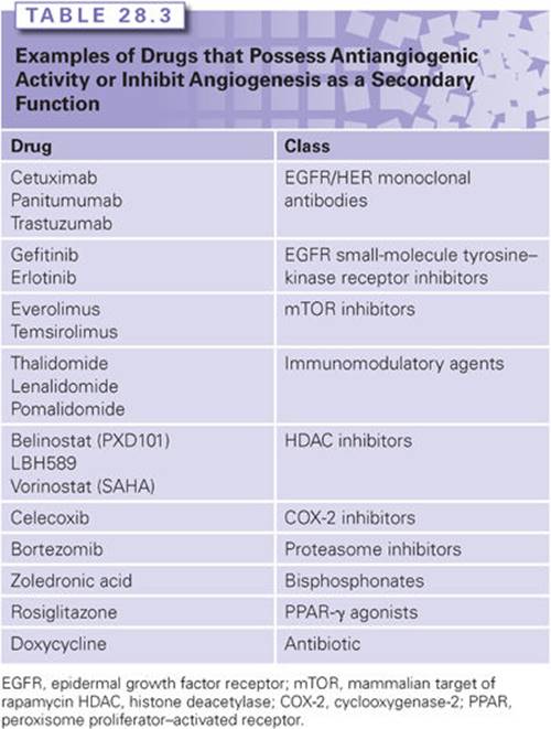
Some drugs possess antiangiogenic properties but with mechanisms that are not completely understood, such as thalidomide and its analogs, lenalidomide and pomalidomide, referred to as immunomodulatory drugs. Thalidomide was originally shown to inhibit angiogenesis by D’Amato et al.50 in 1994 and this was subsequently confirmed in several different in vitro and ex vivo assays.51–54 Interestingly, unlike other mechanisms of action, the antiangiogenic activity of thalidomide is believed to require enzymatic activation. The extent to which the antiangiogenic properties of thalidomide and its analogs play a role in its antimyeloma activity is not clearly understood. Several mechanisms have been proposed that involve the downregulation of cytokines in EC, the inhibition of EC proliferation, the decrease in the level of circulating ECs, or the modulation of adhesion molecules between the multiple myeloma cells and the endogenous bone marrow stromal cells, thereby decreasing the production of VEGF and interleukin 6 (IL-6).55–59 The immunomodulatory agents are discussed in greater detail in another section of this textbook. Examples of the various types of angiogenesis inhibitors are highlighted in Table 28.3.
Drugs with antiangiogenic activity may be classified as either direct or indirect angiogenesis inhibitors. A direct angiogenesis inhibitor blocks vascular ECs from proliferating, migrating, or increasing their survival in response to proangiogenic proteins. They target the activated endothelium directly and inhibit multiple angiogenic proteins. Examples of direct angiogenesis inhibitors include many of the endogenous inhibitors of angiogenesis, such as endostatin, angiostatin, and TSP-1. Indirect angiogenesis inhibitors decrease or block expression of a tumor cell product, neutralize the tumor product itself, or block its receptor on ECs. The limitation to indirect inhibitors is that, over time, tumor cells may acquire mutations that lead to increased expression of other proangiogenic proteins that are not blocked by the indirect inhibitor. This may give the appearance of drug resistance and warrants the addition of a second antiangiogenic agent, one that would target the expression of these upregulated proangiogenic proteins. Examples of drugs that interfere with the angiogenesis-signaling pathway include the anti-VEGF monoclonal antibodies and small-molecule tyrosine–kinase inhibitors. These drugs target the major signaling pathways in tumor angiogenesis: VEGF, PDGF, and their respective receptors, as well as other growth factors and/or signaling pathways.
VEGF (also known as vascular permeability factor) is a potent proangiogenic growth factor and its expression is upregulated by most cancer cell types. It stimulates EC proliferation, migration, and survival as well as induces increased vascular permeability. The different forms of VEGF bind to transmembrane receptor tyrosine kinases (RTK) on ECs: VEGFR1 (Flt-1), VEGFR2 (KDR/Flk-1 or kinase insert domain receptor/fetal liver kinase 1), or VEGFR3 (Flt-4).60 This results in receptor dimerization, activation, and autophosphorylation of the tyrosine–kinase domain, thereby triggering downstream signaling pathways. Other signaling molecules that may represent attractive therapeutic targets include PDGF and the angiopoietins (Ang1, Ang2). PDGF-B/PDGF receptor (R)-β plays an important role in the recruitment of pericytes and maturation of the microvasculature.61 Ang2, which binds the Tie-2 receptor, is mostly expressed in tumor-induced neovasculature, whereby its selective inhibition results in reduced EC proliferation.62 The angiopoietins are also involved in lymphangiogenesis, the formation of new lymphatic vessels, which plays a key role in tumor metastasis. An increased Ang2/Ang1 ratio correlates with tumor angiogenesis and poor prognosis in many cancers, thus making the angiopoietins an attractive therapeutic target. Angiopoietin inhibitors are currently under investigation in the preclinical and clinical setting.
Other strategies for targeting angiogenesis involve the tumor microenvironment. Breakdown of the extracellular matrix is required to allow ECs to migrate into surrounding tissues and proliferate into new blood vessels; thus, drugs that target MMPs, enzymes that catalyze the breakdown of the matrix, can also inhibit angiogenesis. However, clinical development of MMP inhibitors (MMPI) has yielded disappointing results.63–66
Integrins are cell surface adhesion molecules that play an essential role in cell–cell and cell–matrix adhesion as well as in transmitting signals important for cell migration, invasion, proliferation, and survival. The involvement of integrin in tumor angiogenesis was demonstrated in studies that show the β-4 subunit of integrin promoting endothelial migration and invasion.67 Agents that target integrins (inhibitors of αvβ3 and αvβ5) have been evaluated as potential therapeutic options and include etaracizumab, cilengitide, and intetumumab. However, all three integrin inhibitors have proven to be largely ineffective in various early and late stage cancer trials.68–73 In summary, the downstream effects of antiangiogenic agents, in addition to blocking angiogenesis, may involve inducing vessel regression, promoting sensitization to radiotherapy and chemotherapy by depriving ECs of VEGF’s prosurvival signals, and inhibiting the recruitment of proangiogenic bone marrow–derived cells as well as reducing the self-renewal capability of cancer stem cells.74
CLINICAL UTILITY OF APPROVED ANTIANGIOGENIC AGENTS IN CANCER THERAPY
The following section reviews the current FDA-approved angiogenesis inhibitors (Table 28.2). These agents include: (1) the monoclonal anti-VEGF antibodies (bevacizumab and ziv-aflibercept); (2) small-molecule tyrosine–kinase inhibitors (TKI) (sorafenib, sunitinib, pazopanib, vandetanib, axitinib, cabozantinib, and regorafenib); and (3) the mammalian target of rapamycin (mTOR) inhibitors (temsirolimus and everolimus), as examples of drugs that possess antiangiogenic activity. Other approved drugs that also inhibit angiogenesis as a secondary function, such as thalidomide, are discussed in greater detail in another section of this textbook and are presented in Table 28.3.
Anti-VEGF Therapy
Bevacizumab
Bevacizumab is a recombinant humanized anti–VEGF-A monoclonal antibody that received FDA approval in February 2004 for use in combination therapy with fluorouracil-based regimens for metastatic colorectal cancer. Bevacizumab binds VEGF and prevents the interaction of VEGF to its receptors (Flt-1 and KDR) on the surface of ECs. It is the first antiangiogenic agent clinically proven to extend survival following a large, randomized, double-blind, phase III study in which bevacizumab was administered in combination with bolus irinotecan, 5-fluorouracil, and leucovorin (IFL) as first-line therapy for metastatic colorectal cancer (CRC).75 In 2006, its approval extended to first- or second-line treatment of patients with metastatic carcinoma of the colon or rectum. This recommendation is based on the demonstration of a statistically significant improvement in overall survival (OS) in patients receiving bevacizumab plus FOLFOX4 (5-flourouracil, leucovorin, and oxaliplatin) when compared to those receiving FOLFOX4 alone. In January 2013, it was further approved to treat mCRC for second-line treatment when used with fluoropyrimidine-based (combined with irinotecan or oxaliplatin) chemotherapy after disease progression following a first-line treatment with a bevacizumab-containing regimen based on clinical benefits observed in the randomized phase III study (ML18147).76 Despite the benefit in the metastatic setting, the addition of bevacizumab did not improve clinical outcomes in the adjuvant setting in CRC.77,78 In 2006, bevacizumab received an additional approval for use in combination with carboplatin and paclitaxel, and is indicated for first-line treatment of patients with unresectable, locally advanced, recurrent, or metastatic nonsquamous, non–small-cell lung cancer (NSCLC) based on the demonstration of a statistically significant improvement in OS in patients in the bevacizumab arm compared to those receiving chemotherapy alone.79 In February 2008, the FDA granted a conditional, accelerated approval for bevacizumab to be used in combination with paclitaxel for the treatment of patients who have not received chemotherapy for metastatic human epidermal growth factor receptor 2 (HER2)-negative breast cancer. However, additional clinical trials were conducted and the new data showed only a small effect on progression free survival (PFS) without evidence of an improvement in OS or a clinical benefit to patients sufficient to outweigh the risks; thus, the FDA rescinded its approval and removed the breast cancer indication from the drug’s label in November 2011.80–82 This controversial decision continues to be debated with ongoing subgroup analyses to identify patients who would likely benefit from bevacizumab.
Bevacizumab received another accelerated approval as a single agent for patients with glioblastoma multiforme (GBM) with progressive disease following therapy in May 2009. The approval was based on the demonstration of durable objective response rates observed in two single-arm trials, AVF3708g and NCI 06-C-0064E.83 Currently, no data have shown whether bevacizumab improves disease-related symptoms or survival in people previously treated for GBM. Moreover, phase III trials of bevacizumab in newly diagnosed GBM (RTOG 8025 and AVAglio) have shown a 3- to 4-month improvement of PFS, but no OS advantage over the standard of care.84 The AVAglio trial improved patients’ quality of life, whereas the RTOG 0825 did not and instead increased the burden of symptoms with a negative impact on cognition. Although these two studies showed that bevacizumab had a modest benefit as the initial therapy for GBM, it remained effective to treat recurrences where treatment options are limited. In July 2009, bevacizumab was approved for use in combination with IFN-α for the treatment of patients with metastatic renal cell carcinoma (RCC). Results from the AVOREN trial demonstrated a 5-month improvement in median PFS in patients treated with bevacizumab plus IFN-α-2a versus IFN-α-2a plus placebo.85 Another phase III trial (CALGB 90206) of bevacizumab plus IFN-α versus IFN-α monotherapy was conducted in patients with previously untreated, metastatic clear cell RCC. Median PFS was 8.4 months versus 4.9 months in favor of the bevacizumab arm.86 Both studies did not demonstrate a statistically significant advantage in OS.87,88
Clinical studies of bevacizumab in combination with oxaliplatin-containing and 5-fluorouracil–based regimens have shown that combination therapy is well tolerated with toxicity not being substantially greater than that of the chemotherapy alone.89 Side effects included grade 3 hypertension, grade 1 or 2 proteinuria, a slight increase (less than two percentage points) in grade 3 or 4 bleeding, and impaired surgical wound healing in patients who underwent surgery during treatment with bevacizumab. However, potentially life-threatening events (e.g., arterial and venous thromboembolic events, gastrointestinal perforation, hemoptysis, risk of ovarian failure) have occurred in some patients, thus requiring close patient monitoring in individuals who are at greater risk of adverse events.90 In a recent meta-analysis of RCTs, bevacizumab in combination with chemotherapy or biologic therapy, compared with chemotherapy alone, was associated with increased treatment-related mortality.91
Although four phase III randomized studies have demonstrated improvements in PFS for ovarian cancer (OC)—two first-line trials (GOG 218 and ICON7) and two in recurrent OC [platinum-resistant(AURELIA Trial) or platinum-sensitive (OCEANS Trial)]—the role of bevacizumab in OC remains controversial. Bevacizumab is approved for use in combination with chemotherapy in the first- and second-line treatment of advanced OC in Europe, but it is not currently licensed in the United States for this indication. Mature OS data and predictive biomarkers are key to defining the subsets of patients who will most like benefit from this therapy. More recently, a randomized, phase III trial (GOG240) has demonstrated for the first time that bevacizumab can prolong OS and PFS for women with advanced, recurrent, or persistent cervical cancer that was not curable with standard chemotherapy. At the time of writing, there are currently over 400 actively recruiting, ongoing trials investigating the clinical benefits of bevacizumab in combination with chemotherapeutic regimens or as adjuvant therapy in various stages and types of cancer (http://clinicaltrial.gov).
Ziv-aflibercept
Ziv-aflibercept (previously known as aflibercept or VEGFTrap) is a recombinant humanized fusion protein of the extracellular domains of VEGF receptor 1 (VEGFR1) and VEGFR2 with the constant region (Fc) of human immunoglobulin (Ig)G1 that binds to VEGF-A, VEGF-B, PlGF1, and PlGF2, thereby preventing these ligands from binding to and activating their cognate receptors.92 Ziv-aflibercept has a higher VEGF-A binding affinity and more potent blockade of VEGFR1 or VEGFR2 activation than bevacizumab.93 In tumor models, ziv-aflibercept exerts its antiangiogenic effects through regressing tumor vasculature and size, remodeling or normalizing surviving vasculature, and inhibiting ascites formation.94 In August 2012, ziv-aflibercept received regulatory approval for use in combination with 5-fluorouracil, leucovorin, and irinotecan (FOLFIRI) for the treatment of patients with metastatic CRC that is resistant to or that has progressed following treatment with an oxaliplatin-containing regimen. Results from the pivotal phase III VELOUR trial showed that ziv-aflibercept plus FOLFIRI statistically and significantly improved PFS (median PFS, 6.90 versus 4.67 months, respectively), OS (median OS, 13.50 versus 12.06 months, respectively), and overall response rates (19% versus 11.1%, respectively) relative to placebo plus FOLFIRI.95 Toxicities related to ziv-aflibercept were consistent with those expected from the anti-VEGF drug class. The frequency of vascular-related adverse events appeared to be higher with ziv-aflibercept than bevacizumab treatment when compared across trials. Current clinical data are insufficient to directly compare ziv-aflibercept and bevacizumab in the first- or second-line setting for metastatic CRC.
Tyrosine–Kinase Inhibitor Therapy
Sorafenib
Sorafenib is a small-molecule Raf kinase and VEGF receptor kinase (VEGFR2 and VEGFR3) inhibitor. It has been shown to exhibit broad-spectrum effects on multiple targets (PDGF receptor (PDGFR), stem cell factor (c-KIT) receptor, p38) that affect the maintenance of the tumor vasculature and angiogenesis.96 In December 2005, the FDA granted approval for sorafenib, which is considered the first multikinase inhibitor, for the treatment of patients with advanced RCC. Safety and efficacy of sorafenib was proven in the largest randomized phase III study conducted in advanced RCC that showed prolong PFS in favor of sorafenib.97,98 In November 2007, sorafenib was approved for the treatment of patients with unresectable hepatocellular carcinoma (HCC) based on the study results in patients with advanced HCC who had not received previous systemic treatment. Median survival and the time to radiologic progression were nearly 3 months longer for patients treated with sorafenib than for those given placebo.99 In November 2013, sorafenib received a new indication under the FDA’s priority review program for the treatment of locally recurrent or metastatic, progressive differentiated thyroid carcinoma (DTC) refractory to radioactive iodine (RAI) treatment based on positive results from the phase III DECISION trial. Treatment with sorafenib improved PFS (the primary endpoint of the trial) by 41% compared with placebo (10.8 versus 5.8 months, respectively; hazard ratio [HR], 0.587, 95% confidence interval [CI] [0.454 to 0.758]; p <0.0001).100 The overall response rates were 12% for patients who received sorafenib versus 1% for the placebo arm. Although only about 5% to 15% of thyroid cancer patients become refractory to RAI, no standard treatments are available and, thus, sorafenib is the first agent specifically approved for RAI-resistant DTC. Sorafenib was generally well tolerated with a predictable safety profile. Common adverse events include diarrhea, rash/desquamation, fatigue, hand–foot skin reaction, alopecia, and nausea/vomiting. Grade 3/4 adverse events were 38% for sorafenib versus 28% for placebo. Sorafenib-induced hypertension occurred in patients with metastatic RCC. The treatment-related hypertension was noted to be a class effect observed not only with VEGFR inhibitors, but also with the VEGF monoclonal antibody as well.90 No significant relationship between previously described mediators of blood pressure and the magnitude of increase was found in a study evaluating the mechanism of sorafenib-induced hypertension in patients.101
Sunitinib
Sunitinib (SU11248) is a small-molecule, multitargeted TKI that exhibits potent antitumor and antiangiogenic activity and inhibits VEGFR-1, -2, -3, c-KIT, PDGFR; FLT-3; colony-stimulating factor receptor type 1 receptor; and the glial cell line–derived neurotrophic factor receptor. It was rationally designed and chosen for its high bioavailability and its nanomolar-range potency against the antiangiogenic RTKs. Sunitinib received its first U.S. regulatory approval in 2006 for the treatment of gastrointestinal stromal tumor (GIST) after disease progression on, or intolerance to, imatinib and accelerated approval for the treatment of advanced RCC.102 Sunitinib demonstrated significant efficacy (prolonged median time to progression) in imatinib-resistant or -intolerant GIST in a randomized phase III trial.103 The accelerated approval for RCC was based on durable partial responses, with a response rate of 26% to 37%, and a median duration of response of 54 weeks from two phase II, single-arm trials of patients with cytokine-refractory RCC.104 The accelerated approval was converted to regular approval in 2007 following confirmation of an improvement in PFS and OS in a phase III trial of sunitinib for first-line treatment of patients with treatment-naïve, metastatic RCC.105,106 In May 2011, the drug received a new indication for the treatment of progressive, well-differentiated pancreatic neuroendocrine tumors (pNET) in patients with unresectable, locally advanced, or metastatic disease. The randomized phase III trial was discontinued early after the independent data monitoring committee observed more serious adverse events and deaths in the placebo group as well as a difference in PFS favoring sunitinib. The median PFS for patients treated with sunitinib was 10.2 months, compared with 5.4 months for patients treated with placebo (HR, 0.427, 95% CI, 0.271 to 0.673], p <0.001).107 Common adverse effects, including diarrhea, mucositis, asthenia, skin abnormalities, and altered taste, were more common in patients receiving sunitinib. In addition, a decrease in left ventricular ejection fraction and severe hypertension were also more commonly reported in the sunitinib arm. Grade 3 or 4 treatment-emergent adverse events were reported in 56% versus 51% of patients on sunitinib versus placebo, respectively.
Pazopanib
Pazopanib is a second-generation, multitargeted TKI that binds to VEGFR-1, -2, -3, PDGFR-α and -β, c-KIT, and several other key proteins responsible for angiogenesis, tumor growth, and cell survival. Pazopanib exhibited in vivo and in vitro activity against tumor growth, and early clinical trials demonstrated potent antitumor and antiangiogenic activity.108 A phase III clinical trial in treatment-naïve and cytokine-pretreated patients with advanced and/or metastatic RCC showed a significant improvement in PFS and tumor response compared with placebo,109 leading to the approval of pazopanib in the United States in October 2009. A recent, randomized phase III trial (COMPARZ) compared the efficacy and safety of pazopanib and sunitinib as first-line therapy involving patients with metastatic RCC and demonstrated that both pazopanib and sunitinib have similar efficacy, but the safety and quality-of-life profiles favor pazopanib.110 In April 2012, pazopanib was approved for the treatment of patients with metastatic nonadipocytic soft tissue sarcoma who have received prior chemotherapy following a phase III trial that demonstrated a statistically significant improvement in PFS. The median PFS was 4.6 months for patients receiving pazopanib versus 1.6 months for the placebo arm.111 The drug is generally well tolerated, with the most common adverse events being diarrhea, fatigue, anorexia, hypertension, and hair depigmentation, as well as laboratory abnormalities in elevated aspartate aminotransferase and alanine aminotransferase. Pazopanib has shown clinical activity in a variety of tumors, including breast cancer, thyroid cancer, HCC, and cervical cancer.112 Ongoing phase II and III trials are further evaluating pazopanib in these malignancies.
Vandetanib
Vandetanib is an oral, small-molecule TKI that inhibits the activity of RET kinase, VEGFR, epidermal growth factor receptor (EGFR), protein tyrosine kinase 6 (BRK), TIE2, members of the ephrin (EPH) receptors kinase family, and members of the Src family of tyrosine kinases.113 Vandetanib reduced endothelial cell migration, proliferation, survival, and angiogenesis in vitro, and it decreased tumor vessel permeability and inhibited tumor growth and metastasis in vivo. In April 2011, vandetanib received U.S. regulatory approval for the treatment of symptomatic or progressive medullary thyroid cancer (MTC) in patients with unresectable, locally advanced, or metastatic disease. Until the approval of vandetanib, no systemic therapy was approved for the treatment of unresectable MTC, making it the first molecularly targeted agent approved for this disease. Results of a randomized phase III trial of patients with unresectable, locally advanced, or metastatic MTC demonstrated statistically significant and clinically meaningful improvements in PFS for vandetanib compared with placebo (HR, 0.46; 95% CI, 0.31 to 0.69; p <0.001).114Common grade 3 and 4 toxicities (>5%) were diarrhea and/or colitis, hypertension and hypertensive crisis, fatigue, hypocalcemia, rash, and corrected QT interval (QTc) prolongation. Given the toxicity profile, which includes QTc prolongation and sudden death, vandetanib is only available through a restricted distribution program. Vandetanib is also the first targeted drug to show evidence of efficacy in a randomized phase II trial in patients with locally advanced or metastatic differentiated thyroid carcinoma,115 and a phase III trial is currently underway. Early phase studies are also being conducted in solid tumors, including GIST and kidney and pancreatic cancers.
Axitinib
Axitinib is a potent and selective second-generation inhibitor of VEGFR-1, -2, and -3. The in vitro half-maximal inhibitory concentration (IC50) of axitinib is 10-fold lower for the VEGF family of receptors than for other TKIs such as pazopanib, sunitinib, or sorafenib.116 In January 2012, axitinib received approval for the treatment of advanced RCC after the failure of one prior systemic therapy based on a phase III trial (AXIS) comparing the efficacy and safety of axitinib versus sorafenib as a second-line treatment for metastatic RCC.117,118 The median PFS was 6.7 months with axitinib compared to 4.7 months with sorafenib (HR, 0.67; 95% CI, 0.54, 0.81; one-sided p <0.0001). This improvement in PFS was greater in the cytokine-pretreated subgroup in comparison with the sunitinib-pretreated subgroup. The most frequent adverse events with axitinib were diarrhea (all grade), hypertension (all grade), fatigue, decreased appetite, nausea, and dysphonia. Moreover, hypertension, nausea, dysphonia, and hypothyroidism were more common with axitinib, whereas palmar–plantar erythrodysesthesia, alopecia, and rash were more frequent with sorafenib. A phase III trial (AGILE) comparing axitinib with sorafenib as first-line therapy in patients with treatment-naïve metastatic RCC demonstrated no significant difference in median PFS between patients treated with axitinib or sorafenib.119 Additionally, axitinib is being studied as a single agent as well as in combination with chemotherapy across several tumor types including HCC, NSCLC, and pancreatic and thyroid cancers.
Cabozantinib
Cabozantinib (XL184) is a small-molecule TKI with potent activity toward the MET receptor and VEGFR2, as well as a number of other receptor tyrosine kinases, including RET, KIT, AXL, and FLT-3. MET is the only known receptor for hepatocyte growth factor (HGF), and its signaling activity plays a key role in tumorigenic growth, metastasis, and therapeutic resistance. The dysregulated expression and/or activation of MET and HGF have been implicated in the development of numerous human cancers including glioma; melanoma; and hepatocellular, renal, gastric, pancreatic, prostate, ovarian, breast, and lung cancers, and is often correlated with poor prognosis.120 Recent studies have determined that the MET pathway plays an important role in the development of resistance to VEGF pathway inhibition and that the use of VEGFR inhibitors, such as sunitinib, sorafenib, or a VEGFR2-targeting antibody, can result in the development of an aggressive tumor phenotype characterized by increased invasiveness and metastasis.121–123 Thus, there is an advantage to targeting both the MET and VEGF pathways to disrupt angiogenesis, tumorigenesis, and cancer progression. In November 2012, cabozantinib received U.S. regulatory approval for progressive metastatic MTC based on the phase III trial that demonstrated a statistically significant PFS prolongation for the cabozantinib-treatment arm.124 The estimated median PFS was 11.2 months for cabozantinib versus 4.0 months for placebo (HR, 0.28; 95% CI, 0.19 to 0.40; p <0.001). Manageable toxicities included diarrhea, palmar–plantar erythrodysesthesia, decreased weight and appetite, nausea, and fatigue. Cabozantinib has been effective against several solid cancers, including MTC, breast, NSCLC, melanoma, and liver cancer, and is currently being studied in clinical trials in a number of tumor types, with the most significant results observed in the reduction of bone metastatic lesions in castration-resistant prostate cancer.125
Regorafenib
Regorafenib is a small-molecule TKI of multiple membrane-bound and intracellular kinases including RET, VEGFR1, VEGFR2, VEGFR3, KIT, PDGFR-α, PDGFR-β, FGFR1, FGFR2, TIE2, DDR2, TrkA, Eph2A, RAF-1, BRAF, BRAFV600E, SAPK2, PTK5, and Abl pathways.126 Regorafenib is structurally related to sorafenib and differs from the latter by the presence of a fluorine atom in the center phenyl ring, resulting in higher inhibitory potency against various proangiogenic receptors than sorafenib, including VEGFR2 and FGFR1. In September 2012, regorafenib was approved for the treatment of patients with mCRC who have been previously treated with fluoropyrimidine-, oxaliplatin-, and irinotecan-based chemotherapy, with an anti-VEGF therapy, and if KRAS wild type, with an anti-EGFR therapy. The phase III CORRECT trial that resulted in approval of the drug demonstrated a median OS of 6.4 months in the regorafenib group versus 5.0 months in the placebo group (HR, 0.77; 95% CI, 0.64 to 0.94; one-sided p = 0.0052).127 Regorafenib is the first TKI with survival benefits in mCRC that has progressed after all standard therapies. In February 2013, it received another indication for the treatment of patients with locally advanced, unresectable, or metastatic GIST who have been previously treated with imatinib and sunitinib. This was based on positive findings of the phase III GRID trial that demonstrated a median PFS of 4.8 months for regorafenib and 0.9 months for placebo (HR, 0.27, 95% CI, 0.19 to 0.39; p <0.0001).128 In both studies, regorafenib provided significant improvements in PFS to highly refractory patient populations who have progressed on standard treatments. The most common adverse events that were grade 3 or higher and related to regorafenib were hand–foot skin reaction, fatigue, diarrhea, hypertension, and rash or desquamation. Its clinical development as a single agent or in combination with standard chemotherapeutic agents in various malignant tumors is ongoing and includes a phase III trial in patients with HCC whose disease has progressed after treatment with sorafenib.
mTOR Inhibitors
The mTOR pathway is a central component of the PI3K/Akt signaling pathway and a regulator of many biologic processes that are essential for angiogenesis, cell proliferation, and metabolism.129 Inhibition of the mTOR kinase prevents downstream signaling via the Akt pathway, resulting in inhibition of protein translation and cell growth. mTOR plays a key role in angiogenesis and specifically regulates the expression of HIF-1, which is upregulated by the loss of the von Hippel–Lindau gene in RCC. In May 2007, temsirolimus was approved for the treatment of advanced RCC. Efficacy and safety were demonstrated in a phase III study in previously untreated patients (n = 626) with poor risk features of metastatic RCC assigned to one of three treatment arms: IFN-α alone, temsirolimus 25 mg alone, or the combination of temsirolimus (15 mg) and IFN-α.130 Single-agent temsirolimus was associated with a statistically significant improvement in OS when compared with IFN; the addition of temsirolimus to IFN did not improve OS. The results of the phase III INTORSECT trial compared the efficacy of temsirolimus and sorafenib in the second-line treatment of metastatic RCC after disease progression on sunitinib demonstrated that temsirolimus did not improve survival over sorafenib in the second-line setting.131 The significant OS difference in favor of sorafenib (stratified HR, 1.31; 95% CI, 1.05 to 1.63; two-sided p = 0.01) suggested that VEGFR inhibition may be a better option than mTOR inhibitors for patients progressing on sunitinib. The most common adverse reactions that occurred were rash, asthenia, mucositis, nausea, edema, and anorexia. Rare, but serious adverse reactions associated with temsirolimus included interstitial lung disease, bowel perforation, and acute renal failure.
Everolimus (RAD001) was approved in March 2009 for patients with advanced RCC whose disease had progressed on VEGFR-targeted therapy (sunitinib or sorafenib). Efficacy was demonstrated in a phase 3 trial that study met its primary endpoint with a median PFS of 4.9 and 1.9 months in the everolimus and placebo arms, respectively (HR, 0.33; p <0.0001).132 Everolimus is also indicated for subependymal giant cell astrocytoma (SEGA) associated with tuberous sclerosis complex (TSC), renal angiomyolipoma with TSC, progressive neuroendocrine tumors of pancreatic origin, and advanced hormone receptor-positive, HER2-negative breast cancer in combination with exemestane.133 The most common adverse reactions were stomatitis, infections, asthenia, fatigue, cough, and diarrhea. The most common grade 3/4 adverse reactions were infections, dyspnea, fatigue, stomatitis, dehydration, pneumonitis, abdominal pain, and asthenia. Both temsirolimus and everolimus are currently being evaluated in phase I through III studies of various cancer types. By downregulating HIF-1 in the tumor cell, mTOR inhibitors may complement the effects of TKIs at the level of the EC; thus, the combination of mTOR inhibitors with other targeted agents such as bevacizumab or sorafenib/sunitinib are also being investigated.
On the Horizon: Anti-VEGFR2 Monoclonal Antibody
Ramucirumab (IMC-1121B) is a fully human IgG1 monoclonal antibody that binds with high affinity to the extracellular VEGF-binding domain of VEGFR-2. In a phase III trial (REGARD), ramucirumab monotherapy conferred a statistically significant benefit in OS and PFS compared to placebo in patients with advanced gastric or gastroesophageal junction adenocarcinoma in the second-line setting with an acceptable safety profile.134 The survival advantage is the first to be elicited by a single-agent biologic treatment in this setting and, based on these findings, the FDA has assigned a priority review designation for ramucirumab. An ongoing phase III trial (RAINBOW) of ramucirumab in combination with chemotherapy as second-line treatment for patients with advanced gastric cancer is currently underway, and preliminary results demonstrated the trial met both its primary (OS) and secondary (PFS) endpoints. In April 2014, the U.S. FDA approved ramucirumab for use as a single agent for the treatment of patients with advanced or metastatic, gastric or gastroesophageal junction adenocarcinoma with disease progression on or after prior treatment with fluoropyrimidine- or platinum-containing chemotherapy. The recommended ramucirumab dose and schedule is 8 mg/kg administered as a 60-minute intravenous infusion every 2 weeks. The drug also marginally improved survival in the second-line treatment of NSCLC in an ongoing phase III (REVEL) trial.
COMBINATION THERAPIES
Tumor angiogenesis is a highly complex process involving multiple growth factors and their receptor signaling pathways. Based on current evidence, with a few exceptions, effective therapy will probably rely on a combinatorial approach that involves targeting multiple pathways simultaneously. However, a recent study has demonstrated that simultaneous inhibition of the VEGF and EGF pathways in combination with chemotherapy shortens rather than prolongs PFS as compared to inhibition of the VEGF pathway alone in combination with chemotherapy.135 Whether other targeted agents exhibit beneficial effects when combined with VEGF inhibitors remains to be investigated. Moreover, a number of studies have shown that antiangiogenic agents in combination with chemotherapy or radiotherapy result in additive or synergistic effects. Several models have been proposed to explain the mechanism responsible for this potentiation, keying in on the chemosensitizing effects of antiangiogenic therapy.136 One hypothesis is that antiangiogenic therapy may normalize the tumor vasculature, thus resulting in improved oxygenation, better blood perfusion, and consequently, improved delivery of chemotherapeutic drugs.137 A second model suggests that chemotherapy delivered at low doses and at close, regular intervals with no extended drug-free break periods preferentially damages ECs in the tumor neovasculature.138,139 and suppresses circulating endothelial progenitor cells.140,141 This regimen, also called metronomic chemotherapy, sustains antiangiogenic activity and reduces acute toxicity.142 Thus, the efficacy of metronomic chemotherapy may increase when administered in combination with specific antiangiogenic drugs. Another model addresses the use of antiangiogenic drugs to slow down tumor cell repopulation between successive cycles of cytotoxic chemotherapy.143 This model underscores the importance of timing and sequence in achieving the maximal therapeutic benefit from combination therapies. In fact, a preclinical study in murine tumor models demonstrated that the administration of sunitinib markedly reduced chemotherapy-induced bone marrow toxicity, suggesting that the sequential treatment regimen (delivery of antiangiogenics followed by chemotherapy) showed superior survival benefits compared with the simultaneous administration of two drugs.144 Finally, other mechanisms that might also contribute to the synergism include angiogenesis inhibitor–induced tumor blood vessel regression, the prevention of tumor coopting of vessels from surrounding healthy tissues, and the formation of abnormal vessels in the tumor microenvironment.145 Nevertheless, it remains a challenge to determine why bevacizumab has proved largely ineffective as a single agent, whereas VEGF RTK inhibitors have repeatedly failed in randomized phase III trials when used in combination with chemotherapy. Furthermore, an additional challenge is to determine the optimal dose and duration of antiangiogenic drugs as well as the impact of drug sequencing in combination regimens. Studies are warranted to delineate the discrepancy of bevacizumab’s efficacy in the macrometastatic versus micrometastatic disease settings.146,147
BIOMARKERS OF ANTIANGIOGENIC THERAPY
Antiangiogenic therapy has created a need to develop effective biomarkers to assess the activity of these inhibitors. Biomarkers of tumor angiogenesis activity are important to guide clinical development of these agents and to select patients most likely to benefit from this approach. Although there are currently no validated biomarkers for clinically assessing the efficacy of or selecting patients who will respond to antiangiogenic therapies, a number of candidate markers, including tissue, imaging, and circulating biomarkers, are emerging that need to be prospectively validated.148,149 Several avenues are currently being investigated and include tumor biopsy analysis, microvessel density, noninvasive vascular imaging modalities (positron-emission tomography, dynamic contrast-enhanced magnetic resonance imaging), and measuring circulating biomarkers (levels of angiogenic factors in serum, plasma, urine, or circulating ECs and their precursors).150–152 Recent research efforts have focused on identifying genetic and toxicity biomarkers to predict which patients will benefit from anti-VEGF/VEGFR therapy and identify patients at risk of adverse events. The existence of VEGF single-nucleotide polymorphisms (SNP) and their association with clinical outcomes may be predictive of patient response to bevacizumab. A recent study identified a locus in VEGFR1 that correlated with increased VEGFR1 expression and poor bevacizumab treatment outcomes.153 Moreover, a breast cancer study (E2100) reported the VEGF-2578 AA and VEGF-1154 AA genotypes predicted an improved median OS, whereas the VEGF-634 CC and VEGF-1498 TT genotypes predicted protection from grade 3/4 hypertension in the combination-treatment arm.154 The degree of hypertension can serve as a predictive biomarker of survival in patients after bevacizumab or TKI treatment. Although an association between hypertension and anti-VEGF therapy has been described, the clinical implications of this association and the predictive value of hypertension remains to be validated prospectively. A retrospective analysis of hypertension and efficacy outcomes was conducted in seven large phase III trials (n = 6,486 patients) and, in six of seven studies, early treatment-related blood pressure increase was neither predictive of clinical benefit from bevacizumab nor prognostic for the course of the disease.155 However, one study (AVF2107g) showed early increased blood pressure was associated with longer PFS and OS. Because genetics play a significant role in modifying the risk of hypertension,156 it remains to be determined whether polymorphisms in the VEGF/VEGFR pathway may function as potential biomarkers to predict the association between treatment-related hypertension and response to anti-VEGF therapy, as previously implicated in the E2100 trial.154 Other biomarkers of response include elevated VEGF and placental growth factor levels,148,152 whereas biomarkers of resistance, including circulating basic fibroblast growth factor, stromal cell-derived factor 1α, and viable circulating endothelial cells, increased when tumors escaped treatment.157 A first prospective biomarker study (MERiDiAN) in metastatic breast cancer is currently underway to evaluate the impact of bevacizumab in patients stratified for plasma short VEGF-A isoforms. If validated, these findings could help identify which subgroup of patients should receive antiangiogenic therapy and could lead the way to possible future tailoring of individualized antiangiogenic therapy.
RESISTANCE TO ANTIANGIOGENIC THERAPY
Despite a decade of trials with angiogenesis inhibitors, clinical experience reveals that VEGF-targeted therapy often prolongs the survival of cancer patients by only months because tumors elicit evasive resistance.145,158 Resistance to VEGF inhibitors may be observed in late-stage tumors when tumors regrow during treatment after an initial period of growth suppression from these antiangiogenic agents. This resistance involves the reactivation of tumor angiogenesis and increased expression of other proangiogenic factors. As the disease progresses, it is possible that redundant pathways might be implicated, with VEGF being replaced by other angiogenic pathways, warranting the addition of a second angiogenesis inhibitor that would target these secondary growth factors and/or their activated receptor pathways, or the use of a multitargeted TKI antiangiogenic drug (e.g., sunitinib, sorafenib). However, resistance to these drugs eventually occurs, implicating the existence of additional pathways mediating resistance to antiangiogenic therapies. Moreover, tumor cells bearing genetic alterations of the p53 gene may display a lower apoptosis rate under hypoxic conditions, which might reduce their reliance on vascular supply and, therefore, their responsiveness to antiangiogenic therapy.159 The selection and overgrowth of tumor-variant cells that are hypoxia resistant and, thus, less dependent159 on angiogenesis and vasculature remodeling, resulting in vessel stabilization,160 could also explain the resistance to antiangiogenic drugs. Other possible mechanisms for acquired resistance include tumor vessels becoming less sensitive to antiangiogenic agents, tumor regrowth via rebound revascularization, and vessel cooption.161–166 Perhaps one of the most intriguing findings is that, although ECs are assumed to be genetically stable, they may under some circumstances harbor genetic abnormalities and thus acquire resistance as well.167,168
Recent studies report that VEGF-targeted therapies not only induce primary tumor shrinkage and inhibit tumor progression, but can also initiate mechanisms that increase malignancy to promote tumor invasiveness and metastasis.122,123,169 These mechanisms of resistance to antiangiogenic therapy involve tumor- and host-mediated pathways and may allow for differential efficacy in different stages of disease progression.163 Specifically, antiangiogenic drug–resistance mechanisms involve pathways mediated by the tumor, whether intrinsic or acquired in response to therapy or by the host, which is either responding directly to therapy or indirectly to tumoral cues. Taken together, antiangiogenic therapy can enhance tumor invasiveness and metastasis to facilitate and/or accelerate disease in microscopic tumors and, hence, reduce OS benefit. Understanding the mechanisms of resistance, whether intrinsic or acquired, after exposure to antiangiogenic drug treatment is essential for developing strategies that will allow for optimal exploitation of VEGF inhibitors. It is equally important to identify biomarkers of drug resistance and factors mediating this resistance because the development of reliable biomarkers can be invaluable to monitor the development of evasive resistance to angiogenesis inhibitors.
REFERENCES
1. Hanahan D, Weinberg RA. Hallmarks of cancer: the next generation. Cell 2011;144:646–674.
2. Ide AG, Baker NH, Warren SL. Vascularization of the Brown Pearce rabbit epithelioma transplant as seen in the transparent ear chamger. Am J Roentgenol 1939;42:891–899.
3. Algire GH, Chalkley HW, Legallais FY, et al. Vascular reactions of normal and malignant tissues in vivo. I. Vascular reactions of mice to wounds and to normal and neoplastic transplants. J Natl Cancer Inst 1945;6:73–85.
4. Folkman J, Merler E, Abernathy C, et al. Isolation of a tumor factor responsible for angiogenesis. J Exp Med 1971;133:275–288.
5. Folkman J. Tumor angiogenesis: therapeutic implications. N Engl J Med 1971;285:1182–1186.
6. Papetti M, Herman IM. Mechanisms of normal and tumor-derived angiogenesis. Am J Physiol Cell Physiol 2002;282:C947–C970.
7. Hanahan D, Folkman J. Patterns and emerging mechanisms of the angiogenic switch during tumorigenesis. Cell 1996;86:353–364.
8. Black WC, Welch HG. Advances in diagnostic imaging and overestimations of disease prevalence and the benefits of therapy. N Engl J Med 1993;328:1237–1243.
9. Folkman J, Kalluri R. Cancer without disease. Nature 2004;427:787.
10. Weidner N, Semple JP, Welch WR, et al. Tumor angiogenesis and metastasis—correlation in invasive breast carcinoma. N Engl J Med 1991;324:1–8.
11. Holmgren L, O’Reilly MS, Folkman J. Dormancy of micrometastases: balanced proliferation and apoptosis in the presence of angiogenesis suppression. Nat Med 1995;1:149–153.
12. Naumov GN, Bender E, Zurakowski D, et al. A model of human tumor dormancy: an angiogenic switch from the nonangiogenic phenotype. J Natl Cancer Inst 2006;98:316–325.
13. Udagawa T, Fernandez A, Achilles EG, et al. Persistence of microscopic human cancers in mice: alterations in the angiogenic balance accompanies loss of tumor dormancy. Faseb J 2002;16:1361–1370.
14. Holash J, Maisonpierre PC, Compton D, et al. Vessel cooption, regression, and growth in tumors mediated by angiopoietins and VEGF. Science 1999;284:1994–1998.
15. Relf M, LeJeune S, Scott PA, et al. Expression of the angiogenic factors vascular endothelial cell growth factor, acidic and basic fibroblast growth factor, tumor growth factor beta-1, platelet-derived endothelial cell growth factor, placenta growth factor, and pleiotrophin in human primary breast cancer and its relation to angiogenesis. Cancer Res 1997;57:963–969.
16. Carmeliet P, Dor Y, Herbert JM, et al. Role of HIF-1alpha in hypoxia-mediated apoptosis, cell proliferation and tumour angiogenesis. Nature 1998;394:485–490.
17. Folkman J. Endogenous angiogenesis inhibitors. Apmis 2004;112:496–507.
18. Nyberg P, Xie L, Kalluri R. Endogenous inhibitors of angiogenesis. Cancer Res 2005;65:3967–3979.
19. Rak J, Yu JL. Oncogenes and tumor angiogenesis: the question of vascular “supply” and vascular “demand”. Semin Cancer Biol 2004;14:93–104.
20. Bottos A, Bardelli A. Oncogenes and angiogenesis: a way to personalize anti-angiogenic therapy? Cell Mol Life Sci 2013;70:4131–4140.
21. Rak J, Yu JL, Klement G, et al. Oncogenes and angiogenesis: signaling three-dimensional tumor growth. J Investig Dermatol Symp Proc 2000;5:24–33.
22. Langer R, Brem H, Falterman K, et al. Isolations of a cartilage factor that inhibits tumor neovascularization. Science 1976;193:70–72.
23. Langer R, Conn H, Vacanti J, et al. Control of tumor growth in animals by infusion of an angiogenesis inhibitor. Proc Natl Acad Sci U S A 1980;77:4331–4335.
24. Ribatti D. Endogenous inhibitors of angiogenesis: a historical review. Leuk Res 2009;33:638–644.
25. Folkman J. Antiangiogenesis in cancer therapy—endostatin and its mechanisms of action. Exp Cell Res 2006;312:594–607.
26. Karamouzis MV, Moschos SJ. The use of endostatin in the treatment of solid tumors. Expert Opin Biol Ther 2009;9:641–648.
27. Lawler J. Thrombospondin-1 as an endogenous inhibitor of angiogenesis and tumor growth. J Cell Mol Med 2002;6:1–12.
28. Maeshima Y, Manfredi M, Reimer C, et al. Identification of the anti-angiogenic site within vascular basement membrane-derived tumstatin. J Biol Chem 2001;276:15240–15248.
29. Kerbel RS. Vasohibin: the feedback on a new inhibitor of angiogenesis. J Clin Invest 2004;114:884–886.
30. Sato Y. The vasohibin family: a novel family for angiogenesis regulation. J Biochem 2013;153:5–11.
31. Noguera-Troise I, Daly C, Papadopoulos NJ, et al. Blockade of Dll4 inhibits tumour growth by promoting non-productive angiogenesis. Nature 2006;444:1032–1037.
32. Ridgway J, Zhang G, Wu Y, et al. Inhibition of Dll4 signalling inhibits tumour growth by deregulating angiogenesis. Nature 2006;444:1083–1087.
33. Kuhnert F, Kirshner JR, Thurston G. Dll4-Notch signaling as a therapeutic target in tumor angiogenesis. Vasc Cell 2011;3:20.
34. Sund M, Hamano Y, Sugimoto H, et al. Function of endogenous inhibitors of angiogenesis as endothelium-specific tumor suppressors. Proc Natl Acad Sci U S A 2005;102:2934–2939.
35. Dameron KM, Volpert OV, Tainsky MA, et al. Control of angiogenesis in fibroblasts by p53 regulation of thrombospondin-1. Science 1994;265:1582–1584.
36. Zhang L, Yu D, Hu M, et al. Wild-type p53 suppresses angiogenesis in human leiomyosarcoma and synovial sarcoma by transcriptional suppression of vascular endothelial growth factor expression. Cancer Res 2000;60:3655–3661.
37. Sherif ZA, Nakai S, Pirollo KF, et al. Downmodulation of bFGF-binding protein expression following restoration of p53 function. Cancer Gene Ther 2001;8:771–782.
38. Ravi R, Mookerjee B, Bhujwalla ZM, et al. Regulation of tumor angiogenesis by p53-induced degradation of hypoxia-inducible factor 1alpha. Genes Dev 2000;14:34–44.
39. Farhang Ghahremani M, Goossens S, Nittner D, et al. p53 promotes VEGF expression and angiogenesis in the absence of an intact p21-Rb pathway. Cell Death Differ 2013;20:888–897.
40. Teodoro JG, Parker AE, Zhu X, et al. p53-mediated inhibition of angiogenesis through up-regulation of a collagen prolyl hydroxylase. Science 2006;313:968–971.
41. Folkman J. Tumor suppression by p53 is mediated in part by the antiangiogenic activity of endostatin and tumstatin. Sci STKE 2006;2006:pe35.
42. Brouty-Boye D, Zetter BR. Inhibition of cell motility by interferon. Science 1980;208:516–518.
43. Dvorak HF, Gresser I. Microvascular injury in pathogenesis of interferon-induced necrosis of subcutaneous tumors in mice. J Natl Cancer Inst 1989;81:497–502.
44. Sidky YA, Borden EC. Inhibition of angiogenesis by interferons: effects on tumor- and lymphocyte-induced vascular responses. Cancer Res 1987;47:5155–5161.
45. Taylor S, Folkman J. Protamine is an inhibitor of angiogenesis. Nature 1982;297:307–312.
46. Crum R, Szabo S, Folkman J. A new class of steroids inhibits angiogenesis in the presence of heparin or a heparin fragment. Science 1985;230:1375–1378.
47. Ingber D, Fujita T, Kishimoto S, et al. Synthetic analogues of fumagillin that inhibit angiogenesis and suppress tumour growth. Nature 1990;348:555–557.
48. Kerbel RS. Inhibition of tumor angiogenesis as a strategy to circumvent acquired resistance to anti-cancer therapeutic agents. Bioessays 1991;13:31–36.
49. Folkman J. Angiogenesis: an organizing principle for drug discovery? Nat Rev Drug Discov 2007;6:273–286.
50. D’Amato RJ, Loughnan MS, Flynn E, et al. Thalidomide is an inhibitor of angiogenesis. Proc Natl Acad Sci U S A 1994;91:4082–4085.
51. Bauer KS, Dixon SC, Figg WD. Inhibition of angiogenesis by thalidomide requires metabolic activation, which is species-dependent. Biochem Pharmacol 1998;55:1827–1834.
52. Figg WD. The 2005 Leon I. Goldberg Young Investigator Award Lecture: development of thalidomide as an angiogenesis inhibitor for the treatment of androgen-independent prostate cancer. Clin Pharmacol Ther 2006;79:1–8.
53. Kenyon BM, Browne F, D’Amato RJ. Effects of thalidomide and related metabolites in a mouse corneal model of neovascularization. Exp Eye Res 1997;64:971–978.
54. Price DK, Ando Y, Kruger EA, et al. 5’-OH-thalidomide, a metabolite of thalidomide, inhibits angiogenesis. Ther Drug Monit 2002;24:104–110.
55. Dredge K, Marriott JB, Macdonald CD, et al. Novel thalidomide analogues display anti-angiogenic activity independently of immunomodulatory effects. Br J Cancer 2002;87:1166–1172.
56. Gupta D, Treon SP, Shima Y, et al. Adherence of multiple myeloma cells to bone marrow stromal cells upregulates vascular endothelial growth factor secretion: therapeutic applications. Leukemia 2001;15:1950–1961.
57. Ng SS, Gutschow M, Weiss M, et al. Antiangiogenic activity of N-substituted and tetrafluorinated thalidomide analogues. Cancer Res 2003;63:3189–3194.
58. Zhang H, Vakil V, Braunstein M, et al. Circulating endothelial progenitor cells in multiple myeloma: implications and significance. Blood 2005;105:3286–3294.
59. De Sanctis JB, Mijares M, Suarez A, et al. Pharmacological properties of thalidomide and its analogues. Recent Pat Inflamm Allergy Drug Discov 2010;4:144–148.
60. Ferrara N, Gerber HP, LeCouter J. The biology of VEGF and its receptors. Nat Med 2003;9:669–676.
61. Lindahl P, Johansson BR, Leveen P, et al. Pericyte loss and microaneurysm formation in PDGF-B-deficient mice. Science 1997;277:242–245.
62. Oliner J, Min H, Leal J, et al. Suppression of angiogenesis and tumor growth by selective inhibition of angiopoietin-2. Cancer Cell 2004;6:507–516.
63. Fingleton B. MMPs as therapeutic targets—still a viable option? Semin Cell Dev Biol 2008;19:61–68.
64. Roy R, Yang J, Moses MA. Matrix metalloproteinases as novel biomarkers and potential therapeutic targets in human cancer. J Clin Oncol 2009;27:5287–5297.
65. Shi ZG, Li JP, Shi LL, et al. An updated patent therapeutic agents targeting MMPs. Recent Pat Anticancer Drug Discov 2012;7:74–101.
66. Gialeli C, Theocharis AD, Karamanos NK. Roles of matrix metalloproteinases in cancer progression and their pharmacological targeting. FEBS J 2011;278:16–27.
67. Nikolopoulos SN, Blaikie P, Yoshioka T, et al. Integrin beta4 signaling promotes tumor angiogenesis. Cancer Cell 2004;6:471–483.
68. Bradley DA, Daignault S, Ryan CJ, et al. Cilengitide (EMD 121974, NSC 707544) in asymptomatic metastatic castration resistant prostate cancer patients: a randomized phase II trial by the prostate cancer clinical trials consortium. Invest New Drugs 2011;29:1432–1440.
69. Desgrosellier JS, Cheresh DA. Integrins in cancer: biological implications and therapeutic opportunities. Nat Rev Cancer 2010;10:9–22.
70. Hersey P, Sosman J, O’Day S, et al. A randomized phase 2 study of etaracizumab, a monoclonal antibody against integrin alpha(v)beta(3), + or − dacarbazine in patients with stage IV metastatic melanoma. Cancer 2010;116:1526–1534.
71. Heidenreich A, Rawal SK, Szkarlat K, et al. A randomized, double-blind, multicenter, phase 2 study of a human monoclonal antibody to human alphanu integrins (intetumumab) in combination with docetaxel and prednisone for the first-line treatment of patients with metastatic castration-resistant prostate cancer. Ann Oncol 2013;24:329–336.
72. O’Day S, Pavlick A, Loquai C, et al. A randomised, phase II study of intetumumab, an anti-alphav-integrin mAb, alone and with dacarbazine in stage IV melanoma. Br J Cancer 2011;105:346–352.
73. Stupp R, Hegi M, Gorlia T, et al. Standard chemoradiotherapy ± cilengitide in newly diagnosed glioblastoma (GBM): updated results and subgroup analyses of the international randomized phase III CENTRIC trial (EORTC trial #26071-22072/Canadian Brain Tumor Consortium). Program and abstracts presented at: 2013 European Cancer Congress; 2013; Amsterdam.
74. Ellis LM, Hicklin DJ. VEGF-targeted therapy: mechanisms of anti-tumour activity. Nat Rev Cancer 2008;8:579–591.
75. Hurwitz H, Fehrenbacher L, Novotny W, et al. Bevacizumab plus irinotecan, fluorouracil, and leucovorin for metastatic colorectal cancer. N Engl J Med 2004;350:2335–2342.
76. Bennouna J, Sastre J, Arnold D, et al. Continuation of bevacizumab after first progression in metastatic colorectal cancer (ML18147): a randomised phase 3 trial. Lancet Oncol 2013;14:29–37.
77. Allegra CJ, Yothers G, O’Connell MJ, et al. Phase III trial assessing bevacizumab in stages II and III carcinoma of the colon: results of NSABP protocol C-08. J Clin Oncol 2011;29:11–16.
78. de Gramont A, Van Cutsem E, Schmoll HJ, et al. Bevacizumab plus oxaliplatin-based chemotherapy as adjuvant treatment for colon cancer (AVANT): a phase 3 randomised controlled trial. Lancet Oncol 2012;13:1225–1233.
79. Sandler A, Gray R, Perry MC, et al. Paclitaxel-carboplatin alone or with bevacizumab for non-small-cell lung cancer. N Engl J Med 2006;355:2542–2550.
80. Miles DW, Chan A, Dirix LY, et al. Phase III study of bevacizumab plus docetaxel compared with placebo plus docetaxel for the first-line treatment of human epidermal growth factor receptor 2-negative metastatic breast cancer. J Clin Oncol 2010;28:3239–3247.
81. Robert NJ, Dieras V, Glaspy J, et al. RIBBON-1: randomized, double-blind, placebo-controlled, phase III trial of chemotherapy with or without bevacizumab for first-line treatment of human epidermal growth factor receptor 2-negative, locally recurrent or metastatic breast cancer. J Clin Oncol 2011;29:1252–1260.
82. Brufsky AM, Hurvitz S, Perez E, et al. RIBBON-2: a randomized, double-blind, placebo-controlled, phase III trial evaluating the efficacy and safety of bevacizumab in combination with chemotherapy for second-line treatment of human epidermal growth factor receptor 2-negative metastatic breast cancer. J Clin Oncol 2011;29:4286–4293.
83. Cohen MH, Shen YL, Keegan P, et al. FDA drug approval summary: bevacizumab (Avastin) as treatment of recurrent glioblastoma multiforme. Oncologist 2009;14:1131–1138.
84. Soffietti R, Trevisan E, Ruda R. What have we learned from trials on antiangiogenic agents in glioblastoma? Expert Rev Neurother 2014;14:1–3.
85. Escudier B, Pluzanska A, Koralewski P, et al. Bevacizumab plus interferon alfa-2a for treatment of metastatic renal cell carcinoma: a randomised, double-blind phase III trial. Lancet 2007;370:2103–2111.
86. Rini BI, Halabi S, Rosenberg JE, et al. Bevacizumab plus interferon alfa compared with interferon alfa monotherapy in patients with metastatic renal cell carcinoma: CALGB 90206. J Clin Oncol 2008;26:5422–5428.
87. Escudier B, Bellmunt J, Negrier S, et al. Phase III trial of bevacizumab plus interferon alfa-2a in patients with metastatic renal cell carcinoma (AVOREN): final analysis of overall survival. J Clin Oncol 2010;28:2144–2150.
88. Rini BI, Halabi S, Rosenberg JE, et al. Phase III trial of bevacizumab plus interferon alfa versus interferon alfa monotherapy in patients with metastatic renal cell carcinoma: final results of CALGB 90206. J Clin Oncol 2010;28:2137–2143.
89. Hurwitz H, Saini S. Bevacizumab in the treatment of metastatic colorectal cancer: safety profile and management of adverse events. Semin Oncol 2006;33:S26–S34.
90. Chen HX, Cleck JN. Adverse effects of anticancer agents that target the VEGF pathway. Nat Rev Clin Oncol 2009;6:465–477.
91. Ranpura V, Hapani S, Wu S. Treatment-related mortality with bevacizumab in cancer patients: a meta-analysis. JAMA 2011;305:487–494.
92. Holash J, Davis S, Papadopoulos N, et al. VEGF-Trap: a VEGF blocker with potent antitumor effects. Proc Natl Acad Sci U S A 2002;99:11393–11398.
93. Papadopoulos N, Martin J, Ruan Q, et al. Binding and neutralization of vascular endothelial growth factor (VEGF) and related ligands by VEGF Trap, ranibizumab and bevacizumab. Angiogenesis 2012;15:171–185.
94. Gaya A, Tse V. A preclinical and clinical review of aflibercept for the management of cancer. Cancer Treat Rev 2012;38:484–493.
95. Van Cutsem E, Tabernero J, Lakomy R, et al. Addition of aflibercept to fluorouracil, leucovorin, and irinotecan improves survival in a phase III randomized trial in patients with metastatic colorectal cancer previously treated with an oxaliplatin-based regimen. J Clin Oncol2012;30:3499–3506.
96. Wilhelm SM, Carter C, Tang L, et al. BAY 43-9006 exhibits broad spectrum oral antitumor activity and targets the RAF/MEK/ERK pathway and receptor tyrosine kinases involved in tumor progression and angiogenesis. Cancer Res 2004;64:7099–7109.
97. Escudier B, Eisen T, Stadler WM, et al. Sorafenib in advanced clear-cell renal-cell carcinoma. N Engl J Med 2007;356:125–134.
98. Escudier B, Eisen T, Stadler WM, et al. Sorafenib for treatment of renal cell carcinoma: Final efficacy and safety results of the phase III treatment approaches in renal cancer global evaluation trial. J Clin Oncol 2009;27:3312–3318.
99. Llovet JM, Ricci S, Mazzaferro V, et al. Sorafenib in advanced hepatocellular carcinoma. N Engl J Med 2008;359:378–390.
100. Brose MS, Nutting C, Jarzab B, et al. Sorafenib in locally advanced or metastatic patients with radioactive iodine refractory differentiated thyroid cancer: the phase III DECISION trial. J Clin Oncol 2013;31.
101. Veronese ML, Mosenkis A, Flaherty KT, et al. Mechanisms of hypertension associated with BAY 43-9006. J Clin Oncol 2006;24:1363–1369.
102. Goodman VL, Rock EP, Dagher R, et al. Approval summary: sunitinib for the treatment of imatinib refractory or intolerant gastrointestinal stromal tumors and advanced renal cell carcinoma. Clin Cancer Res 2007;13:1367–1373.
103. Demetri GD, van Oosterom AT, Garrett CR, et al. Efficacy and safety of sunitinib in patients with advanced gastrointestinal stromal tumour after failure of imatinib: a randomised controlled trial. Lancet 2006;368:1329–1338.
104. Motzer RJ, Michaelson MD, Redman BG, et al. Activity of SU11248, a multitargeted inhibitor of vascular endothelial growth factor receptor and platelet-derived growth factor receptor, in patients with metastatic renal cell carcinoma. J Clin Oncol 2006;24:16–24.
105. Motzer RJ, Hutson TE, Tomczak P, et al. Sunitinib versus interferon alfa in metastatic renal-cell carcinoma. N Engl J Med 2007;356:115–124.
106. Motzer RJ, Hutson TE, Tomczak P, et al. Overall survival and updated results for sunitinib compared with interferon alfa in patients with metastatic renal cell carcinoma. J Clin Oncol 2009;27:3584–3590.
107. Raymond E, Dahan L, Raoul JL, et al. Sunitinib malate for the treatment of pancreatic neuroendocrine tumors. N Engl J Med 2011;364:501–513.
108. Kumar R, Knick VB, Rudolph SK, et al. Pharmacokinetic-pharmacodynamic correlation from mouse to human with pazopanib, a multikinase angiogenesis inhibitor with potent antitumor and antiangiogenic activity. Mol Cancer Ther 2007;6:2012–2021.
109. Sternberg CN, Davis ID, Mardiak J, et al. Pazopanib in locally advanced or metastatic renal cell carcinoma: results of a randomized phase III trial. J Clin Oncol 2010;28:1061–1068.
110. Motzer RJ, Hutson TE, Cella D, et al. Pazopanib versus sunitinib in metastatic renal-cell carcinoma. N Engl J Med 2013;369:722–731.
111. van der Graaf WT, Blay JY, Chawla SP, et al. Pazopanib for metastatic soft-tissue sarcoma (PALETTE): a randomised, double-blind, placebo-controlled phase 3 trial. Lancet 2012;379:1879–1886.
112. Schutz FA, Choueiri TK, Sternberg CN. Pazopanib: Clinical development of a potent anti-angiogenic drug. Crit Rev Oncol Hematol 2011;77:163–171.
113. Thornton K, Kim G, Maher VE, et al. Vandetanib for the treatment of symptomatic or progressive medullary thyroid cancer in patients with unresectable locally advanced or metastatic disease: U.S. Food and Drug Administration drug approval summary. Clin Cancer Res2012;18:3722–3730.
114. Wells SA, Jr., Robinson BG, Gagel RF, et al. Vandetanib in patients with locally advanced or metastatic medullary thyroid cancer: a randomized, double-blind phase III trial. J Clin Oncol 2012;30:134–141.
115. Leboulleux S, Bastholt L, Krause T, et al. Vandetanib in locally advanced or metastatic differentiated thyroid cancer: a randomised, double-blind, phase 2 trial. Lancet Oncol 2012;13:897–905.
116. Gross-Goupil M, Francois L, Quivy A, et al. Axitinib: a review of its safety and efficacy in the treatment of adults with advanced renal cell carcinoma. Clin Med Insights Oncol 2013;7:269–277.
117. Rini BI, Escudier B, Tomczak P, et al. Comparative effectiveness of axitinib versus sorafenib in advanced renal cell carcinoma (AXIS): a randomised phase 3 trial. Lancet 2011;378:1931–1939.
118. Motzer RJ, Escudier B, Tomczak P, et al. Axitinib versus sorafenib as second-line treatment for advanced renal cell carcinoma: overall survival analysis and updated results from a randomised phase 3 trial. Lancet Oncol 2013;14:552–562.
119. Hutson TE, Lesovoy V, Al-Shukri S, et al. Axitinib versus sorafenib as first-line therapy in patients with metastatic renal-cell carcinoma: a randomised open-label phase 3 trial. Lancet Oncol 2013;14:1287–1294.
120. Graveel CR, Tolbert D, Vande Woude GF. MET: a critical player in tumorigenesis and therapeutic target. Cold Spring Harb Perspect Biol 2013;5.
121. Shojaei F, Lee JH, Simmons BH, et al. HGF/c-Met acts as an alternative angiogenic pathway in sunitinib-resistant tumors. Cancer Res 2010;70:10090–10100.
122. Ebos JM, Lee CR, Cruz-Munoz W, et al. Accelerated metastasis after short-term treatment with a potent inhibitor of tumor angiogenesis. Cancer Cell 2009;15:232–239.
123. Paez-Ribes M, Allen E, Hudock J, et al. Antiangiogenic therapy elicits malignant progression of tumors to increased local invasion and distant metastasis. Cancer Cell 2009;15:220–231.
124. Elisei R, Schlumberger MJ, Muller SP, et al. Cabozantinib in progressive medullary thyroid cancer. J Clin Oncol 2013;31:3639–3646.
125. Smith DC, Smith MR, Sweeney C, et al. Cabozantinib in patients with advanced prostate cancer: results of a phase II randomized discontinuation trial. J Clin Oncol 2013;31:412–429.
126. Strumberg D, Schultheis B. Regorafenib for cancer. Expert Opin Investig Drugs 2012;21:879–889.
127. Grothey A, Van Cutsem E, Sobrero A, et al. Regorafenib monotherapy for previously treated metastatic colorectal cancer (CORRECT): an international, multicentre, randomised, placebo-controlled, phase 3 trial. Lancet 2013;381:303–312.
128. Demetri GD, Reichardt P, Kang YK, et al. Efficacy and safety of regorafenib for advanced gastrointestinal stromal tumours after failure of imatinib and sunitinib (GRID): an international, multicentre, randomised, placebo-controlled, phase 3 trial. Lancet 2013;381:295–302.
129. Gibbons JJ, Abraham RT, Yu K. Mammalian target of rapamycin: discovery of rapamycin reveals a signaling pathway important for normal and cancer cell growth. Semin Oncol 2009;36 Suppl 3:S3–S17.
130. Hudes G, Carducci M, Tomczak P, et al. Temsirolimus, interferon alfa, or both for advanced renal-cell carcinoma. N Engl J Med 2007;356:2271–2281.
131. Hutson TE, Escudier B, Esteban E, et al. Randomized phase III trial of temsirolimus versus sorafenib as second-line therapy after sunitinib in patients with metastatic renal cell carcinoma. J Clin Oncol 2014;32:760–767.
132. Motzer RJ, Escudier B, Oudard S, et al. Efficacy of everolimus in advanced renal cell carcinoma: a double-blind, randomised, placebo-controlled phase III trial. Lancet 2008;372:449–456.
133. Lebwohl D, Anak O, Sahmoud T, et al. Development of everolimus, a novel oral mTOR inhibitor, across a spectrum of diseases. Ann N Y Acad Sci 2013;1291:14–32.
134. Fuchs CS, Tomasek J, Yong CJ, et al. Ramucirumab monotherapy for previously treated advanced gastric or gastro-oesophageal junction adenocarcinoma (REGARD): an international, randomised, multicentre, placebo-controlled, phase 3 trial. Lancet 2014;383:31–39.
135. Tol J, Koopman M, Cats A, et al. Chemotherapy, bevacizumab, and cetuximab in metastatic colorectal cancer. N Engl J Med 2009;360:563–572.
136. Kerbel RS. Antiangiogenic therapy: a universal chemosensitization strategy for cancer? Science 2006;312:1171–1175.
137. Jain RK. Normalization of tumor vasculature: an emerging concept in antiangiogenic therapy. Science 2005;307:58–62.
138. Browder T, Butterfield CE, Kraling BM, et al. Antiangiogenic scheduling of chemotherapy improves efficacy against experimental drug-resistant cancer. Cancer Res 2000;60:1878–1886.
139. Klement G, Baruchel S, Rak J, et al. Continuous low-dose therapy with vinblastine and VEGF receptor-2 antibody induces sustained tumor regression without overt toxicity. J Clin Invest 2000;105:R15–R24.
140. Bertolini F, Paul S, Mancuso P, et al. Maximum tolerable dose and low-dose metronomic chemotherapy have opposite effects on the mobilization and viability of circulating endothelial progenitor cells. Cancer Res 2003;63:4342–4346.
141. Mancuso P, Colleoni M, Calleri A, et al. Circulating endothelial-cell kinetics and viability predict survival in breast cancer patients receiving metronomic chemotherapy. Blood 2006;108:452–459.
142. Kerbel RS, Kamen BA. The anti-angiogenic basis of metronomic chemotherapy. Nat Rev Cancer 2004;4:423–436.
143. Hudis CA. Clinical implications of antiangiogenic therapies. Oncology (Williston Park) 2005;19:26–31.
144. Zhang D, Hedlund EM, Lim S, et al. Antiangiogenic agents significantly improve survival in tumor-bearing mice by increasing tolerance to chemotherapy-induced toxicity. Proc Natl Acad Sci U S A 2011;108:4117–4122.
145. Kerbel RS. Tumor angiogenesis. N Engl J Med 2008;358:2039–2049.
146. Mountzios G, Pentheroudakis G, Carmeliet P. Bevacizumab and micrometastases: Revisiting the preclinical and clinical rollercoaster. Pharmacol Ther 2014;141:117–124.
147. Ebos JM, Kerbel RS. Antiangiogenic therapy: impact on invasion, disease progression, and metastasis. Nat Rev Clin Oncol 2011;8:210–221.
148. Jain RK, Duda DG, Willett CG, et al. Biomarkers of response and resistance to antiangiogenic therapy. Nat Rev Clin Oncol 2009;6:327–338.
149. Murukesh N, Dive C, Jayson GC. Biomarkers of angiogenesis and their role in the development of VEGF inhibitors. Br J Cancer 2010;102:8–18.
150. Davis DW, McConkey DJ, Abbruzzese JL, et al. Surrogate markers in antiangiogenesis clinical trials. Br J Cancer 2003;89:8–14.
151. Wehland M, Bauer J, Magnusson NE, et al. Biomarkers for anti-angiogenic therapy in cancer. Int J Mol Sci 2013;14:9338–9364.
152. Lambrechts D, Lenz HJ, de Haas S, et al. Markers of response for the antiangiogenic agent bevacizumab. J Clin Oncol 2013;31:1219–1230.
153. Lambrechts D, Claes B, Delmar P, et al. VEGF pathway genetic variants as biomarkers of treatment outcome with bevacizumab: an analysis of data from the AViTA and AVOREN randomised trials. Lancet Oncol 2012;13:724–733.
154. Schneider BP, Wang M, Radovich M, et al. Association of vascular endothelial growth factor and vascular endothelial growth factor receptor-2 genetic polymorphisms with outcome in a trial of paclitaxel compared with paclitaxel plus bevacizumab in advanced breast cancer: ECOG 2100. J Clin Oncol2008;26:4672–4678.
155. Hurwitz HI, Douglas PS, Middleton JP, et al. Analysis of early hypertension and clinical outcome with bevacizumab: results from seven phase III studies. Oncologist 2013;18:273–280.
156. Levy D, Ehret GB, Rice K, et al. Genome-wide association study of blood pressure and hypertension. Nat Genet 2009;41:677–687.
157. Batchelor TT, Sorensen AG, di Tomaso E, et al. AZD2171, a pan-VEGF receptor tyrosine kinase inhibitor, normalizes tumor vasculature and alleviates edema in glioblastoma patients. Cancer Cell 2007;11:83–95.
158. Sennino B, McDonald DM. Controlling escape from angiogenesis inhibitors. Nat Rev Cancer 2012;12:699–709.
159. Yu JL, Rak JW, Coomber BL, et al. Effect of p53 status on tumor response to antiangiogenic therapy. Science 2002;295:1526–1528.
160. Glade Bender J, Cooney EM, Kandel JJ, et al. Vascular remodeling and clinical resistance to antiangiogenic cancer therapy. Drug Resist Updat 2004;7:289–300.
161. Bergers G, Hanahan D. Modes of resistance to anti-angiogenic therapy. Nat Rev Cancer 2008;8:592–603.
162. Crawford Y, Ferrara N. Tumor and stromal pathways mediating refractoriness/resistance to anti-angiogenic therapies. Trends Pharmacol Sci 2009;30:624–630.
163. Ebos JM, Lee CR, Kerbel RS. Tumor and host-mediated pathways of resistance and disease progression in response to antiangiogenic therapy. Clin Cancer Res 2009;15:5020–5025.
164. Kerbel RS, Yu J, Tran J, et al. Possible mechanisms of acquired resistance to anti-angiogenic drugs: implications for the use of combination therapy approaches. Cancer Metastasis Rev 2001;20:79–86.
165. Shojaei F, Ferrara N. Role of the microenvironment in tumor growth and in refractoriness/resistance to anti-angiogenic therapies. Drug Resist Updat 2008;11:219–230.
166. Sweeney CJ, Miller KD, Sledge GW Jr. Resistance in the anti-angiogenic era: nay-saying or a word of caution? Trends Mol Med 2003;9:24–29.
167. Hida K, Hida Y, Amin DN, et al. Tumor-associated endothelial cells with cytogenetic abnormalities. Cancer Res 2004;64:8249–8255.
168. Streubel B, Chott A, Huber D, et al. Lymphoma-specific genetic aberrations in microvascular endothelial cells in B-cell lymphomas. N Engl J Med 2004;351:250–259.
169. Loges S, Mazzone M, Hohensinner P, et al. Silencing or fueling metastasis with VEGF inhibitors: antiangiogenesis revisited. Cancer Cell 2009;15:167–170.