
Dean E. Brenner, Scott M. Lippman, and Susan T. Mayne
WHY CANCER PREVENTION AS A CLINICAL ONCOLOGY DISCIPLINE
Until recently, clinical oncology has been defined as a medical specialty that attempts to intervene in order to slow or reverse the final stage of the cancer process—the clonally derived, genomically damaged, invasive cell mass. Cancer is a long process, a stepwise carcinogenic progression that encompasses critical molecular events that culminate in the loss of key cellular control homeostatic functions (e.g., control of proliferation, apoptosis, invasion, angiogenesis).1 These events occur prior to and during the morphologic changes that have historically defined neoplasia. Morphologic changes, such as subtle increases in cellular proliferation that progress to early and late precancerous lesions containing dysplastic cells, characterize the carcinogenesis process (Fig. 33.1).1–3 Opportunities for intervention in this process can include diverse, nonpharmacologic approaches (e.g., obesity management via diet/lifestyle interventions) or pharmacologic interventions (e.g., drugs or nutrients/nonnutrient substances used as drugs) aimed at delaying or reversing the carcinogenesis process prior to or following the appearance of early morphologic changes. Cancer screening and early detection strategies (e.g., surveillance endoscopy, fecal occult blood testing, mammography) identify not only those individuals with early stage, curable malignant transformations, but also those individuals with noninvasive neoplasias who are at risk for progression to transformed invasive malignancies.

Recognizing that cancer is a continuum, oncologists are increasingly expected to be knowledgeable about a diverse array of cancer-related topics including lifestyle behaviors such as diet and exercise, risk assessment, screening, other preventive interventions, in addition to current treatments for advanced malignancy. The understanding, use, and management of interventions designed to delay or reverse the carcinogenesis process have become integral components of the this role.4
DEFINING CANCER RISK–REDUCING AGENTS (CHEMOPREVENTION)
Cancer risk reduction, commonly referred to as chemoprevention, is the use of a range of interventions from drugs to isolated dietary components to whole-diet modulation to block, reverse, or prevent the development of invasive cancer.5,6 Human cancer risk reduction asserts that one can intervene at many steps in the carcinogenic process, which occurs over many years. This prolonged latency provides opportunities to intervene at many time points and at multiple events in the carcinogenic process. Successful deployment of cancer risk–reducing agent interventions requires evidence of reduced cancer-associated incidence and/or mortality.
The concept of field carcinogenesis was first described in the early 1950s as field cancerization in squamous cell carcinomas of the head and neck, and subsequently ascribed to many epithelial sites. The field carcinogenesis concept is that patients have a wide surface area of precancerous or cancerous tissue change that can be detected at the gross (oral premalignant lesions, polyps), microscopic (metaplasia, dysplasia), and/or molecular (gene loss or amplification) levels. Recent molecular studies detecting profound genetic alterations in histologically normal tissue from high-risk individuals have provided strong support for the field carcinogenesis concept. The implication of the field effect is that multifocal, genetically distinct, and clonally related premalignant lesions can progress over a broad tissue region.1 The essence of cancer risk reduction, then, is intervention within the multistep carcinogenic process and throughout a wide field.
IDENTIFYING POTENTIAL CANCER RISK–REDUCING AGENTS
Cancer risk–reducing agent identification results from the synthesis of data from population, basic, translational, and clinical sciences. Findings from all of these disciplines are combined to contribute to the identification of agents with the potential to delay or reverse the carcinogenesis process (see Fig. 33.1).
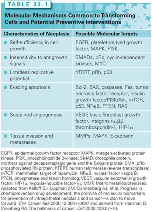
The Hanahan and Weinberg hallmarks of malignant transformation—self-sufficiency in growth signals, insensitivity to growth-inhibitory signals, evasion of apoptosis, limitless replication potential, sustained angiogenesis, and tissue invasion and metastasis7—reflect the loss of cellular signaling control. The molecular damage that results in transformation is triggered by a large array of genetic and environmental stressors such as chronic inflammation, oxidation, inherited genetic mutations, and exogenous environmental exposures. Many such signaling intermediates have common functions in multiple organ sites (see Fig. 33.1). The complexity and overlap of signal transduction pathways suggests that single molecular therapeutic/preventive targets may have limited effectiveness. Interventions aimed at preventing the occurrence of or overcoming the effects of molecular defects in multiple pathways or targets may be required to arrest or reverse carcinogenesis. Using the Hanahan and Weinberg hallmarks, examples of possible targets are shown in Table 33.1.
PRECLINICAL DEVELOPMENT OF CANCER RISK–REDUCING AGENTS
Similar to the development of therapeutic interventions, the assessment of efficacy and toxicity of single chemically synthesized entities, agents designed in silico, botanicals, nutrients/nonnutrient substances used as drugs for cancer risk–reducing agent efficacy proceeds through a translational paradigm that identifies efficacy in cell culture models, in live animal models, and in humans. Preclinical models that simulate the carcinogenesis process in target epithelia identify molecular biomarkers for modulation by interventions. These models can be used to identify potential toxicity of interventions and to assess the effect of interventions on the development and progression of preneoplasia/neoplasia.8
The U.S. National Cancer Institute’s (NCI) PREVENT Cancer Preclinical Drug Development Program is a prime example of a rational strategy to select promising agents for clinical trials through a stepwise approach of preclinical in vitro testing followed by in vivo screening.8–10 This system involves several phases: biochemical prescreening assays, in vitro efficacy models, in vivo short-term screening, animal efficacy testing, and preclinical toxicology testing.
Biochemical Prescreening Assays
Prescreening assays are a series of short-term, mechanistic assays developed to evaluate the ability of a test compound to modulate biochemical events presumed to be mechanistically linked to carcinogenesis.8 These in vitro assays are rapidly completed for potential cancer risk–reducing agents. Examples of such assays include carcinogen-DNA binding, prostaglandin synthesis inhibition, glutathione–S-transferase inhibition, and ornithine decarboxylase inhibition.
In Vitro Efficacy Models
In vitro assays test the cancer risk–reducing activity of a screened compound in four epithelial cell systems (primary rat tracheal epithelial cells, human lung tumor [A427] cells, mouse mammary organ cultures [MMOC], and human foreskin epithelial cells). The assays measure the ability of potential cancer risk–reducing agents to reverse transformation in normal epithelial cells exposed to carcinogens. For example, after treatment with a carcinogen such as 7,12-demethylbenz(a)anthracene, MMOCs develop lesions similar to alveolar nodules that are considered precancerous in mouse mammary glands in vivo.11Pretreatment of organ cultures before carcinogen exposure measures the effect of cancer risk–reducing agents in the initiation stage of carcinogenesis, whereas treatment after carcinogen exposure measures activity during tumor promotion. Three of these assays (using rat tracheal epithelial, A427, and MMOC cells) have shown predictive values of 76% to 83% for cancer risk–reducing agent efficacy in in vivo models.8
Preclinical In Vivo Models for Cancer Risk–Reducing Agent Efficacy Testing
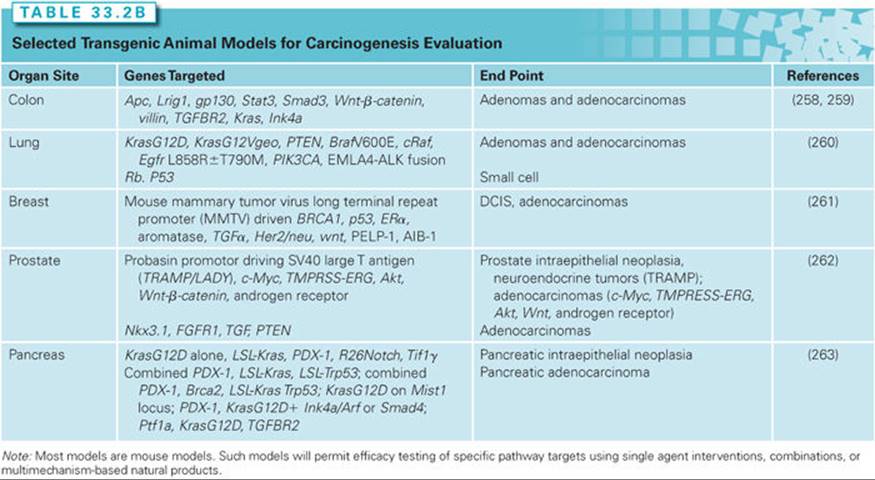
Animal models remain a crucial link in the efficacy assessment of cancer risk–reducing agents for epithelial cancer. Chemical carcinogenesis models provide the reproducible development of tumors in animals following the administration of a known chemical initiator or combination initiator/promoter and have been the primary in vivo screening tool for cancer risk–reducing agents (Table 33.2A).12,13Carcinogenesis models employing genetically engineered mice permit the interrogation of targeted pathways and the corresponding efficacy of cancer risk–reducing agents. Although useful for mechanistic studies, knockout or genetic mutational models create accelerated neoplastic progression that does not accurately recapitulate the more complex, stepwise, human carcinogenesis process. Recombinant alleles can be driven by the addition of drug-sensitive regulatory elements, such as tetracycline or tamoxifen analogs. The drug-sensitive regulatory elements achieve temporal control over a gene promoter through the administration of the drug that binds to the regulatory element. Such a system permits the inhibition or overexpression of the organ-specific gene using Cre recombinase, a site-specific DNA recombinase that targets DNA regions flanked by loxP sequences. Tables 33.2A and 2B list representative organ-specific chemical and transgenic mouse models that may be used for cancer risk–reducing agent testing.

CLINICAL DEVELOPMENT OF CANCER RISK–REDUCING AGENTS
Special Features of Cancer Risk–Reducing Agent Development
The clinical efficacy assessment of cancer risk–reducing agents employs phased testing (phase I to III) models used for development of drugs14 but with crucial differences in study design and end points. Special features for the clinical development of cancer risk–reducing agents create the following challenges to be overcome: (1) the need for large therapeutic index (doses associated with potential toxicity of an intervention need to substantially exceed doses aimed at delaying or reversing transformation) for use in individuals who are asymptomatic yet may benefit from an extended (years) treatment course; (2) the long latency to malignant transformation (an assessment of effectiveness based on the reduction in cancer incidence requires studies lasting for years and involving thousands of participants); (3) adherence (once-daily dosing regimens using interventions that have sufficiently long half-lives may minimize the impact of a missed dose yet maintain the biologic impact on the physiologic target; minimal toxicity and strong psychological commitment to preventive goals also enhance adherence15); and (4) complex risk assessment for cancer (individuals with highly penetrant but infrequent, germ-line genetic susceptibility to breast and colon cancers16,17 are excellent candidates for cancer risk–reducing agents and are likely to accept some toxicity for reduced cancer risk). For individuals at more modestly increased risk (e.g., long-term, current smokers; persons with a family history of cancer; women with mammographically dense breasts), quantitative risk assessment algorithms may be useful in the future to identify optimal cancer risk–reducing agents. The refinement of cancer risk calculators for breast,18 colon,19 and prostate cancer20 promises to appropriately select high-risk individuals for cancer risk–reducing agents such that anticipated benefits exceed potential risks.
Biomarkers as Cancer Risk–Reducing Agent Targets and Efficacy End Points
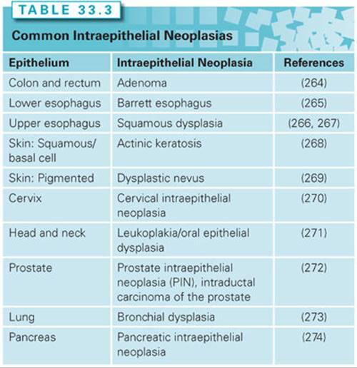
A biomarker is a characteristic that is measured and evaluated as an indicator of normal biologic processes, pathogenic processes, or pharmacologic responses to therapeutic interventions.21 A surrogate end point for cancer prevention assumes that a measured biologic feature will predict the presence or future development of a cancer outcome.22 Biomarkers enable a reduction in the size and duration of an intervention trial by replacing a rare or distal end point with a more frequent, proximate end point.23 Intraepithelial neoplasia has served and continues to serve as a biomarker for invasive malignancy (Table 33.3). Although many advocate the use of intraepithelial neoplasia-based biomarkers as regulatory surrogate end points, others caution that intraepithelial neoplasias may not serve as sufficiently robust surrogate biomarkers for cancer incidence or mortality.24
In order to be useful as end points for cancer risk–reducing agent efficacy testing as regulatory end points, any biomarker must have statistical accuracy, precision, and effectiveness of results24 that demonstrate prediction of a harddisease end point—cancer incidence or mortality. An independent validation data set must address defined standards of validation that minimize bias in the study design and the populations studied.25 The biomarker must be generalizable to the specific clinical or screening population (Table 33.4).

Phases of Cancer Risk–Reducing Agent Development
Phase I cancer risk–reducing agent trials define an optimal cancer risk–reducing agent dose. An optimal cancer risk–reducing agent dose is one that is usually nontoxic, scheduled once daily, and modulates a tissue, cellular, or serum biomarker of drug activity (e.g., the dose of aspirin that inhibits prostaglandin production in a target tissue site). The definition of a maximum tolerated dose is not an essential end point of a phase I cancer risk–reducing agent trial. Higher, yet nontoxic doses may lower cancer risk–reducing agent efficacy. For example, β-carotene at high doses has pro-oxidant activity and may enhance the carcinogenesis process, whereas at low doses, it is a potent antioxidant and differentiating agent.26
Phase II cancer risk–reducing agent trials begin to define cancer risk–reduction efficacy. These short-term (6 months to 1 year) treatment periods gather evidence of risk reduction by assessing drug effects on tissue, cellular, or blood surrogate markers of carcinogenesis. Phase IIa trials are nonrandomized, biomarker modulation trials. Phase IIb trials are randomized, placebo-controlled trials of several hundred subjects testing, for example, whether a risk-reducing agent reduces recurrence of a previously resected intraepithelial neoplastic lesion as the primary end point. Cellular dynamic (e.g., proliferation, apoptotic index), biochemical, or molecular (e.g., p53, cyclin D) end points may be used as secondary end points. Preoperative or window of opportunity trials enroll subjects for brief study periods prior to obtaining tissue by a planned resection of an invasive neoplasm. Such designs permit the exploration of biomarker modulation in the invasive neoplasm and in contiguous epithelial fields proximal and distal to the invasive neoplasm.27
Phase III cancer risk–reducing agent trials define reduction in a hard cancer end point such as cancer incidence or mortality. Such trials, using large, higher risk populations in a randomized, double-blinded intervention, are designed to identify a standard of preventive care for a given risk population. For example, trials of tamoxifen for the reduction of breast cancer incidence,28,29 finasteride for the reduction of prostate cancer incidence,30 and β-carotene for the reduction of lung cancer incidence31 serve as examples of well-conducted, definitive phase III cancer risk–reducing agent clinical trials.
Some investigators consider randomized, controlled clinical trials with an end point sufficient for regulatory review as phase III. Using such a definition, a clinical trial with an end point of reduction in adenoma recurrence is considered a phase III trial. Other investigators define phase III cancer risk–reducing agent trials as randomized, controlled clinical trials with a cancer incidence or mortality end point. This controversy causes confusion in the literature. For the purpose of clarity in this textbook, the latter definition of phase III trial is used—a prospective, randomized, controlled clinical trial with a cancer incidence or mortality end point. Randomized, controlled clinical trials with a surrogate biomarker end point such as an intraepithelial neoplasia (e.g., adenoma) are defined as phase IIb cancer risk–reducing agent trials.
MICRONUTRIENTS
Definition
Micronutrients comprise a large, diverse group of molecules typically ingested as part of the diet that play roles in normal human biology. This group of compounds has been investigated extensively as cancer risk–reducing agents in purified forms (i.e., supplements), as components of multiagent cocktails, and occasionally, as components of food extracts/other mixtures. Although retinoids are not micronutrients per se, they are related to retinol (vitamin A) and share certain properties with carotenoids, which are diet derived and, therefore, included along with micronutrients.
Retinoids, Carotenoids, and Antioxidant Nutrients
Overview and Mechanisms
Retinoids are the natural derivatives and synthetic analogs of vitamin A.32 Cancer risk–reduction intervention studies have evaluated the parent compound (retinol, typically given as retinyl acetate or palmitate), naturally occurring retinoids such as all-trans–retinoic acid (ATRA) and 13-cis-retinoic acid (13cRA), and also synthetic retinoids such as etretinate and fenretinide (4-hydroxy[phenyl]retinamide [4HPR]). These agents have been of interest for cancer risk reduction for decades. Mechanistically, retinoids have been shown to modulate cellular growth and differentiation, as well as apoptosis.32 A large body of research indicates that retinoids have activity in the promotion and progression phases of carcinogenesis, including extensive evidence of efficacy in the setting of premalignant lesions, leading to their evaluation in human Phase III trials. Nuclear retinoic acid receptors mediate many of the retinoid-signaling effects; however, retinoids interact with other signaling pathways, such as estrogen signaling in breast cancer.33
Carotenoids are a group of naturally occurring plant pigments, only some of which are found in appreciable levels in the human diet and human tissues, including beta-carotene, alpha-carotene, lycopene, lutein, and β-cryptoxanthin.34 Of these, the most widely studied carotenoids for cancer risk reduction are beta-carotene and lycopene. Beta-carotene has the highest pro–vitamin A activity of the carotenoids, but alpha-carotene and β-cryptoxanthin also possess pro–vitamin A activity. Other carotenoids, such as lycopene, do not possess vitamin A activity but are known to have potent antioxidant activity, particularly with regard to singlet oxygen quenching.35 Furthermore, eccentric cleavage products of beta-carotene,36 as well as other non-pro–vitamin A carotenoids such as lycopene (e.g., apocarotenals, apocarotenoic acids) appear to be biologically active and may also act via retinoid-signaling pathways.37
Because of the known antioxidant function of carotenoids, they are often studied for risk-reducing efficacy in combination with other antioxidant nutrients, especially vitamins E and C and selenium (sometimes as a cocktail, versus placebo). Thus, we will consider this group of nutrients first, followed by other micronutrients that are thought to act via different mechanisms and/or pathways.
Epidemiology
A large body of literature indicates that people who consume greater amounts of carotenoids from foods (primarily fruits and vegetables) and people with higher serum or plasma levels of various carotenoids have a lower risk for various cancers.38 In particular, according to the systematic review of the literature conducted by the World Cancer Research Fund/American Institute for Cancer Research,38 foods containing carotenoids are “probably” associated with lower risks of cancers of the mouth/pharynx/larynx and lung; foods containing beta-carotene are “probably” associated with lower risks of cancers of the esophagus; and foods containing lycopene are “probably” associated with lower risks of prostate cancer. Preformed retinol intake is inconsistently associated with risks of various cancers but that, in part, likely reflects confounding, because dietary sources of preformed retinol primarily include foods of animal origin (e.g., liver, eggs, milk). Vitamin C in the diet comes primarily from the consumption of fruits and vegetables; therefore, vitamin C and carotenoids often trend together in epidemiologic findings. Vitamin E, in contrast, is found in different foods, especially nuts, seeds, and vegetable oils; intake and blood concentrations are somewhat inconsistently associated with cancer risk.39 Selenium, being a trace mineral, is difficult to measure in the diet, but higher selenium status has been associated with a lower risk of certain cancers, although the results are not entirely consistent.39
Preclinical In Vivo Models
In preclinical models, retinoids induce differentiation as well as arrest proliferation33 of various cancers, making them attractive agents for cancer risk reduction.40 The International Agency for Research on Cancer reviewed the preclinical research involving beta-carotene, concluding that there was “sufficient” evidence of cancer preventive activity, particularly involving mouse skin tumor models and the hamster buccal pouch model.41 Notably, there was inconsistent evidence of efficacy in respiratory tract models. Lycopene has been evaluated in numerous cell culture systems and in a variety of models of prostate carcinogenesis, including chemically induced, orthotopic implantation, transgenic, and xenotransplantation, with mixed evidence of efficacy.42 Evidence, primarily from cell culture studies, suggests that lycopene metabolites may be at least partially responsible for anticarcinogenic activity.37
Clinical Trials: Retinoids, Carotenoids, and Antioxidant Nutrients
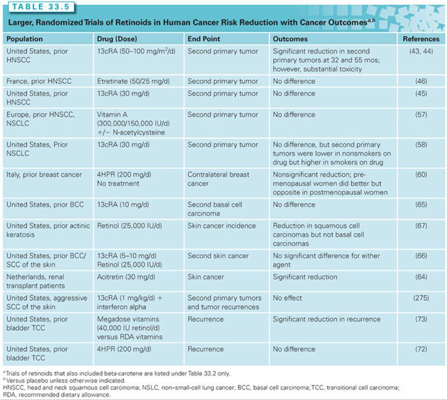
Retinoids, carotenoids, and antioxidant nutrients have been evaluated in the setting of preneoplasia/neoplasia in many different organ sites, as will be discussed. Of the many clinical trials of retinoids and carotenoids/other antioxidant nutrients, key trials with cancer incidence/recurrence as primary outcomes are tabulated (see Tables 33.5 and 33.6) and reviewed.

Clinical Efficacy in the Upper Airway
Many trials of cancer risk–reducing agents have been done in the setting of squamous cell carcinomas of the head and neck, in large part because of the substantial clinical problem of relatively high rates of recurrences and second primary tumors in curatively treated cancer patients. Early work demonstrated that high-dose 13cRA (50 to 100 mg/m2 per day) produced no significant differences in disease recurrence (local, regional, or distant) but significantly lowered the rate of second primary invasive neoplasms,43 with the benefits persisting for at least 5 years.44 Substantial retinoid toxicity, however, including skin dryness and peeling, cheilitis, conjunctivitis, and hypertriglyceridemia, was evident in a large proportion of patients. Subsequent trials thus used lower doses of retinoids (13cRA or a synthetic retinoid, etretinate), but failed to show efficacy in reducing second primary tumor formation (Table 33.5).45,46
Supplemental beta-carotene has also been studied as a single agent and in combination with other agents for the prevention of second primary cancers of the mouth and throat (Table 33.6). One trial of beta-carotene alone observed no harm or benefit47; another observed nonsignificantly fewer second head and neck cancers but more lung cancers48; and a third gave beta-carotene with α-tocopherol (400 IU per day).49 In the third trial, beta-carotene was discontinued early due to adverse findings from lung cancer prevention trials (see the following); however, after a median follow-up of 6.5 years, all-cause mortality was increased, which the authors attributed to the supplemental α-tocopherol. As will be discussed later, adverse effects of antioxidant nutrients are not limited to head and neck cancer patients; potential mechanisms for adverse effects are discussed further.
Clinical Efficacy in the Lung (Lower Airway)
Reversal of Metaplasia or Dysplasia. Active smokers and recent quitters have multiple preinvasive metaplastic and dysplastic lesions in the pulmonary tree. Most of these lesions resolve upon smoking cessation, but some remain and progress to invasive neoplasms. Unfortunately, micronutrient or retinoid interventions have not demonstrated preventive efficacy in most rigorous trials in patients with early lesions. For example, a US trial randomized 755 asbestos workers to receive beta-carotene (50 mg per day) and retinol (25,000 IU every other day) versus placebo; sputum atypia was not reduced after 5 years.50 As another example, eligible smokers with lung metaplasia or dysplasia were randomized to 6 months of 13cRA or placebo. The extent of metaplasia decreased similarly (in approximately 50% of subjects) in both study arms.51 Only smoking cessation was associated with a significant reduction in the metaplasia index during the 6-month intervention.
Prevention of Invasive Neoplasms. Large, phase III efficacy trials of beta-carotene plus other micronutrients for primary prevention of lung cancer have been completed, as summarized in Table 33.6. The Alpha-Tocopherol, Beta-carotene (ATBC) Trial involved 29,133 men from Finland who were heavy cigarette smokers at entry.52 In a two-by-two factorial design, participants were randomized to receive either supplemental α-tocopherol, beta-carotene, the combination, or placebo. Unexpectedly, participants receiving beta-carotene (alone or in combination with α-tocopherol) had a statistically significant 18% increase in lung cancer incidence and an 8% increase in total mortality relative to participants receiving placebo. α-Tocopherol had no effect.
The finding of an increased incidence of lung cancer in the beta-carotene–supplemented smokers was replicated in the Carotene and Retinol Efficacy Trial (CARET), a large randomized trial of supplemental beta-carotene plus retinol versus placebo in asbestos workers and smokers.53 This trial was terminated early, but, at the time of termination, overall lung cancer incidence was increased by 28% in the supplemented subjects and total mortality was also increased by 17%. In contrast, the Physicians’ Health Study (PHS) of supplemental beta-carotene versus placebo in 22,071 male US physicians reported no significant effect—positive or negative—of 12 years of supplementation of beta-carotene on total cancer, lung cancer, or cardiovascular disease (see Table 33.6).54 Two other trials involving supplemental beta-carotene alone (the Women’s Health Study55) or with other antioxidant nutrients (the Medical Research Council/British Heart Foundation Heart Protection Study56) on overall cancer incidence also failed to observe efficacy.
Prevention of Second Primary Invasive Neoplasms. EUROSCAN was a multicenter trial employing a two-by-two factorial design to test retinyl palmitate and N-acetylcysteine (also a compound with known antioxidant activity) in preventing second primary invasive neoplasms in patients with early stage cancers of the head and neck or lung. None of the interventions reduced second airway primary invasive neoplasms.57 The Lung Intergroup Trial randomized patients with surgically resected lung cancer to 13cRA versus placebo and found no significant differences between the two arms in second primary tumors.58 Notably, smoking status modified the effect of the 13cRA intervention, which was harmful in current smokers yet beneficial in former smokers.
Thus, phase III trials of both carotenoids/antioxidants and retinoids indicate that these agents overall do not reduce the risk of developing invasive lung cancers, nor do they prevent the development of second primary invasive neoplasms. However, the finding that former smokers seemed to benefit from both 13cRA58 and beta-carotene53 is intriguing. Mechanistic work suggests this interaction is real rather than chance (see the following), suggesting that (1) risk-reduction in smokers is especially challenging,59 and (2) trials in former smokers may merit consideration.
Clinical Efficacy in the Breast
Moon et al.40 first showed that fenretinide was a promising cancer risk–reducing agent for the breast, having a high therapeutic index and synergistic interaction with tamoxifen in mammary carcinogenesis model studies. This laboratory work led to a large-scale randomized trial of fenretinide (versus no treatment) for 5 years to prevent contralateral breast cancer in women aged 30 to 70 years with a history of resected early breast cancer and no prior adjuvant therapy.60 The intervention produced no significant overall effect, although fenretinide reduced contralateral and ipsilateral breast cancer rates in premenopausal women, with an opposite (adverse) trend observed in postmenopausal women. The reduced incidence of second breast cancer in premenopausal patients persisted with longer follow-up.61
Retinoid X receptor (RXR)–selective retinoids are also being evaluated in preclinical and clinical studies. Ongoing work suggests that combination treatment may represent a promising new strategy to suppress both estrogen receptor–negative and estrogen receptor–positive breast tumors, and the combination of retinoids with antiestrogens may be particularly effective.62
Clinical Efficacy in the Skin
Retinoids have been widely studied for cancer risk–reducing efficacy in skin. Early work was done in patients who have substantial skin cancer risk either due to xeroderma pigmentosum or medication-induced immunosuppression for transplants. For example, 13cRA reduced skin cancer by 63% in patients with xeroderma pigmentosum; however, severe, acute mucocutaneous toxicity with the 13cRA occurred.63 Also, the preventive effect of the retinoid was lost after stopping retinoid therapy. In renal transplant patients, acitretin (30 mg per day) reduced the numbers of premalignant lesions, the number of patients with skin cancer, and the cumulative number of skin cancers.64
In lower risk populations, low-dose 13cRA (10 mg per day)65 and retinol or 13cRA alone did not reduce the recurrence of basal or squamous cell skin cancers,66 although retinol alone reduced squamous but not basal cell carcinomas in patients with prior actinic keratoses.67
Beta-carotene (50 mg per day), in a randomized trial did not reduce the recurrence of nonmelanoma skin cancers.68 Consistent with findings in the lung, the risk was increased by 44% in current smokers randomized to beta-carotene but not in never smokers randomized to beta-carotene as compared with placebo. Supplemental beta-carotene (30 mg per day) also did not prevent basal cell carcinoma or squamous cell carcinoma of the skin in an Australian trial.69
Clark et al.70 randomized patients with a history of nonmelanoma skin cancer to 200 μg per day selenium or placebo.70 Selenium did not reduce the incidence of second skin cancers; a further report of this trial with longer follow-up71 indicated that there was instead a significant increase in total nonmelanoma skin cancer (hazard ratio [HR] = 1.17; 95% confidence interval [CI], 1.02 to 1.34) and squamous cell skin cancer (HR = 1.25; 95% CI, 1.03 to 1.51).
Clinical Efficacy in the Bladder
A trial of fenretinide (200 mg per day orally for 12 months) versus placebo was conducted for preventing tumor recurrence in patients with nonmuscle-invasive bladder transitional cell carcinoma after transurethral resection with or without adjuvant intravesical bacillus Calmette-Guérin; recurrence rates were similar in both groups.72 Another trial randomized 65 patients with biopsy-confirmed transitional cell carcinoma of the bladder to a multivitamin (recommended dietary allowance [RDA] levels) alone or supplemented with 40,000 IU retinol, 100 mg pyridoxine, 2,000 mg ascorbic acid, 400 U of α-tocopherol, and 90 mg zinc.73 The 5-year estimate of tumor recurrence was 91% in the RDA arm versus 41% in the higher-dose nutrient arm (p = 0.0014).
Clinical Efficacy in the Cervix
Randomized trials include four with beta-carotene (alone or with other antioxidant nutrients), and five with retinoids. Only one of these trials, involving ATRA,74 found a significant treatment effect. This trial administered a 0.372% ATRA solution by collagen sponge in a cervical cap delivery system. There was a higher complete response rate in the ATRA group (43%) than the placebo group (27%; p = 0.041) among the 141 patients with moderate dysplasia; no significant differences in dysplasia regression rates between the two study arms were detected in patients with severe dysplasia. The investigators experienced substantial losses to follow-up in this patient population.
Clinical Efficacy in the Esophagus and Stomach
Certain regions of China (Huixian and Linxian) have strikingly high incidence rates of esophageal and gastric cancers. Two trials were done in Linxian County; one was a general population trial that tested the efficacy of four different nutrient combinations at inhibiting the development of esophageal and gastric cancers.75 Those who were given the combination of beta-carotene, vitamin E, and selenium had a 13% reduction in total cancer deaths, a 4% reduction in esophageal cancer deaths, and a 21% reduction in gastric cancer deaths (see Table 33.6). None of the other nutrient combinations reduced gastric or esophageal cancer deaths significantly in this trial. The treatment benefit has been shown to persist for 10 years postintervention, with greater efficacy seen in participants under age 55 years.76 This finding stands in contrast to most other antioxidant nutrient supplement intervention trials, suggesting that the applicability of these results for populations with adequate nutritional status and for other tumor sites may be limited.77
The other Linxian trial evaluated a multivitamin/multimineral preparation plus beta-carotene (15 mg per day) in residents with esophageal dysplasia.78 There was no clear evidence of efficacy, although confidence intervals were wide.
Clinical Efficacy in the Colon/Rectum
Of the randomized trials aimed at the prevention of recurrent colorectal adenomas with micronutrients that have been completed, some used beta-carotene alone79 or with other nonmicronutrient interventions.80 Others evaluated beta-carotene with and without supplemental vitamins C and E.81 None of the trials observed benefit with supplementation. A subsequent report from one trial noted that alcohol intake and cigarette smoking modified the efficacy of beta-carotene.82 Among nonsmokers and nondrinkers, beta-carotene was associated with a significant decrease in the risk of one or more recurrent adenomas (relative risk [RR] = 0.56). Among persons who smoked and also drank more than one alcoholic drink per day, beta-carotene significantly increased the risk of recurrent adenoma (RR = 2.07).
Clinical Efficacy in the Prostate
Because oxidative stress may play a role in the etiology of prostate cancer, several antioxidant nutrients, including vitamin E, selenium, and lycopene, have been of interest for preventing prostate cancer. The largest trial to date of these nutrients is the Selenium and Vitamin E Cancer Prevention Trial (SELECT), which tested selenium and vitamin E in a two-by-two factorial design for the primary prevention of prostate cancer. Despite preliminary indications of prostate cancer risk–reducing efficacy for selenium (from a trial of selenium for skin cancer70) and vitamin E (from a trial of vitamin E to prevent lung cancer83), there was no evidence of efficacy.84 With extended follow-up, the nonsignificant adverse effect of vitamin E became significantly adverse.85 Negative/neutral findings also were reported for vitamins E and C and prostate and total cancer in the PHS II randomized controlled trial.86
The carotenoid lycopene has generated much interest with regard to prostate cancer risk, and several intervention trials have been conducted based on lycopene supplements. These studies have been small, short term, based on intermediate end points, and often lack adequate control groups. The use of a tomato sauce–based intervention is arguably a better approach to evaluate, based on animal data indicating that tomato powder (which includes lycopene along with other phytochemicals), but not lycopene alone, was effective at inhibiting prostate carcinogenesis.87
Mechanisms for Ineffective Retinoid and Carotenoid Cancer Risk–Reducing Activity
There are now a number of trials demonstrating that supplemental beta-carotene/retinoids given to current smokers can produce increases rather than reductions in cancer incidence. In tobacco users, beta-carotene and other carotenoids may produce oxidative carotenoid breakdown products that alter retinoid metabolism and signaling pathways, along with pro-oxidation.88 For retinoids such as 13cRA, smoking may induce genetic and epigenetic changes in the lung that affect retinoid activity; for example, tobacco smoking can affect RAR-β expression.58 The adverse effects of supplemental nutrients are not limited to smokers; α-tocopherol increased rather than reduced prostate cancer in SELECT (which had relatively few smokers) and selenium increased prostate cancer among men without a baseline selenium deficiency.89 This may be a consequence of the relatively high doses used in SELECT,77 but certainly calls into question the notion that reducing oxidative stress is a pivotal cancer risk–reduction strategy, even in nonsmokers.
It has become clear that reactive oxygen species (ROS), such as hydrogen peroxide, can act as important physiologic regulators of intracellular signaling pathways.90,91 Data in mouse models have shown that vitamin E accelerates lung tumor growth by disrupting the ROS–p53 axis, potentially by removing oxidative damage to DNA, which can serve as a potent stimulus for p53 activation.92 Although some of the large cancer risk–reducing trials may have failed in their primary objective, they may indirectly contribute to a clearer understanding of cancer biology, leading to the recognition that the role of oxidative stress and ROS in human disease is much more nuanced than originally hypothesized.93
As for the retinoids, these agents are generally too toxic to be used as single agents for risk-reducing efficacy; however, a major area of ongoing research is examining retinoids (low doses) given in combination with other agents, especially those that regulate the epigenome, such histone deacetylase (HDAC) inhibitors.33
Folic Acid and Other B Vitamins
Overview and Mechanisms. Folate is a water-soluble B vitamin found in foods, whereas folic acid is the synthetic form found in supplements and fortified foods. Adequate folate is critical for DNA methylation, repair, and synthesis.94,95 The methylation status of genes can play a key role in gene silencing and gene expression, lending plausibility to the idea that folate could be a key nutrient in regulating cell growth and proliferation.
Epidemiology. Epidemiologic studies have linked low folate intake with higher risk of several cancers, most notably colorectal cancer.96 Long-term use of multivitamin supplements, which are a major source of folate and other B vitamins, has been associated with a reduction in the risk of colon cancer in some studies, including recent (postfortification) findings.97–99 Supporting an anticancer role of folate is that genotypes for methylene tetrahydrofolate reductase, an enzyme known to be involved in folate metabolism, predict the risk of colon cancer dependent on folate intake or status.100 Vitamin B6 has been less studied in relation to cancer than folate, but some epidemiologic studies suggest that vitamin B6 may be important for colorectal cancer.101,102 A higher risk of cancer related to deficiencies of these vitamins has been suggested for alcohol drinkers.103
Clinical Trials: Folic Acid and B Vitamins. Risk-reducing efficacy for supplemental folic acid has been primarily evaluated in the setting of prevention of recurrent colorectal adenomas (i.e., in patients with prior adenomas). Of six randomized trials of folic acid, two small trials reported suggestions of benefit of folic acid supplementation.104,105 However, benefits were not observed in two much larger trials, the Aspirin/Folate Polyp Prevention Study (AFPPS) (dose: 1 mg of folic acid daily)106 and the United Kingdom Colorectal Adenoma Prevention (ukCAP) trial (dose: 500 μg of folic acid daily).107 AFPPS found indications of an increased risk for advanced lesions and multiple adenomas with prolonged treatment and follow-up. A third large trial, the Nurses Health Study/Health Professionals Follow-up Study (NHS/HPFS) folic acid polyp prevention trial, showed no overall risk reduction.108 The most recent trial, done in a Chinese population >50 years of age,109 reported that 1 mg folic acid per day reduced sporadic colorectal adenomas when compared to no intervention (not a placebo-controlled study). One possible explanation for the discrepancy of the Chinese trial versus North American and European trials is the baseline plasma folate status. In the Chinese trial, the mean baseline folate concentration of 5 ng/mL109 was half of the reported 10 ng/mL in a United States trial,106 where folate fortification of the food supply occurs.
Calcium and Vitamin D
Overview and Mechanisms. There are two major forms of vitamin D: ergocalciferol (D2) and cholecalciferol (D3). Vitamin D2 is absorbed through dietary sources such as fortified milk products, and D3 is synthesized via ultraviolet (UV) B light isomerization of 7-dehydrocholestrol in the epidermis.110 Vitamin D3 is converted to calcitriol (1, 25-[OH]2 D3) in a two-step process requiring both hepatic and renal hydroxylation. Calcitriol binds to the vitamin D receptor, which translocates to the nucleus and binds to multiple gene promoter sites. Through this mechanism, vitamin D regulates cytoplasmic signaling pathways that impact cellular differentiation and growth through proteins such as Ras and mitogen-activated protein kinase (MAPK), protein lipase A, prostaglandins, cyclic adenosine monophosphate (AMP), protein kinase A, and phosphatidyl inositol 3 kinase.110 1,25(OH)2D3 regulates cellular proliferation and apoptosis. For example, 1,25(OH)2D3 can induce cleavage of caspase 3, poly (ADP-ribose) polymerase (PARP), and MAPK, leading to apoptosis. 1,25(OH)2D3 inhibits the expression and phosphorylation of Akt, a key regulator of cellular proliferation. The differentiation properties of 1,25(OH)2D3 are mediated through transcriptional activation of the CDK inhibitor p21. The effects of vitamin D on multiple signal transduction pathways operational in cancer cells are reviewed by Deeb et al.111
Epidemiology. Observational epidemiologic studies have shown a relatively consistent inverse association between low calcium intake, including that from supplements, and increased colorectal and colon cancer risk.112,113 Vitamin D exposure is typically assessed by measuring 25(OH)vitamin D in plasma because exposure is derived not only from diet and supplements, but also from cutaneous synthesis following dermal exposure to UV radiation. A large number of observational studies have evaluated the association between vitamin D status and cancer risk, as systematically reviewed by the Agency for Healthcare Research and Quality (AHRQ).114 The evidence is inconsistent for most cancer sites, with the exception of studies showing that individuals with lower blood vitamin D levels have a higher risk of colorectal cancer or adenoma. Although some observational studies have reported that higher serum vitamin D is associated with lower breast cancer risk, the association is inconsistent.114,115 Also, there are some studies suggesting high serum vitamin D is associated with increases in certain cancers, particularly pancreatic cancer.116
Clinical Trials: Calcium and Vitamin D
Clinical Efficacy in the Colon. Baron et al.117 randomized subjects with a recent history of colorectal adenomas to either calcium carbonate (1,200 mg per day of elemental calcium) or placebo. Results showed significant benefit for the calcium arm (adjusted RR = 0.81; 95% CI, 0.67 to 0.99; p = 0.04). In a smaller, similar study of calcium gluconolactate and carbonate (2 g elemental calcium daily), the adjusted odds ratio (OR) for adenoma recurrence was 0.66 (95% CI, 0.38 to 1.17; p = 0.16) for calcium treatment,118 and while not statistically significant, it was similar to the data of Baron et al.117
In the largest trial of calcium and vitamin D with primary cancer end points (e.g., colon, breast), the US Women’s Health Initiative (WHI) evaluated the combination of 400 IU of vitamin D per day plus 1,000 mg of calcium per day in 36,282 postmenopausal women. For colon cancer, there was no benefit observed,119 although the mean baseline intake of calcium was already very high (more than 1,151 mg per day). With regard to vitamin D as a single agent, there was also no suggestion of benefit for colon cancer incidence in a 5-year British trial of vitamin D (100,000 IU every 4 months) that reported colon cancer incidence,120 although this was not a primary end point.
Clinical Efficacy in the Breast. Vitamin D has received considerable attention for a possible role in the prevention of breast cancer,121 although no trials have yet investigated vitamin D as a single agent for breast cancer risk reduction. The large WHI trial gave a combination of calcium and vitamin D, as noted previously, and there was no significant effect of this combination on breast cancer risk (HR, 0.96; 95% CI, 0.85 to 1.09).122 Lappe et al.123conducted a trial that examined the relation between calcium plus vitamin D (1,100 IU per day) supplementation (versus calcium alone or placebo) in 1,179 healthy postmenopausal women in Nebraska.123 Although fracture was the primary outcome of the trial, total cancer incidence was reportedly lower in the calcium plus vitamin D group, although the number of end points was very small (n = 50 total cancers observed during the follow-up). An ongoing randomized trial of vitamin D and omega-3 fatty acids (the VITamin D and OmegA-3 TriaL [VITAL]) among 20,000 participants is expected to provide more definitive data on a possible role of vitamin D in the prevention of breast and other cancers.124
Summary and Conclusion: Micronutrients
Certain agents, including the retinoids, beta-carotene, folic acid, calcium plus vitamin D, vitamin E, and selenium, have received substantial attention for a possible role in reducing the risk of cancer in humans. As reviewed herein, some of the trials have observed statistically significant reductions in the risk of the primary end point (e.g., retinoids in skin carcinogenesis models, calcium in colorectal adenomas, antioxidant nutrients in Linxian, China, for gastric cancer prevention), whereas others have observed statistically significant increases in the risk of the primary end points (beta-carotene and retinoid lung cancer prevention trials in smokers, vitamin E and prostate cancer, selenium and nonmelanoma skin cancer). Considering the completed trials, there is clear evidence against the general use of nutrient supplements for cancer prevention, which is the conclusion also reached by the World Cancer Research Fund/American Institute for Cancer Research.38 Note that there is no evidence that food sources of these nutrients increase risk.
Having noted that, there are other key themes emerging from this growing body of research. One such theme is that nutrient supplementation may be of benefit to some but not all. One such population that may benefit includes persons who are low in the nutrient of interest at baseline.77 This was initially suggested in the Linxian Country trial (done in a micronutrient-deficient population), with growing support from subgroup analyses of several completed trials.77 However, the hypothesis that nutrient supplementation can reduce cancer risk in subgroups selected based on inadequate nutritional status has, to date, not been formally evaluated in intervention trials.
Another consistent theme is that lifestyle factors (e.g., smoking) and genetics (polymorphisms) may determine who is most likely to benefit from supplementation. Trial data will likely be increasingly mined to identify genetic profiles associated with both better outcomes (risk prediction) and response to intervention.125,126 Ultimately, a more personalized approach to cancer risk reduction may emerge, consistent with the movement toward a more personalized approach for cancer treatment.
Finally, nearly all of these trials initiate intervention with older adults (who are more likely to develop cancer end points during the follow-up); but, animal models suggest that the timing of exposure may likely be quite relevant. For example, folic acid may protect against initiation, but may also promote the proliferation of existing neoplasms.127 Thus, the dose, form (food versus supplement), timing, and nutritional and lifestyle characteristics may all be relevant in affecting the efficacy of risk-reducing interventions involving nutrients and related substances. Further research, drawing upon newer tools now available through the field of nutritional genomics, will be needed to gain greater clarity on the heterogeneous biologic effects observed in nutrient-based risk reduction.
ANTI-INFLAMMATORY DRUGS
Mechanism
Nonsteroidal anti-inflammatory drugs (NSAIDs) represent a class of drugs that reduce cellular inflammation through multiple mechanisms, the most prominent of them being the modulation of eicosanoid metabolism.128 Eicosanoids are metabolites of dietary fatty acids, primarily linoleic acid. Linoleic acid is metabolized to arachidonic acid, which is stored in the lipid membrane and, once mobilized from the membrane, further metabolized by prostaglandin-H synthases (PGHS) 1 and 2 to PGD2, PGE2, PGF2α, PGI2, or thromboxane A2 (TxA2) by specific synthases. Leukotriene pathways involve the conversion of arachidonic acid to leukotriene A4 by 5-lipoxygenase and subsequent hydrolysis of leukotriene A4 to other downstream leukotrienes. Newly formed prostaglandins function primarily through binding to prostaglandin receptors (EP receptors), releasing coupled G-proteins to elicit responses in the same or neighboring cells.129
Prostaglandins (PG) play crucial roles in controlling cellular proliferation, apoptosis, cellular invasiveness, and angiogenesis and in modulating immunosuppression.129 Because PGE2 is the most abundant PG in tumors, reducing local concentrations of PGE2 may be a pivotal cancer preventive strategy.129
PGHS-independent mechanisms of NSAID action may, at least in part, explain NSAID preventive efficacy.130 A diverse group of NSAIDs inhibit apoptosis via multiple mechanisms. Among the more prominent of these mechanisms is the inhibition of cyclic guanosine monophosphate (cGMP) phosphodiesterase activity, attenuation of beta-catenin mRNA through suppressing transcription of the CTNNB1gene, and activation of c-Jun N-terminal kinase 1.130 NSAIDs activate peroxisome proliferator–activated receptor (PPAR)γ, leading to increased E-cadherin expression and reduced colony formation in vitro, while reducing PPARδ, leading to reduced resistance to apoptosis.130 Selective cyclooxygenase 2 (COX-2) inhibitors inhibit Akt signaling and induce apoptosis of human colorectal and prostate cancer cells in vitro in a COX-2–independent manner via the inhibition of phosphoinositide-dependent kinase-1 (PDK-1). NSAIDs inhibit nuclear factor kappa B (NF-κB) at pharmacologic concentrations and key cellular proliferation signaling intermediates such as activator protein 1 (AP-1) and other intermediates of the MAPK pathway.130 The impact of NSAIDs on carcinogenic events driven by these upstream pathways in humans as opposed to preliminary in vitro or in vivo models remains unclear.
Epidemiology
Pooled analyses of 34 controlled trials of aspirin 75 mg to 100 mg daily (69,224 participants), conducted primarily for cardiovascular disease reduction, observed reduced cancer deaths (OR, 0.63; 95% CI, 0.49 to 0.82). Most of the benefit occurred after 5 years follow-up.131 In a pooled analysis of 150 case control and 45 cohort studies, in addition to a reduced risk of death from colorectal cancer (OR, 0.58; 95% CI, 0.44 to 0.78), chronic and frequent (once daily or more) use of aspirin also reduced the risk of death from esophageal (OR, 0.58; 95% CI, 0.44 to 0.76), gastric (OR, 0.61; 95% CI, 0.40 to 0.93), and breast (OR, 0.81; 95% CI, 0.72 to 0.93) cancers.132 An analysis of 662,624 men and women enrolled in the American Cancer Society’s Cancer Prevention Study II found that aspirin taken at least 16 times per month over a 6-year period conferred a 40% reduced risk of colorectal cancer mortality.133 Both the 46,363 male patients of the Health Professional Study134 and the 82,911 patients of the Nurse’s Health Study135suggest that prolonged use (>10 years) of 325 mg of aspirin twice weekly or more reduces colorectal cancer risk (RR, 0.77; 95% CI, 0.67 to 0.88, from the Nurse’s Health Study). Daily NSAID intake is associated with a 40% reduction (OR, 0.56; 95% CI, 0.43 to 0.73) in the risk of esophageal adenocarcinoma.136
Evidence in Preclinical In Vivo Carcinogenesis Models
NSAIDs, including aspirin, indomethacin, piroxicam, sulindac, ibuprofen, and ketoprofen, suppress colonic tumorigenesis induced chemically (1,2-dimethylhydrazine or its metabolites) or transgenically (Min+).137,138 The selective COX-2 inhibitors were the most efficacious colon tumorigenesis inhibitors in both chemical and transgenic rodent models.139,140 In preclinical models, NSAIDs affect the onset and progression of cancers in the stomach, skin, breast, lung, prostate, and urinary bladder, although the evidence is more limited than for colon cancers.141
Clinical Trials
Key clinical trials of NSAIDs for the prevention of colorectal cancer are summarized in Table 33.7. Sulindac reduced the size and number of preexisting adenomas in patients with familial adenomatous polyposis but did not suppress the development of new adenomas,142 whereas the selective COX-2 inhibitor, celecoxib, suppressed the development of new adenomatous polyps in patients with familial adenomatous polyposis.143 Although these results are promising, reports of invasive neoplasms developing in familial adenomatous polyposis patients being treated with sulindac144 raise the question of whether NSAIDs preferentially alter the formation or regression of those adenomas less likely to progress to invasive adenocarcinomas, as compared to those more likely to progress.
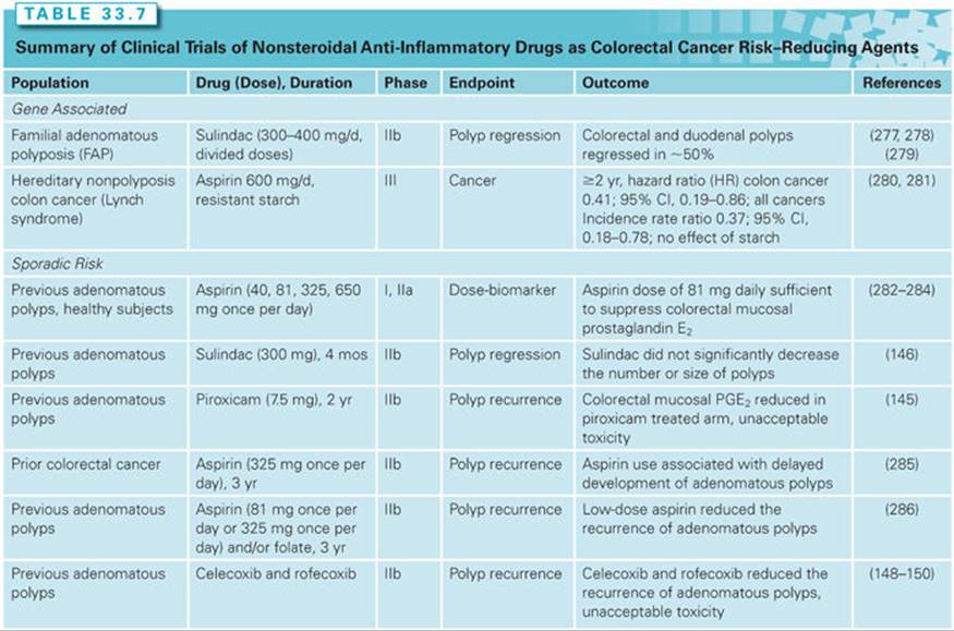
Randomized, double-blinded placebo controlled trials of NSAIDs as cancer risk–reducing agents for colorectal adenocarcinoma (see Table 33.7) have confirmed that aspirin suppresses adenoma recurrence in patients previously treated for adenomas or for cancer. Neither sulindac nor piroxicam alone suppressed adenoma formation in high-risk, sporadic populations at tolerable doses.145,146 Sulindac, in combination with difluoromethylornithine, has potent colorectal anticarcinogenesis effects.147 Selective COX-2 inhibitors (celecoxib, rofecoxib) reduce the recurrence of adenomas by one-third in all patients previously treated for adenomas and by one-half in patients with previously resected large (≥1 cm) adenomas,148–150 but they are too toxic as cancer risk–reducing agents due to their cardiovascular toxicity.151,152 Although most NSAIDs (piroxicam, indomethacin) have sufficient gastrointestinal (GI) toxicity to reduce their acceptability as cancer risk–reducing agents,153,154 the long-term administration of low-dose aspirin in vascular prevention trials demonstrates acceptable GI toxicity.155
Up to 40% of individuals screened for colorectal neoplasms will have an adenomatous polyp detected and removed, yet only 10% of these lesions will progress to invasive neoplasms. To date, prospective NSAID trials of only 2 to 3 years cannot substitute for cancer incidence or mortality end points. Given the 10-year latency between adenoma formation and a cancer event, prospective trials sufficiently powered to detect colorectal cancer incidence end points are unlikely in the future.156 Alternatively, a follow-up of patients randomized on trials of aspirin in the prevention of vascular events in the 1980s and 1990s offers secondary analysis opportunities. In a pooled analysis of three prospective vascular end point cohort studies, 20-year low-dose aspirin treatments reduced cancer deaths from all solid tumors (OR, 0.69; CI, 0.54 to 0.88) and from lung and esophageal adenocarcinomas (OR, 0.66; CI, 0.56 to 0.77).155 Despite this, the U.S. Preventive Services Task Force (USPSTF) does not recommend the use of aspirin or NSAIDs as cancer risk–reducing agents for normal risk populations, preferring adherence to colorectal cancer screening recommendations (fecal occult blood testing and endoscopy).153,154,157
Minimal prospective cancer risk reduction data are available at other epithelial organ sites. Ketorolac, given as a 1% rinse solution, did not reduce the size or histology of leukoplakia lesions.158 Celecoxib reduces the Ki67 labeling index and increases the expression of nuclear survivin without significantly changing the cytoplasmic survivin in bronchial biopsies of smokers.159 Cancer prevention trials of aspirin as interventions for delaying progression from intraepithelial neoplasias in other epithelial sites remain ongoing for the lower esophagus.136 No prospective, randomized trials or data are available for breast, prostate, or gynecologic cancer prevention.
EPIGENETIC TARGETING AGENTS (SELECTIVE ESTROGEN RECEPTOR MODULATORS, 5α-STEROID REDUCTASE INHIBITORS, POLYAMINE INHIBITORS)
Posttranslational pathway targets remain a fertile source of chemopreventive strategies. Phase III data support cancer risk–reduction agent efficacy of selective estrogen receptor modulators (SERM) and 5α-steroid reductase inhibitors for breast and prostate cancer prevention, respectively. Inhibitors of the polyamine pathway may be useful preventives for colorectal cancer.
Selective Estrogen Receptor Modulators
Mechanism
SERMs function as estrogen receptor (ER) agonists and antagonists depending on the SERM structure and target tissue. Predominant ERα receptors occur in the human uterus, cortical bone, and the liver; whereas predominant ERβ receptors occur in blood vessels, cancellous bone, the whole brain, and immune cells.160,161 During carcinogenesis, the amount of ERα increases while the amount of ERβ decreases in breast tissues.162 Ideally, a desirable SERM for cancer prevention will function as an antiestrogen in the breast and uterus, but a partial estrogen agonist in skeletal, cardiovascular, central nervous system (CNS), GI tract, and vaginal tissues. In addition, an ideal SERM will not have procoagulant effects and will not cause perimenopausal symptoms such as hot flashes.162
Tamoxifen. Tamoxifen is a triphenylethylene compound developed for the treatment of ER-positive breast cancer in the 1960s and 1970s.163,164 Tamoxifen inhibits the initiation and promotion phases of breast carcinogenesis in the dimethylbenzanthracene chemical carcinogenesis model.164,165 When tamoxifen binds to ERβ, which then binds to an AP-1 type gene promoter, it functions as an estrogen agonist. When bound to ERα, which binds to an estrogen response element (ERE) target gene promoter, tamoxifen functions as an estrogen antagonist.162,166 Tamoxifen has estrogen antagonist effects in the human breast; partial estrogen agonist effects in bone, the cardiovascular system, and CNS; and predominant estrogen agonist effects in the uterus, liver, and vagina. The estrogen agonist effects in the liver and uterus result in tamoxifen’s toxicities of thromboembolism and endometrial cancer, respectively. The clinical finding that tamoxifen reduces the incidence of contralateral second primary breast cancers during adjuvant treatment regimens catalyzed the push for its development as a cancer risk–reduction agent.167,168
Raloxifene. The benzothiophene structure of raloxifene confers a different tissue-specific ER-binding profile than the triphenylethylene tamoxifen. Raloxifene has greater estrogen agonist activity in bone but reduced estrogen agonist activity in the uterus. Raloxifene was studied for the treatment and prevention of osteoporosis in a large, pivotal trial (the Multiple Outcomes of Raloxifene Evaluation [MORE]) and was found to reduce the rate of vertebral fracture as compared to placebo in postmenopausal women.169
Lasofoxifene and Arzoxifene. Lasofoxifene and arzoxifene are third-generation SERMs developed as more potent blockers of bone resorption with the goal of reducing the risk of fractures, breast cancer, and heart disease while minimizing the SERM-induced risk of endometrial hyperplasia in postmenopausal women. Both agents proved potent in vitro and in preliminary clinical trials for bone fracture prevention.170–173
Selective Estrogen Receptor Modulators as Risk-Reducing Agents for Breast Cancer Prevention
Efficacy. Table 33.8 summarizes the phase III data for SERM-based breast cancer–risk reduction. In a systematic review of MEDLINE and Cochrane databases through December, 2012, the USPSTF identified seven trials of tamoxifen or raloxifene that showed a reduced incidence of invasive breast cancer by 7 to 9 cases in 1,000 women over 5 years compared to placebo.174 Tamoxifen is more effective than raloxifene; it reduces breast cancer incidence more than raloxifene by 5 cases in 1,000 women. Both drugs reduce the incidence of ER-positive breast cancer, but neither reduces the risk of ER-negative breast cancer. Neither drug reduced breast cancer–specific or all cause mortality rates. Based on benefit–risk models, women with estimated 5-year risks of breast cancer of 3% or greater are likely to benefit from treatment.175 Using similar analysis methods as the USPSTF, the American Society of Clinical Oncology recommends the use of tamoxifen (20 mg per day orally for 5 years) or raloxifene (60 mg per day orally for 5 years) “in premenopausal women who are age ≥35 years with a 5-year projected absolute breast cancer risk ≥1.66% according to the NCI Breast Cancer Risk Assessment Tool (or equivalent measures), or with lobular carcinoma in situ.”176 Tamoxifen reduces the risk of in situ (preinvasive) breast neoplasms (lobular carcinoma in situ, ductal carcinoma in situ) by 50%.177,178 The reduction during treatment persists for at least 5 years after treatment.177,178 Raloxifene does not reduce the risk of in situ breast neoplasms.
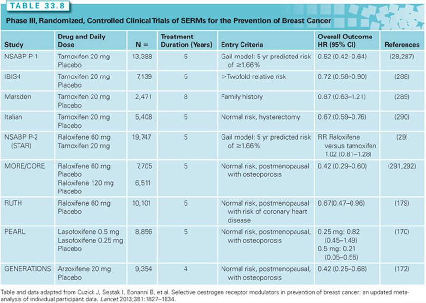
Data from two trials designed to evaluate the safety and efficacy of lasofoxifene (PEARL)170,171 and arzoxifene (GENERATIONS)172,173 as bone fracture preventives have been analyzed for breast cancer–risk reduction. Their effect at reducing breast cancer incidence was captured in secondary analyses (see Table 33.8). Neither lasofoxifene nor arzoxifene have been evaluated in phase III randomized controlled breast cancer prevention trials. Arzoxifene development has been discontinued in the United States.
Toxicity Profiles. Tamoxifen causes a twofold increase in the risk of endometrial adenocarcinoma (RR, 2.13; 95% CI, 1.36 to 3.32) and is related to more benign gynecologic conditions, uterine bleeding, and surgical procedures than the placebo controls, whereas raloxifene did not increase the risk for endometrial cancer or uterine bleeding.174 Tamoxifen causes a twofold increase in thromboembolic events (RR, 1.93; 95% CI, 1.41 to 2.64), whereas raloxifene causes a 60% increase in risk of venous thromboembolism (RR, 1.60; 95% CI, 1.15 to 2.23).174 Raloxifene does not differ from tamoxifen in risk of fractures, other cancers, or cardiovascular events.177 Raloxifene’s lower risk of endometrial adenocarcinomas compared to tamoxifen needs to be weighed against the increased risk of stroke seen in in the MORE/CORE trials (see Table 33.8).179 Raloxifene’s effectiveness in the community may also be compromised by its poor bioavailability (2%) due to rapid phase II enzyme metabolism in the gut and liver,180 whereas tamoxifen is more bioavailable and has active metabolites that permit a prolonged drug effect. Missed raloxifene doses may potentially compromise efficacy and prevention outcomes in widespread, community use.
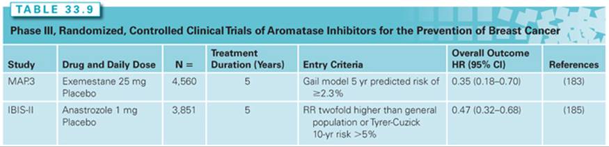
Aromatase Inhibitors. In adjuvant clinical trials for breast cancer, aromatase inhibitors (anastrozole, exemestane, letrozole) given after 5 years of tamoxifen enhance the reduction of breast cancer recurrence in the contralateral breast compared to tamoxifen alone.181 In a phase I cancer risk–reducing agent trial, letrozole reduced the Ki-67 proliferation index of breast epithelial cells aspirated from high-risk women.182Exemestane reduced the overall risk of ER-positive invasive breast cancer (Table 33.9). It did not reduce the risk of noninvasive breast neoplasms or ER-negative breast cancer.183 Exemestane has no increased risk of venous thromboembolism, endometrial cancer, fracture, or cataract,183 but losses in bone mineral density and cortical thickness of the distal tibia and radius occurred after 2 years of treatment despite calcium and vitamin D supplementation.184 The results of the International Breast Cancer Intervention Study II (IBIS-II), comparing anastrozole with placebo, are similar to those reported for exemestane.185Compared to exemestane, anastrozole decreases the incidence of ductal carcinoma in situ (DCIS), whereas exemestane does not. Neither aromatase inhibitor increased survival compared to placebo controls. The American Society of Clinical Oncology recommends exemestane for breast cancer prevention in addition to tamoxifen and raloxifene.
Use Counseling. Despite the widespread evidence of breast cancer preventive efficacy for tamoxifen and raloxifene, only 3% to 20% of eligible high-risk women agree to take tamoxifen for primary prevention.186 The low willingness of eligible women to take tamoxifen for 5 years demonstrates the issue of risk benefit for cancer risk–reducing agents. Women with high short-term risk (5 year Gail risk of >3%)—for example, those with ER-positive atypical hyperplasia, lobular carcinoma in situ, and the majority of non–high-grade ductal carcinoma in situ lesions—have an acceptable risk to benefit ratio and are the most likely to benefit from a 5-year cancer risk–reducing agent intervention with a SERM.175,176 The toxicity profile of aromatase inhibitors differs from SERMs. Although aromatase inhibitors may have a more favorable risk to benefit profile than SERMs, long-term outcomes and toxicity experience for aromatase inhibitor risk-reducing agent intervention are not available to date. In the National Surgical Adjuvant Breast and Bowel Project, tamoxifen-treated women with a BRCA2 mutation but not a BRCA1 mutation had reduced cancer incidence,187 but subsequent data from another group have found reduced cancer risk in women with both BRCA mutations.188 Data remain insufficient to recommend the use of SERMs for risk reduction in women with BRCA mutations.
5α-Steroid Reductase Inhibitors
Mechanism
Prostate cancers require androgens to proliferate and evade apoptosis. The primary nuclear androgen responsible for the maintenance of epithelial function is dihydrotestosterone. The testes and adrenal gland synthesize dihydrotestosterone by the conversion of testosterone by 5α-steroid reductase types 1 and 2 isozymes. Dihydrotestosterone binds to intracellular androgen receptors to form a complex that binds to DNA hormone response elements controlling cellular proliferation and apoptosis. Finasteride, a selective, competitive inhibitor of type 2 5α-steroid reductase,189 inhibits proliferation in the transformed prostate cell. In the 3,2′-dimethyl-4-aminobiphenyl (DMAB), methylnitrosourea (MNU), and testosterone chemical carcinogenesis models in rats, finasteride reduces prostate tumor incidence by close to six-fold. Finasteride appears to be more effective in the promotion phase of prostate carcinogenesis.190 Dutasteride inhibits both 5α-steroid reductase inhibitor190 types 1 and 2 isoforms and has similar anticarcinogenesis activity in preclinical models to finasteride.
Cancer Risk–Reducing Agent Activity
Randomized, placebo-controlled cancer incidence end point risk-reducing agent clinical trials demonstrated that finasteride and dutasteride reduced the incidence of prostate cancer by approximately 22% (Table 33.10).30,191,192Patients who are treated with either drug yet progress to transformed neoplasms develop more tumors of a high Gleason grade (7 to 10) compared to the placebo arm (22%). After 18 years of follow-up, no significant differences in overall survival or survival after prostate cancer diagnosis were found in the finasteride-treated group compared to the placebo-treated group.193 Sexual function side effects (e.g., erectile dysfunction, loss of libido, gynecomastia) were more common in the finasteride- or dutasteride-treated groups.30,192

The 5α-steroid reductase inhibitors, finasteride and dutasteride, prevent or delay carcinogenesis progression in the prostate, yet progression of high-grade lesions is unaffected. Use of finasteride for a period of 7 years reduced the incidence of prostate cancer but did not significantly affect mortality.193 Increasing the diagnosis of low-grade prostate cancer through prostate-specific antigen (PSA) testing or intervention with a drug with a minimal toxicity profile without reducing mortality is of no benefit and “all forms of therapy cause considerable burden to the patient and to society.”193
SIGNAL TRANSDUCTION MODIFIERS
Both cancer therapy and cancer prevention have investigated drugs that modify specific targets in signal transduction pathways. Although the emphasis in drug development has focused on cancer treatment, interventions aimed at modulating signal transduction pathways promise new approaches to interventions in the carcinogenic process. Because of the complexity of signaling systems, the inhibition of single targets may not be effective or may cause unacceptable toxicity.
Difluoromethylornithine
Mechanism
Polyamines (spermidine, spermine, and the diamine, putrescine) are required to maintain cellular growth and function.194 In mammalian cells, polyamine inhibition by genetic mutation or pharmaceutical agents is associated with virtual cessation in cellular growth. Difluoromethylornithine (DFMO) is an enzyme-activated irreversible inhibitor of ornithine decarboxylase (which is transactivated by the c-MYConcogene and cooperates with the RAS oncogene in malignant transformation).195
Evidence in Preclinical In Vivo Carcinogenesis Models
Extensive preclinical data has found that DFMO prevents tumor promotion in a variety of systems, including skin, mammary, colon, cervical, and bladder carcinogenesis models.194 Synergistic or additive activity with retinoids, butylated hydroxyanisole, tamoxifen, piroxicam, and fish oil has been demonstrated with low concentrations of DFMO.194
Clinical Trials
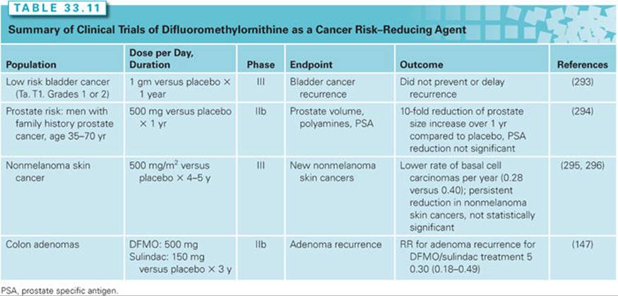
In phase I prevention trials, DFMO at a dose of 0.5 mg/m2 per day reduced tissue polyamines in the colon and skin196,197 and causes regression of cervical intraepithelial neoplasia when used topically,198 but does not reduce tissue polyamines or other biomarkers of cellular proliferation in the human breast.199 As a single agent, DFMO has anticarcinogenic activity for nonmelanoma skin cancers, primarily basal cell carcinoma. In combination with an NSAID (sulindac), DFMO reduced adenoma recurrences, suggesting a synergistic reduction of colorectal cancer risk (Table 33.11). Preliminary data suggest some cancer risk–reducing agent activity for the lower esophagus and the prostate (see Table 33.11).200
Statins
Mechanism
Statins are hydroxyl-3-methylglutaryl coenzyme A (HMG-CoA) reductase inhibitors that inhibit the conversion of HMG-CoA to mevalonate, a cholesterol precursor. The statins are a class of medications with similar structures but with variable moieties that can result in hydrophilic forms (e.g., pravastatin, rosuvastatin) and lipophilic forms (e.g., lovastatin, simvastatin, fluvastatin, atorvastatin).201 Statins decrease the risk of cancer in preclinical studies by inhibiting RAS- and RHO-mediated cell proliferation, upregulating cell cycle inhibitors (e.g., p21 and p27), and inducing apoptosis of transformed cells and the inhibition of angiogenesis.202
Evidence in Preclinical In Vivo Carcinogenesis Models
Lipophilic statins delay progression of pancreatic intraepithelial neoplasias and the growth of pancreatic carcinoma xenografts. Atorvastatin alone and in combination with NSAIDs reduced colonic adenoma and adenocarcinoma incidence and multiplicity by half in rodent transgenic and chemical carcinogenesis models. Lovastatin reduced lung adenoma multiplicity but not incidence.201
Clinical Trials
Although several large trials of pravastatin or simvastatin on cardiovascular disease risk with cancer as secondary end points have shown no benefit for reducing cancer risk with follow-ups between 18 months to 4 years, these trials were not adequately powered to examine cancer end points.201 Several case control studies evaluating statin effects have shown a significant association with lower risk of colorectal adenocarcinoma with odds ratios ranging from 0.53 to 0.91 for arzoxifene. A secondary analysis of a celecoxib prevention trial demonstrated no statin protection against colorectal neoplasms.203 The Women’s Health Initiative (prospective longitudinal cohort of 159,319 women) found that lovastatin was associated with a lower risk of developing colorectal cancer (HR = 0.62; 95% CI, 0.39 to 0.99).204Prospective longitudinal studies have shown mixed results. The PHS reported statin use was inversely associated with prostate cancer (adjusted RR, 0.51),205 whereas the Nurse’s Health Study showed no association with risk of breast cancer.206 Interventional trials to determine statin preventive efficacy for colon and breast cancer are ongoing.201 Statins may be effective risk-reducing agents in individuals with the A/A variant of the predominant T/T genotype of rs12654264 of the HMG-CoA reductase gene.207
Bisphosphonates
Mechanism
Bisphosphonates are pyrophosphate analogs with a central phosphorus-carbon-phosphorus bond that resists bone degradation preventing bone loss and fractures. Second- and third-generation amino bisphosphonates (pamidronic, alendronic, risedronic, ibandronic, and zoledronic acids) inhibit farnesyl diphosphate synthase downstream of HMG-CoA reductase, leading to decreased posttranslational prenylation of GTP-binding proteins such as RAS and Rho. Amino bisphosphonates inhibit cell proliferation, angiogenesis, and cell cycle arrest while inducing apoptosis.201
Evidence in In Vivo Preclinical and Clinical Models
HER2-transgenic mice treated with zoledronic acid had increased tumor-free survival and overall survival. Zoledronic acid suppressed bone, lung, and liver metastases when treated prior to an injection of breast cancer cells.201 The short-term use of bisphosphonates is associated with reduced breast cancer incidence in case control studies208,209 and prospective cohort studies (HR = 0.68; 95% CI, 0.52 to 0.88 in a prospective cohort study).210 Randomized trials of amino-bisphosphonates in postmenopausal women with breast cancer treated for 1 year have found a reduction of breast cancer risk in the contralateral breast (HR = 0.39; 95% CI, 0.18 to 0.88).211 Case control data suggesting a bisphosphonate-associated reduction in colorectal cancer risk have not been confirmed by prospective cohort studies (i.e., Women’s Health Initiative, Nurse’s Health Study).201
Metformin
Mechanism
Metformin, an oral antidiabetic drug in the biguanide class, is the first-line drug of choice for the treatment of type 2 diabetes.212 Cancers are more common in diabetics and obese individuals than their normal weight and normoglycemic counterparts, leading to the hypothesis that elevated serum insulin concentrations promote cancer risk.213,214 Insulin and insulin-like growth factors (IGF1 and 2) stimulate cellular DNA synthesis, proliferation, and tumor growth through phosphoinositide-3 kinase (PI3K), mammalian target of rapamycin (mTOR), and the RAS-MAPK signaling pathways.213 Metformin activates the adenosine monophosphate-activated protein kinase (AMPK) via LKB1, a protein-threonine kinase that has tumor-suppressor activity.201 Metformin anticarcinogenesis activity appears to be broad and includes downregulation of erbB-2 and epidermal growth factor receptor (EGFR) expression, inhibiting the phosphorylation of erbB family members, IGF1R, Akt, mTOR, and STAT3 in vivo. Low doses of metformin inhibit the self-renewal/proliferation of cancer stem cells in breast, colon, and pancreatic models.215,216
Evidence in Preclinical In Vivo Carcinogenesis Models
Metformin reduces tobacco carcinogen–induced tumors in mice, and pancreatic premalignant and malignant tumors in hamsters.201 However, metformin’s anticarcinogenic activity appears dependent on the dose and the induced carcinogenesis process. Metformin promoted carcinogenesis in MNU-induced rat breast cancers, MMTV-Neu ER-negative breast cancers, OH-BBN induced bladder cancer, and Min+mouse intestinal tumors using nonobese rodents.217 Metformin cancer risk–reducing agent effects may be limited to obesity- and diabetes-associated carcinogenesis mechanisms.
Clinical Trials
Two large retrospective cohort studies have shown that metformin therapy is associated with a reduced risk of solid tumors by 25% to 30%.201 The Women’s Health Imitative observed a lower incidence of invasive breast cancer in metformin-treated women with type 2 diabetes mellitus.218 Phase II window of opportunity randomized trials have shown reduced proliferation and increased apoptosis in resected tissue of breast cancer patients.201
Diet-Derived Natural Products
Mechanism
Polyphenolic phytochemicals, such as curcumin, resveratrol, epigallocatechin gallate (EGCG), genistein, and ginger, are attractive as cancer risk–reducing agents for their low toxicity and multimechanism anticarcinogenic properties. They have anti-inflammatory activity, in part through scavenging of ROS, modulation of protein kinase signal transduction pathways (e.g., STAT-3, HER2/neu, MAPK, and Akt), and downstream inhibition of eicosanoid synthesis potentially due to upstream inhibition of NFκ-B and PPAR or direct blockade or inhibition of eicosanoid-metabolizing enzymes.219–221 Curcumin, and presumably other polyphenolics, downregulate stem cell driver signaling systems Wnt, Hedgehog, and Notch with subsequent reductions of breast, pancreatic, and colonic stem cell self-renewal.222,223
Omega-3 fatty acids (derived from marine products) compete with omega-6 fatty acid substrates for eicosanoid-metabolizing enzymes with subsequent tissue reduction of these inflammatory mediators.224,225These fatty acids have other diverse anticarcinogenic mechanisms (e.g., G-protein inhibition, changes in membrane physical characteristics that alter transmembrane signaling protein dynamics) that make them attractive as cancer risk–reducing agents.226,227
Whole berries, black raspberries, and strawberries contain mixtures of multiple anticarcinogenic compounds such as ellagic acid, anthocyanins, and tocopherols.228 Research-grade berries are grown in a standardized cultivation environment and assayed for key components to ensure year-to-year reproducibility despite yearly climatologic variation. Berries have potent stabilization of methylation properties in addition to the expected anti-inflammatory and antioxidative properties associated with the prominent components.229,230
Preclinical and Clinical Anticarcinogenesis Efficacy. Diverse diet-derived natural products have moderate-to-strong anticarcinogenic effects in both chemical and transgenic rodent carcinogenesis models (Table 33.12). Phase I clinical trials of curcumin detected little parent compound in plasma or tissues, raising the possibility of biologically active conjugates or deconjugation at the target site.231–233Resveratrol’s plasma bioavailability exceeds that of curcumin and ginger, and partitions into human colon tissue at 10-fold concentrations compared to plasma.219,234,235 No natural products have been studied in large prospective, cancer incidence risk–reduction trials. Using intraepithelial biomarker end points in human phase II trials, berry formulations reduce esophageal dysplasia and oral leukoplakia.236,237Curcumin reduces the number of colon aberrant crypt foci in human smokers.238
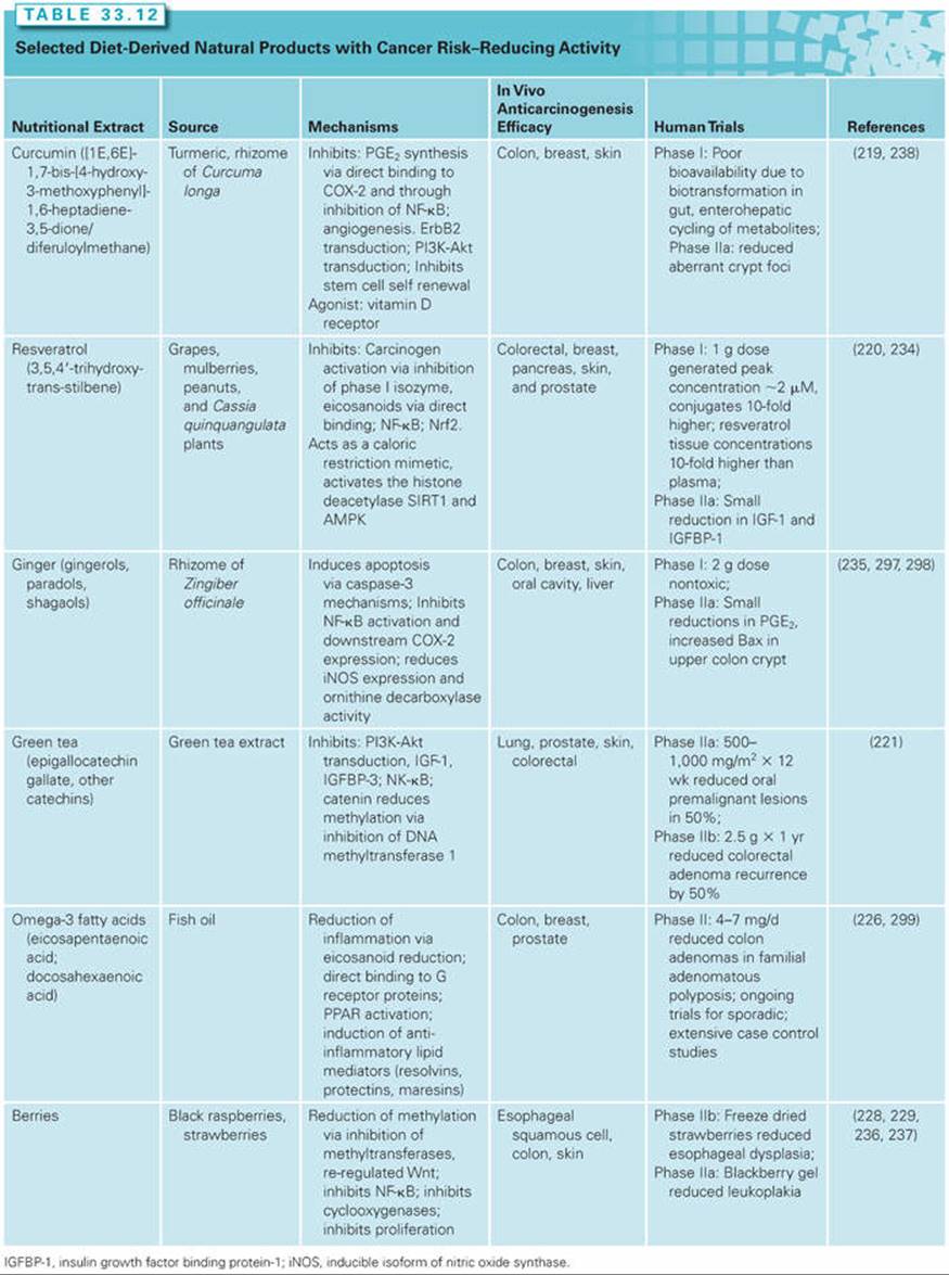
ANTI-INFECTIVES
Many infectious agents are known causes of human cancers, including the human hepatitis viruses, hepatitis B virus (HBV) and hepatitis C virus (HCV) for hepatocellular carcinoma239; Helicobacter pylori for gastric adenocarcinoma240; human papilloma viruses (HPV) for cervical, anal, vulva, penis, and oral cavity and pharynx carcinomas241; herpes virus-8 for Kaposi sarcoma242; Epstein-Barr virus for Burkitt and other lymphomas243; liver flukes for cholangiocarcinoma244; and schistosomes for bladder carcinoma.245 The success of the HPV vaccine at reducing the incidence of intraepithelial neoplasia of the cervix is one example that demonstrates the potential of immunochemoprevention for epithelial targets for which an etiologic agent can be identified.
Helicobacter pylori
Intestinal-type gastric adenocarcinoma arises through a multistep process that begins with chronic gastritis initiated by H. pylori, progressing through gastric mucosal atrophy, intestinal metaplasia to dysplasia, and ultimately, to adenocarcinoma.246 H. pylori infects 50% of the world’s population.240 Infection occurs early in life, remains quiescent, and may be associated with chronic gastritis of variable intensity but with minimal symptoms. Although the majority of H. pylori organisms remain in the gastric mucous layer, 10% adhere to the gastric mucosa through adhesion BabA, an outer membrane protein that binds to the Lewis-B histo-blood group antigen.240 Progression to atrophic gastritis and peptic ulcer disease (occurs in 10% to 15% of infected individuals) requires other bacterial and host cofactors.245,246 Infection with H. pylori is associated with an OR of 2.7 to 6.0 for gastric cancer; CagAincreases this risk by 20- to 40-fold. The risk of developing gastric adenocarcinoma with an H. pylori infection is estimated to be 1% to 3%.245,246
The eradication of H. pylori with antibiotics and anti-inflammatory agents—for example, amoxicillin, metronidazole, and bismuth subsalicylate—increases the rate of regression of nonmetaplastic gastric atrophy and intestinal metaplasia in geographically diverse regions.247,248 A combination of a 2-week course of a proton pump inhibitor (omeprazole) and an antibiotic (amoxicillin) reduced the risk of gastric cancer in a high-risk population in China (OR = 0.61; (95% CI, 0.36 to 0.96) for 14.7 years after the treatment.249 The sequence of giving proton pump inhibitors and antibiotic therapy does not alter the treatment outcome. In addition to contributing to gastric cancer risk, H. pyloriinfections may also contribute to pancreatic cancer risk.250 Because H. pylori infections are so widespread, mass eradication campaigns in high-risk regions are being considered.251 However, complicating this is that H. pyloriinfections have also been associated with a reduced risk of both esophageal adenocarcinoma and gastric cardia carcinoma.252
MULTIAGENT APPROACHES TO CANCER RISK REDUCTION
In the transition to molecularly targeted interventions, combinations of targets that logically address critical carcinogenic pathways may have greater efficacy than single agents. For example, previously demonstrated interactive signaling of EGFRs and COX-2 experiments in Min+ mice253 demonstrates cancer preventive synergism. Combining atorvastatin with selective or nonselective COX inhibitors enhanced the inhibition of azoxymethane-induced colon carcinogenesis in F344 rats and reduced the dose of the combined drugs required to achieve a reduction of colon carcinogenesis.254
DFMO plus sulindac inhibited adenoma formation in a phase IIb trial of 375 patients with a prior history of adenomas followed for 36 months (see Table 33.11).147 Cardiovascular-adverse outcomes were higher in DFMO/sulindac-treated patients who had preexisting high baseline cardiovascular risk; however, the cardiovascular-adverse events were similar to placebo in moderate or low cardiovascular risk patients.255 Using IGF-1 as a biomarker, Guerrieri-Gonzaga et al.256 showed that the combination of low-dose tamoxifen with low-dose fenretinide is safe but not synergistic. As more data accumulate from in vivo models, combined drugs aimed at specific targets in coordinated signaling pathways will enter clinical biomarker-based trials. Optimal doses, toxicity, and biomarker modulation data will select those combinations useful for risk reduction trials and, ultimately, generalized use in at-risk populations.
REFERENCES
1. Vogelstein B, Papadopoulos N, Velculescu VE, et al. Cancer genome landscapes. Science 2013;339:1546–1558.
2. Fearon ER, Vogelstein B. A genetic model for colorectal tumorigenesis. Cell 1990;61:759–767.
3. Sidransky D. Emerging molecular markers of cancer. Nat Rev Cancer 2002;2:210–219.
4. Lippman SM, Levin B, Brenner DE, et al. Cancer prevention and the American Society of Clinical Oncology. J Clin Oncol 2004;22:3848–3851.
5. Wattenberg L. Chemoprevention of cancer. Cancer Res 1985;45:1–8.
6. Greenwald P, Kelloff G. The role of chemoprevention in cancer control. IARC Scientific Publications (Lyon) 1996;139:13–22.
7. Hanahan D, Weinberg RA. The hallmarks of cancer. Cell 2000;100:57–70.
8. Steele VE, Boone CW, Lubet RA, et al. Preclinical drug development paradigms for chemopreventives. Hematol Oncol Clin North Am 1998;12:943–961.
9. Perloff M, Steele VE. Early-phase development of cancer prevention agents: challenges and opportunities. Cancer Prev Res (Phila) 2013;6:379–383.
10. National Cancer Institute. PREVENT Cancer Preclinical Drug Development Program. National Cancer Institute Web site. http://prevention.cancer.gov/programs-resources/programs/prevent. Accessed December 7, 2013.
11. Mehta RG, Naithani R, Huma L, et al. Efficacy of chemopreventive agents in mouse mammary gland organ culture (MMOC) model: a comprehensive review. Curr Med Chem. 2008;15:2785–2825.
12. Hoenerhoff MJ, Hong HH, Ton TV, et al. A review of the molecular mechanisms of chemically induced neoplasia in rat and mouse models in National Toxicology Program bioassays and their relevance to human cancer. Toxicol Pathol 2009;37:835–848.
13. Steele VE, Lubet RA. The use of animal models for cancer chemoprevention drug development. Semin Oncol 2010;37:327–338.
14. Shureiqi I, Reddy P, Brenner DE. Chemoprevention: general perspective. Crit Rev Oncol Hematol 2000;33:157–167.
15. Becker M. Adherence to prescribed therapies. Med Care 1985;23:539–554.
16. Miki Y, Swensen J, Shattuck-Eidens D, et al. A strong candidate for the breast and ovarian cancer susceptibility gene BRCA1. Science 1994;266:66–71.
17. Powell SM, Petersen GM, Krush AJ, et al. Molecular diagnosis of familial adenomatous polyposis. N Engl J Med 1993;329:1982–1987.
18. Meads C, Ahmed I, Riley RD. A systematic review of breast cancer incidence risk prediction models with meta-analysis of their performance. Breast Cancer Res Treat 2012;132:365–377.
19. Kastrinos F, Steyerberg EW, Balmana J, et al. Comparison of the clinical prediction model PREMM(1,2,6) and molecular testing for the systematic identification of Lynch syndrome in colorectal cancer. Gut 2013;62:272–279.
20. Ankerst DP, Boeck A, Freedland SJ, et al. Evaluating the PCPT risk calculator in ten international biopsy cohorts: results from the Prostate Biopsy Collaborative Group. World J Urol 2012;30:181–187.
21. National Institutes of Health, U.S. Food and Drug Administration. Biomarkers and Surrogate Endpoints: Advancing Clinical Research and Applications. Bethesda, MD: National Insitutes of Health; 1999.
22. Schatzkin A, Freedman LS, Schiffman MH, et al. Validation of intermediate end points in cancer research. J Natl Cancer Inst 1990;82:1746–1752.
23. Prentice R. Surrogate endpoints in clinical trials: definition and operational criteria. Statistics Med 1989;8:431–440.
24. Ransohoff DF. Rules of evidence for cancer molecular-marker discovery and validation. Nat Rev Cancer 2004;4:309–314.
25. Pepe MS, Feng Z, Janes H, et al. Pivotal evaluation of the accuracy of a biomarker used for classification or prediction: standards for study design. J Natl Cancer Inst 2008;100:1432–1438.
26. Pryor WA, Stahl W, Rock CL. Beta carotene: from biochemistry to clinical trials. Nutr Rev 2000;58:39–53.
27. Brenner DE, Hawk E. Trials and tribulations of interrogating biomarkers to define efficacy of cancer risk reductive interventions. Cancer Prev Res (Phila) 2013;6:71–73.
28. Fisher B, Costantino J, Wickerham D, et al. Tamoxifen for prevention of breast cancer: report of the National Surgical Adjuvant Breast and Bowel Project P-1 study. J Natl Cancer Inst 1998;90:1371–1388.
29. Vogel VG, Costantino JP, Wickerham DL, et al. Update of the National Surgical Adjuvant Breast and Bowel Project Study of Tamoxifen and Raloxifene (STAR) P-2 Trial: Preventing breast cancer. Cancer Prev Res (Phila) 2010;3:696–706.
30. Thompson IM, Goodman PJ, Tangen CM, et al. The influence of finasteride on the development of prostate cancer. N Engl J Med 2003;349:215–224.
31. Omenn G, Goodman G, Thornquist M, et al. Effects of a combination of beta carotene and vitamin A on lung cancer and cardiovascular disease. N Engl J Med 1996;334:1150–1155.
32. Lotan R. Retinoids in cancer chemoprevention. Faseb J 1996;10:1031–1039.
33. Tang X-H, Gudas LJ. Retinoids, retinoic acid receptors, and cancer. Annu Rev Pathol 2011;6:345–364.
34. Khachik F, Beecher, G, Smith JC Jr. Lutein, lycopene, and their oxidative metabolites in chemoprevention of cancer. J Cell Biochem 1995;22:236–246.
35. Di Mascio P, Kaiser S, Sies H. Lycopene as the most efficient biological carotenoid singlet oxygen quencher. Arch Biochem Biophys 1989;274:532–538.
36. Eroglu A, Hruszkewycz DP, dela Sena C, et al. Naturally occurring eccentric cleavage products of provitamin A beta-carotene function as antagonists of retinoic acid receptors. J Biol Chem 2012;287:15886–15895.
37. Ford NA, Erdman JW Jr. Are lycopene metabolites metabolically active? Acta Biochim Pol 2012;59:1–4.
38. World Cancer Research Fund, American Institute for Cancer Research. Food, Nutrition, Physical Activity, and the Prevention of Cancer: A Global Perspective. Washington, DC: AICR; 2007.
39. Panel on Dietary Antioxidants and Related Compounds, Subcommittees on Upper Reference Levels of Nutrients and Interpretation and Uses of DRIs, Standing Committee on the Scientific Evaluation of Dietary Reference Intakes, Food and Nutrition Board, Institute of Medicine. Dietary Reference Intakes for Vitamin C, Vitamin E, Selenium, and Carotenoids. Washington, DC: National Academy Press; 2000.
40. Moon RC, Mehta RG, Rao KVN. Retinoids and cancer in experimental animals. In: Sporn MB, Roberts AB, Goodman DS, eds., The Retinoids. 2nd ed. New York: Raven Press; 1994: 573–595.
41. International Agency for Research on Cancer World Health Organization. Carotenoids. Lyon: International Agency for Research on Cancer; 1998.
42. Holzapfel NP, Holzapfel BM, Champ S, et al. The potential role of lycopene for the prevention and therapy of prostate cancer: from molecular mechanisms to clinical evidence. Int J Mol Sci 2013;14:14620–14646.
43. Hong WK, Lippman SM, Itri LM, et al. Prevention of second primary tumors with 13cRA in squamous-cell carcinoma of the head and neck. N Engl J Med 1990;323:795–801.
44. Benner SE, Pajak TF, Lippman SM, et al. Prevention of second primary tumors with isotretinoin in patients with squamous cell carcinoma of the head and neck: long term follow-up. J Natl Cancer Inst 1994;86:140–141.
45. Khuri FR, Lee JJ, Lippman SM, et al. Randomized phase III trial of low-dose isotretinoin for prevention of second primary tumors in stage I and II head and neck cancer patients. J Natl Cancer Inst 2006;98:441–450.
46. Bolla M, Lefur R, Ton Van J, et al. Prevention of second primary tumours with etretinate in squamous cell carcinoma of the oral cavity and oropharynx. Results of a multicentric double-blind randomised study. Eur J Cancer 1994;30A:767–772.
47. Toma S, Bonelli L, Sartoris A, et al. beta-carotene supplementation in patients radically treated for stage I-II head and neck cancer: results of a randomized trial. Oncol Rep 2003;10:1895–1901.
48. Mayne ST, Cartmel B, Baum M, et al. Randomized trial of supplemental beta-carotene to prevent second head and neck cancer. Cancer Res 2001;61:1457–1463.
49. Bairati I, Meyer F, Jobin E, et al. Antioxidant vitamins supplementation and mortality: a randomized trial in head and neck cancer patients. Int J Cancer 2006;119:2221–2224.
50. McLarty JW, Holiday DB, Girard WM, et al. Beta-carotene, vitamin A and lung cancer chemoprevention: results of an intermediate endpoint study. Am J Clin Nutr 1995;62:1431S–1438S.
51. Lee JS, Lippman SM, Benner SE, et al. Randomized placebo-controlled trial of isotretinoin in chemoprevention of bronchial squamous metaplasia. J Clin Oncol 1994;12:937–945.
52. The Alpha-Tocopherol Beta Carotene Cancer Prevention Study Group. The effect of vitamin E and beta carotene on the incidence of lung cancer and other cancers in male smokers. N Engl J Med 1994;330:1029–1035.
53. Omenn GS, Goodman G, Thornquist M, et al. Chemoprevention of lung cancer: the beta-Carotene and Retinol Efficacy Trial (CARET) in high-risk smokers and asbestos-exposed workers. IARC Sci Publ 1996; 67–85.
54. Hennekens CH, Buring JE, Manson JE, et al. Lack of effect of long-term supplementation with beta carotene on the incidence of malignant neoplasms and cardiovascular disease. N Engl J Med 1996;334:1145–1149.
55. Lee IM, Cook NR, Manson JE, et al. Beta-carotene supplementation and incidence of cancer and cardiovascular disease: the Women’s Health Study. J Natl Cancer Inst 1999;91:2102–2106.
56. Heart Protection Study Collaborative Group. MRC/BHF Heart Protection Study of antioxidant vitamin supplementation in 20,536 high-risk individuals: a randomised placebo-controlled trial. Lancet 2002;360:23–33.
57. van Zandwijk N, Dalesio O, Pastorino U, et al. EUROSCAN, a randomized trial of vitamin A and N-acetylcysteine in patients with head and neck cancer or lung cancer. For the European Organization for Research and Treatment of Cancer Head and Neck and Lung Cancer Cooperative Groups. J Natl Cancer Inst 2000;92:977–986.
58. Lippman SM, Lee JJ, Karp DD, et al. Randomized phase III intergroup trial of isotretinoin to prevent second primary tumors in stage I non-small-cell lung cancer. J Natl Cancer Inst 2001;93:605–618.
59. Mayne ST, Lippman SM. Cigarettes: a smoking gun in cancer chemoprevention. J Natl Cancer Inst 2005;97:1319–1321.
60. Veronesi U, De Palo G, Marubini E, et al. Randomized trial of fenretinide to prevent second breast malignancy in women with early breast cancer. J Natl Cancer Inst 1999;91:1847–1856.
61. De Palo G, Mariani L, Camerini T, et al. Effect of fenretinide on ovarian carcinoma occurrence. Gynecol Oncol 2002;86:24–27.
62. Uray IP, Brown PH. Chemoprevention of hormone receptor-negative breast cancer: new approaches needed. Recent Results Cancer Res 2011;188:147–162.
63. Kraemer KH, DiGiovanna JJ, Moshell AN, et al. Prevention of skin cancer in xeroderma pigmentosum with the use of oral isotretinoin. N Engl J Med 1988;318:1633–1637.
64. Bouwes Bavinck JN, Tieben LM, Van Der Woude FJ, et al. Prevention of skin cancer and reduction of keratotic skin lesions during acitretin therapy in renal transplant recipients: a double-blind, placebo-controlled study. J Clin Oncol 1995;13:1933–1938.
65. Tangrea JA, Edwards BK, Taylor PR, et al. Long-term therapy with low-dose isotretinoin for prevention of basal cell carcinoma: a multicenter clinical trial Isotretinoin-Basal Cell Carcinoma Study Group. J Natl Cancer Inst 1992;84:328–332.
66. Levine N, Moon TE, Cartmel B, et al. Trial of retinol and isotretinoin in skin cancer prevention: a randomized, double-blind, controlled trial. Southwest Skin Cancer Prevention Study Group. Cancer Epidemiol Biomarkers Prev 1997;6:957–961.
67. Moon TE, Levine N, Cartmel B, et al. Effect of retinol in preventing squamous cell skin cancer in moderate-risk subjects: a randomized, double-blind, controlled trial. Southwest Skin Cancer Prevention Study Group. Cancer Epidemiol Biomarkers Prev 1997;6:949–956.
68. Greenberg ER, Baron JA, Stukel TA, et al. A clinical trial of beta carotene to prevent basal-cell and squamous-cell cancers of the skin. The Skin Cancer Prevention Study Group. N Engl J Med 1990;323:789–895.
69. Green A, Williams G, Neale R, et al. Daily sunscreen application and betacarotene supplementation in prevention of basal-cell and squamous-cell carcinomas of the skin: a randomised controlled trial. Lancet 1999;354:723–729.
70. Clark LC, Combs GF Jr, Turnbull BW, et al. Effects of selenium supplementation for cancer prevention in patients with carcinoma of the skin. A randomized controlled trial. Nutritional Prevention of Cancer Study Group. JAMA 1996;276:1957–1963.
71. Duffield-Lillico AJ, Slate EH, Reid ME, et al. Nutritional Prevention of Cancer Study Group. Selenium supplementation and secondary prevention of nonmelanoma skin cancer in a randomized trial. J Natl Cancer Inst 2003;95:1477–1481.
72. Sabichi AL, Lerner SP, Atkinson EN, et al. Phase III prevention trial of fenretinide in patients with resected non-muscle-invasive bladder cancer. Clin Cancer Res 2008;14:224–229.
73. Lamm DL, Riggs DR, Shriver JS, et al. Megadose vitamins in bladder cancer: a double-blind clinical trial. J Urol 1994;151:21–26.
74. Meyskens FL Jr, Surwit E, Moon TE, et al. Enhancement of regression of cervical intraepithelial neoplasia II (moderate dysplasia) with topically applied all-trans-retinoic acid: a randomized trial. J Natl Cancer Inst 1994;86:539–543.
75. Blot WJ, Li JY, Taylor PR, et al. Nutrition intervention trials in Linxian, China: supplementation with specific vitamin/mineral combinations, cancer incidence, and disease-specific mortality in the general population. J Natl Cancer Inst 1993;85:1483–1492.
76. Qiao YL, Dawsey SM, Kamangar F, et al. Total and cancer mortality after supplementation with vitamins and minerals: follow-up of the Linxian General Population Nutrition Intervention Trial. J Natl Cancer Inst 2009;101:507–518.
77. Mayne ST, Ferrucci LM, Cartmel B. Lessons learned from randomized clinical trials of micronutrient supplementation for cancer prevention. Annu Rev Nutr 2012;32:369–390.
78. Li JY, Taylor PR, Li B, et al. Nutrition intervention trials in Linxian, China: multiple vitamin/mineral supplementation, cancer incidence, and disease-specific mortality among adults with esophageal dysplasia. J Natl Cancer Inst 1993;85:1492–1498.
79. Kikendall JW, Mobarhan S, Nelson R, et al. Oral beta carotene does not reduce the recurrence of colorectal adenomas [Abstract]. Am J Gastroenterol 1991;36:1356.
80. MacLennan R, Macrae F, Bain C, et al. Randomized trial of intake of fat, fiber, and beta carotene to prevent colorectal adenomas. J Natl Cancer Inst 1995;87:1760–1766.
81. Greenberg ER, Baron JA, Tosteson TD, et al. A clinical trial of antioxidant vitamins to prevent colorectal adenoma. Polyp Prevention Study Group. N Engl J Med 1994;331:141–147.
82. Baron JA, Cole BF, Mott L, et al. Neoplastic and antineoplastic effects of beta-carotene on colorectal adenoma recurrence: results of a randomized trial. J Natl Cancer Inst 2003;95:717–722.
83. Heinonen OP, Albanes D, Virtamo J, et al. Prostate cancer and supplementation with alpha-tocopherol and beta-carotene: incidence and mortality in a controlled trial. J Natl Cancer Inst 1998;90:440–446.
84. Lippman SM, Klein EA, Goodman PJ, et al. Effect of selenium and vitamin E on risk of prostate cancer and other cancers: the Selenium and Vitamin E Cancer Prevention Trial (SELECT). JAMA 2009;301:39–51.
85. Klein EA, Thompson IM Jr, Tangen CM, et al. Vitamin E and the risk of prostate cancer: the Selenium and Vitamin E Cancer Prevention Trial (SELECT). JAMA 2011;306:1549–1556.
86. Gaziano JM, Glynn RJ, Christen WG, et al. Vitamins E and C in the prevention of prostate and total cancer in men: the Physicians’ Health Study II randomized controlled trial. JAMA 2009;301:52–62.
87. Boileau TW, Liao Z, Kim S, et al. Prostate carcinogenesis in N-methyl-N-nitrosourea (NMU)-testosterone-treated rats fed tomato powder, lycopene, or energy-restricted diets. J Natl Cancer Inst 2003;95:1578–1586.
88. Goralczyk R. Beta-carotene and lung cancer in smokers: review of hypotheses and status of research. Nutr Cancer 2009;61:767–774.
89. Kristal AR, Darke AK, Morris JS, et al. Baseline Selenium Status and Effects of Selenium and Vitamin E Supplementation on Prostate Cancer Risk. J Natl Cancer Inst 2014:106:djt456.
90. Stone JR, Yang S. Hydrogen peroxide: a signaling messenger. Antioxid Redox Signal 2006;8:243–270.
91. Finkel T. Signal transduction by reactive oxygen species. J Cell Biol 2011;194:7–15.
92. Sayin V, Ibrahim M, Larsson E, et al. Antioxidants accelerate lung cancer progression in mice. Sci Transl Med 2014;6:ra15.
93. Mayne ST. Oxidative stress, dietary antioxidant supplements, and health: is the glass half full or half empty? Cancer Epidemiol Biomarkers Prev 2013;22:2145–2147.
94. Duthie SJ, Narayanan S, Blum S, et al. Folate deficiency in vitro induces uracil misincorporation and DNA hypomethylation and inhibits DNA excision repair in immortalized normal human colon epithelial cells. Nutr Cancer 2000;37:245–251.
95. Blount BC, Mack MM, Wehr CM, et al. Folate deficiency causes uracil misincorporation into human DNA and chromosome breakage: implications for cancer and neuronal damage. Proc Natl Acad Sci U S A 1997;94:3290–3295.
96. Giovannucci E. Epidemiologic studies of folate and colorectal neoplasia: a review. J Nutr 2002;132:2350S–2355S.
97. White E, Shannon JS, Patterson RE. Relationship between vitamin and calcium supplement use and colon cancer. Cancer Epidemiol Biomarkers Prev 1997;6:769–774.
98. Jacobs EJ, Connell CJ, Patel AV, et al. Multivitamin use and colon cancer mortality in the Cancer Prevention Study II cohort (United States). Cancer Causes Control 2001;12:927–934.
99. Gibson TM, Weinstein SJ, Pfeiffer RM, et al. Pre- and postfortification intake of folate and risk of colorectal cancer in a large prospective cohort study in the United States. Am J Clin Nutr 2011;94:1053–1062.
100. Chen J, Giovannucci E, Kelsey, K, et al. A methylenetetrahydrofolate reductase polymorphism and the risk of colorectal cancer. Cancer Res 1996;56:4862–4864.
101. Larsson S, Giovannucci, E, Wolk A. Vitamin B6 intake, alcohol consumption, and colorectal cancer: a longitudinal population-based cohort of women. Gastroenterology 2005;128:1830–1837.
102. Wei E, Giovannucci E, Selhub J, et al. Plasma vitamin B6 and the risk of colorectal cancer and adenoma in women. J Natl Cancer Inst 2005;97:684–692.
103. Giovannucci E, Rimm EB, Ascherio A, et al. Alcohol, low-methionine-low-folate diets, and risk of colon cancer in men. J Natl Cancer Inst 1995;87:265–273.
104. Jaszewski R, Misra S, Tobi M, et al. Folic acid supplementation inhibits recurrence of colorectal adenomas: a randomized chemoprevention trial. World J Gastroenterol 2008;14:4492–4498.
105. Paspatis GA, Karamanolis DG. Folate supplementation and adenomatous colonic polyps. Dis Colon Rectum 1994;37:1340–1341.
106. Cole BF, Baron JA, Sandler RS, et al. Folic acid for the prevention of colorectal adenomas: a randomized clinical trial. JAMA 2007;297:2351–2359.
107. Logan RF, Grainge MJ, Shepherd VC, et al. Aspirin and folic acid for the prevention of recurrent colorectal adenomas. Gastroenterology 2008;134:29–38.
108. Wu K, Platz EA, Willett WC, et al. A randomized trial on folic acid supplementation and risk of recurrent colorectal adenoma. Am J Clin Nutr 2009;90:1623–1631.
109. Gao Q-Y, Chen H-M, Chen Y-X, et al. Folic acid prevents the initial occurrence of sporadic colorectal adenoma in Chinese older than 50 years of age: a randomized clinical trial. Cancer prevention research (Phila) 2013;6:744–752.
110. Ramnath N, Kim S, Christensen PJ. Vitamin D and lung cancer. Expert Rev Respir Med 2011;5:305–309.
111. Deeb KK, Trump DL, Johnson CS. Vitamin D signalling pathways in cancer: potential for anticancer therapeutics. Nat Rev Cancer 2007;7:684–700.
112. McCullough M, Robertson AS, Rodriguez C, et al. Calcium, vitamin D, dairy products, and risk of colorectal cancer in the cancer prevention study II nutrition cohort (United States). Cancer Causes Control 2003;14:1–12.
113. Wu K, Willett WC, Fuchs CS, et al. Calcium intake and risk of colon cancer in women and men. J Natl Cancer Inst 2002;94:437–446.
114. Chung M, Balk EM, Brendel M, et al. Vitamin D and Calcium: A Systematic Review of Health Outcomes. Evidence Report No. 183 (Prepared by the Tufts Evidence-based Practice Center). Rockville, MD: Agency for Healthcare Research and Quality; 2009.
115. World Health Organization, International Agency for Research on Cancer. Vitamin D and Cancer. Working Group Reports, Volume 5. Lyon, France: IARC; 2008.
116. Stolzenberg-Solomon RZ, Jacobs EJ, Arslan AA, et al. Circulating 25-hydroxyvitamin D and risk of pancreatic cancer: Cohort Consortium Vitamin D Pooling Project of Rarer Cancers. Am J Epidemiol 2010;172:81–93.
117. Baron J, Beach M, Mandel JS, et al. Calcium supplements for the prevention of colorectal adenomas. The Calcium Polyp Prevention Study Group. N Engl J Med 1999;340:101–107.
118. Bonithon-Kopp C, Kronborg O, Giacosa A, et al. Calcium and fibre supplementation in prevention of colorectal adenoma recurrence: a randomized intervention trial. Lancet 2000;356:1300–1306.
119. Wactawski-Wende J, Kotchen JM, Anderson GL, et al. Calcium plus vitamin D supplementation and the risk of colorectal cancer. N Engl J Med 2006;354:684–696.
120. Trivedi DP, Doll R, Khaw KT. Effect of four monthly oral vitamin D3 (cholecalciferol) supplementation on fractures and mortality in men and women living in the community: randomised double blind controlled trial. BMJ 2003;326:469.
121. Cui Y, Rohan TE. Vitamin D, calcium, and breast cancer risk: a review. Cancer Epidemiol Biomarkers Prev 2006;15:1427–1437.
122. Chlebowski RT, Johnson KC, Kooperberg C, et al. Calcium plus vitamin D supplementation and the risk of breast cancer. J Natl Cancer Inst 2008;100:1581–1591.
123. Lappe JM, Travers-Gustafson D, Davies KM, et al. Vitamin D and calcium supplementation reduces cancer risk: results of a randomized trial. Am J Clin Nutr 2007;85:1586–1591.
124. Manson JE, Bassuk SS, Lee IM, et al. The VITamin D and OmegA-3 TriaL (VITAL): rationale and design of a large randomized controlled trial of vitamin D and marine omega-3 fatty acid supplements for the primary prevention of cancer and cardiovascular disease. Contemp Clin Trials 2012;33:159–171.
125. Wu X, Spitz MR, Lee JJ, et al. Novel susceptibility loci for second primary tumors/recurrence in head and neck cancer patients: large-scale evaluation of genetic variants. Cancer Prev Res (Phila) 2009;2:617–624.
126. Platz EA. Is prostate cancer prevention with selenium all in the genes? Cancer Prev Res (Phila) 2010;3:576–578.
127. Miller JW, Ulrich CM. Folic acid and cancer—where are we today? Lancet 2013;381:974–976.
128. Thun MJ, Henley SJ, Patrono C. Nonsteroidal anti-inflammatory drugs as anticancer agents: mechanistic, pharmacologic, and clinical issues. J Natl Cancer Inst 2002;94:252–266.
129. Wang D, Mann JR, DuBois RN. The role of prostaglandins and other eicosanoids in the gastrointestinal tract. Gastroenterology 2005;128:1445–1461.
130. Gurpinar E, Grizzle WE, Piazza GA. COX-Independent Mechanisms of Cancer Chemoprevention by Anti-Inflammatory Drugs. Front Oncol 2013;3:181.
131. Rothwell PM, Wilson M, Price JF, et al. Effect of daily aspirin on risk of cancer metastasis: a study of incident cancers during randomised controlled trials. Lancet 2012;379:1591–1601.
132. Rothwell PM, Price JF, Fowkes FG, et al. Short-term effects of daily aspirin on cancer incidence, mortality, and non-vascular death: analysis of the time course of risks and benefits in 51 randomised controlled trials. Lancet 2012;379:1602–1612.
133. Thun MJ, Namboodiri MM, Heath C Jr. Aspirin use and reduced risk of fatal colon cancer. N Engl J Med 1991;325:1593–1596.
134. Chan AT, Giovannucci EL, Meyerhardt JA, et al. Aspirin dose and duration of use and risk of colorectal cancer in men. Gastroenterology 2008;134:21–28.
135. Chan AT, Giovannucci EL, Meyerhardt JA, et al. Long-term use of aspirin and nonsteroidal anti-inflammatory drugs and risk of colorectal cancer. JAMA 2005;294:914–923.
136. Liao LM, Vaughan TL, Corley DA, et al. Nonsteroidal anti-inflammatory drug use reduces risk of adenocarcinomas of the esophagus and esophagogastric junction in a pooled analysis. Gastroenterology 2012;142:442–452.
137. Pollard M, Luckert PH. Effect of indomethacin on intestinal tumor induced in rats by the acetate derivative of dimethylnitrosamine. Science 1981;214:558–559.
138. Jacoby RF, Marshall DJ, Newton MA, et al. Chemoprevention of spontaneous intestinal adenomas in the Apc Min mouse model by the nonsteroidal anti-inflammatory drug piroxicam. Cancer Res 1996;56:710–714.
139. Kawamori T, Rao C, Seibert K, et al. Chemopreventive effect of celecoxib, a specific cyclooxygenase-2 inhibitor on colon carcinogenesis. Cancer Res 1998;58:409–412.
140. Oshima M, Dinchuk JE, Kargman SL. Suppression of intestinal polyposis in Apc delta 716 knockout ice by inhibition of cyclooxygenase 2 (COX-2). Cell 1996;87:803–809.
141. Anderson WF, Umar A, Viner JL, et al. The role of cyclooxygenase inhibitors in cancer prevention. Curr Pharm Des 2002;8:1035–1062.
142. Giardiello FM, Yang VW, Hylind LM, et al. Primary chemoprevention of familial adenomatous polyposis with sulindac. N Engl J Med 2002;346:1054–1059.
143. Steinbach G, Lynch PM, Phillips RK. The effect of celecoxib, a cyclooxygenase-2 inhibitor, in familial adenomatous polyposis. N Engl J Med 2000;342:1946–1952.
144. Thorson AG, Lynch HT, Smyrk TC. Rectal cancer in FAP patient after sulindac. Lancet 1994;343:180.
145. Calaluce R, Earnest DL, Heddens D, et al. Effects of piroxicam on prostaglandin E2 levels in rectal mucosa of adenomatous polyp patients: a randomized phase IIb trial. Cancer Epidemiol Biomarkers Prev 2000;9:1287–1292.
146. Ladenheim J, Garcia G, Titzer D, et al. Effects of sulindac on sporadic colonic polyps. Gastroenterology 1995;108:1083–1087.
147. Meyskens FL Jr, McLaren CE, Pelot D, et al. Difluoromethylornithine plus sulindac for the prevention of sporadic colorectal adenomas: a randomized placebo-controlled, double-blind trial. Cancer Prev Res (Phila) 2008;1:32–38.
148. Arber N, Eagle CJ, Spicak J, et al. Celecoxib for the prevention of colorectal adenomatous polyps. N Engl J Med 2006;355:885–895.
149. Bertagnolli MM, Eagle CJ, Zauber AG, et al. Celecoxib for the prevention of sporadic colorectal adenomas. N Engl J Med 2006;355:873–884.
150. Baron JA, Sandler RS, Bresalier RS, et al. A randomized trial of rofecoxib for the chemoprevention of colorectal adenomas. Gastroenterology 2006;131:1674–1682.
151. Bresalier RS, Sandler RS, Quan H, et al. Cardiovascular events associated with rofecoxib in a colorectal adenoma chemoprevention trial. N Engl J Med 2005;352:1092–1102.
152. Solomon SD, McMurray JJ, Pfeffer MA, et al. Cardiovascular risk associated with celecoxib in a clinical trial for colorectal adenoma prevention. N Engl J Med 2005;352:1071–1080.
153. Rostom A, Dube C, Lewin G, et al. Nonsteroidal anti-inflammatory drugs and cyclooxygenase-2 inhibitors for primary prevention of colorectal cancer: a systematic review prepared for the U.S. Preventive Services Task Force. Ann Intern Med 2007;146:376–389.
154. Dube C, Rostom A, Lewin G, et al. The use of aspirin for primary prevention of colorectal cancer: a systematic review prepared for the U.S. Preventive Services Task Force. Ann Intern Med 2007;146:365–375.
155. Rothwell PM, Fowkes FG, Belch JF, et al. Effect of daily aspirin on long-term risk of death due to cancer: analysis of individual patient data from randomised trials. Lancet 2011;377:31–41.
156. Rothwell PM. Aspirin in prevention of sporadic colorectal cancer: current clinical evidence and overall balance of risks and benefits. Recent Results Cancer Res 2013;191:121–142.
157. U.S. Preventive Services Task Force. Routine aspirin or nonsteroidal anti-inflammatory drugs for the primary prevention of colorectal cancer: U.S. Preventive Services Task Force recommendation statement. Ann Intern Med 2007;146:361–364.
158. Mulshine JL, Atkinson JC, Greer RO, et al. Randomized, double-blind, placebo-controlled phase IIb trial of the cyclooxygenase inhibitor ketorolac as an oral rinse in oropharyngeal leukoplakia. Clin Cancer Res 2004;10:1565–1573.
159. Mao JT, Fishbein MC, Adams B, et al. Celecoxib decreases Ki-67 proliferative index in active smokers. Clin Cancer Res 2006;12:314–320.
160. Bord S, Horner A, Beavan S, et al. Estrogen receptors alpha and beta are differentially expressed in developing human bone. J Clin Endocrinol Metab 2001;86:2309–2314.
161. Kuiper GG, Carlsson B, Grandien K, et al. Comparison of the ligand binding specificity and transcript tissue distribution of estrogen receptors alpha and beta. Endocrinology 1997;138:863–870.
162. Fabian CJ, Kimler BF. Selective estrogen-receptor modulators for primary prevention of breast cancer. J Clin Oncol 2005;23:1644–1655.
163. Jordan VC. SERMs: meeting the promise of multifunctional medicines. J Natl Cancer Inst 2007;99:350–356.
164. Jordan VC. Tamoxifen (ICI46,474) as a targeted therapy to treat and prevent breast cancer. Br J Pharmacol 2006;147:S269–S276.
165. Jordan VC. Effect of tamoxifen (ICI 46,474) on initiation and growth of DMBA-induced rat mammary carcinomata. Eur J Cancer 1976;12:419–424.
166. Jordan VC. Chemoprevention of breast cancer with selective oestrogen-receptor modulators. Nat Rev Cancer 2007;7:46–53.
167. Cuzick J, Baum M. Tamoxifen and contralateral breast cancer. Lancet 1985;2:282.
168. Fisher B, Redmond C. New perspective on cancer of the contralateral breast: a marker for assessing tamoxifen as a preventive agent. J Natl Cancer Inst 1991;83:1278–1280.
169. Ettinger B, Black DM, Mitlak BH, et al. Reduction of vertebral fracture risk in postmenopausal women with osteoporosis treated with raloxifene: results from a 3-year randomized clinical trial. Multiple Outcomes of Raloxifene Evaluation (MORE) Investigators. JAMA1999;282:637–645.
170. LaCroix AZ, Powles T, Osborne CK, et al. Breast cancer incidence in the randomized PEARL trial of lasofoxifene in postmenopausal osteoporotic women. J Natl Cancer Inst 2010;102:1706–1715.
171. Cummings SR, Ensrud K, Delmas PD, et al. Lasofoxifene in postmenopausal women with osteoporosis. N Engl J Med 2010;362:686–696.
172. Cummings SR, McClung M, Reginster JY, et al. Arzoxifene for prevention of fractures and invasive breast cancer in postmenopausal women. J Bone Miner Res 2011;26:397–404.
173. Powles TJ, Diem SJ, Fabian CJ, et al. Breast cancer incidence in postmenopausal women with osteoporosis or low bone mass using arzoxifene. Breast Cancer Res Treat 2012;134:299–306.
174. Nelson HD, Smith ME, Griffin JC, et al. Use of medications to reduce risk for primary breast cancer: a systematic review for the U.S. Preventive Services Task Force. Ann Intern Med 2013;158:604–614.
175. Moyer VA. Medications for risk reduction of primary breast cancer in women: U.S. Preventive Services Task Force recommendation statement. Ann Intern Med 2013;159:698–708.
176. Visvanathan K, Hurley P, Bantug E, et al. Use of pharmacologic interventions for breast cancer risk reduction: American Society of Clinical Oncology clinical practice guideline. J Clin Oncol 2013;31:2942–2962.
177. Vogel VG, Costantino JP, Wickerham DL, et al. Carcinoma in situ outcomes in National Surgical Adjuvant Breast and Bowel Project Breast Cancer Chemoprevention Trials. J Natl Cancer Inst Monogr 2010;2010:181–186.
178. Cuzick J, Sestak I, Bonanni B, et al. Selective oestrogen receptor modulators in prevention of breast cancer: an updated meta-analysis of individual participant data. Lancet 2013;381:1827–1834.
179. Barrett-Connor E, Mosca L, Collins P, et al. Effects of raloxifene on cardiovascular events and breast cancer in postmenopausal women. N Engl J Med 2006;355:125–137.
180. Snyder KR, Sparano N, Malinowski JM. Raloxifene hydrochloride. Am J Health Syst Pharm 2000;57:1669–1675.
181. Goss PE, Ingle JN, Martino S, et al. A randomized trial of letrozole in postmenopausal women after five years of tamoxifen therapy for early-stage breast cancer. N Engl J Med 2003;349:1793–1802.
182. Fabian CJ, Kimler BF, Zalles CM, et al. Reduction in proliferation with six months of letrozole in women on hormone replacement therapy. Breast Cancer Res Treat 2007;106:75–84.
183. Goss PE, Ingle JN, Ales-Martinez JE, et al. Exemestane for breast-cancer prevention in postmenopausal women. N Engl J Med 2011;364:2381–2391.
184. Cheung AM, Tile L, Cardew S, et al. Bone density and structure in healthy postmenopausal women treated with exemestane for the primary prevention of breast cancer: a nested substudy of the MAP.3 randomised controlled trial. Lancet Oncol 2012;13:275–284.
185. Cuzick J, Sestak I, Forbes JF, et al. Anastrozole for prevention of breast cancer in high-risk postmenopausal women (IBIS-II): an international, double-blind, randomised placebo-controlled trial. Lancet 2014;383:1041–1048.
186. Waters EA, McNeel TS, Stevens WM, et al. Use of tamoxifen and raloxifene for breast cancer chemoprevention in 2010. Breast Cancer Res Treat 2012;134:875–880.
187. King MC, Wieand S, Hale K, et al. Tamoxifen and breast cancer incidence among women with inherited mutations in BRCA1 and BRCA2: National Surgical Adjuvant Breast and Bowel Project (NSABP-P1) Breast Cancer Prevention Trial. JAMA 2001;286:2251–2256.
188. Gronwald J, Tung N, Foulkes WD, et al. Tamoxifen and contralateral breast cancer in BRCA1 and BRCA2 carriers: an update. Int J Cancer 2006;118:2281–2284.
189. Hess-Wilson JK, Knudsen KE. Endocrine disrupting compounds and prostate cancer. Cancer Lett 2006;241:1–12.
190. Andriole G, Bostwick D, Civantos F, et al. The effects of 5alpha-reductase inhibitors on the natural history, detection and grading of prostate cancer: current state of knowledge. J Urol 2005;174:2098–2104.
191. Thompson IM, Tangen CM, Goodman PJ, et al. Chemoprevention of prostate cancer. J Urol 2009;182:499–507.
192. Andriole GL, Bostwick DG, Brawley OW, et al. Effect of dutasteride on the risk of prostate cancer. N Engl J Med 2010;362:1192–1202.
193. Thompson IM Jr, Goodman PJ, Tangen CM, et al. Long-term survival of participants in the prostate cancer prevention trial. N Engl J Med 2013;369:603–610.
194. Gerner EW, Meyskens FL Jr. Polyamines and cancer: old molecules, new understanding. Nat Rev Cancer 2004;4:781–792.
195. Meyskens FL Jr, Gerner EW. Development of difluoromethylornithine (DFMO) as a chemoprevention agent. Clin Cancer Res 1999;5:945–951.
196. Love R, Carbone P, Verma A, et al. Randomized phase I chemoprevention dose seeking study of alpha-difluoromethylornithine. J Natl Cancer Inst 1993;85:732–737.
197. Alberts DS, Dorr RT, Einspahr JG, et al. Chemoprevention of human actinic keratoses by topical 2-(difluoromethyl)-dl-ornithine. Cancer Epidemiol Biomarkers Prev 2000;9:1281–1286.
198. Meyskens FL Jr, Surwit E, Moon TE, et al. Enhancement of regression of cervical intraepithelial neoplasia II (moderate dysplasia) with topically applied all-trans-retinoic acid: randomized trial. J Natl Cancer Inst 1994;86:539–543.
199. Fabian CJ, Kimler BF, Brady DA, et al. A phase II breast cancer chemoprevention trial of oral alpha-difluoromethylornithine: breast tissue, imaging, and serum and urine biomarkers. Clin Cancer Res 2002;8:3105–3117.
200. Jeter JM, Alberts DS. Difluoromethylornithine: the proof is in the polyamines. Cancer Prev Res (Phila) 2012;5:1341–1344.
201. Gronich N, Rennert G. Beyond aspirin-cancer prevention with statins, metformin and bisphosphonates. Nat Rev Clin Oncol 2013;10:625–642.
202. Moyad MA. Why a statin and/or another proven heart healthy agent should be utilized in the next major cancer chemoprevention trial: part II. Urologic Oncol 2004;22:472–477.
203. Bertagnolli MM, Hsu M, Hawk ET, et al. Statin use and colorectal adenoma risk: results from the adenoma prevention with celecoxib trial. Cancer Prev Res (Phila) 2010;3:588–596.
204. Simon MS, Rosenberg CA, Rodabough RJ, et al. Prospective analysis of association between use of statins or other lipid-lowering agents and colorectal cancer risk. Ann Epidemiol 2012;22:17–27.
205. Platz EA, Leitzmann MF, Visvanathan K, et al. Statin drugs and risk of advanced prostate cancer. J Natl Cancer Inst 2006;98:1819–1825.
206. Eliassen AH, Colditz GA, Rosner B, et al. Serum lipids, lipid-lowering drugs, and the risk of breast cancer. Arch Intern Med 2005;165:2264–2271.
207. Lipkin SM, Chao EC, Moreno V, et al. Genetic variation in 3-hydroxy-3-methylglutaryl CoA reductase modifies the chemopreventive activity of statins for colorectal cancer. Cancer Prev Res (Phila) 2010;3:597–603.
208. Rennert G. Bisphosphonates: beyond prevention of bone metastases. J Natl Cancer Inst 2011;103:1728–1729.
209. Rennert G, Pinchev M, Rennert HS. Use of bisphosphonates and risk of postmenopausal breast cancer. J Clin Oncol 2010;28:3577–3581.
210. Chlebowski RT, Chen Z, Cauley JA, et al. Oral bisphosphonate use and breast cancer incidence in postmenopausal women. J Clin Oncol 2010;28:3582–3590.
211. Monsees GM, Malone KE, Tang MT, et al. Bisphosphonate use after estrogen receptor-positive breast cancer and risk of contralateral breast cancer. J Natl Cancer Inst 2011;103:1752–1760.
212. Kirpichnikov D, McFarlane SI, Sowers JR. Metformin: an update. Ann Intern Med 2002;137:25–33.
213. Pollack MN. Insulin, insulin-like growth factors, insulin resistance, and neoplasia. Am J Clin Nutr 2007;86:s820–s822.
214. Evans JM, Donnelly LA, Emslie-Smith AM, et al. Metformin and reduced risk of cancer in diabetic patients. BMJ 2005;330:1304–1305.
215. Zhu P, Davis M, Blackwelder A, et al. Metformin selectively targets tumor initiating cells in erbB-2 overexpressing breast cancer models. Cancer Prev Res (Phila) 2014;7:199–210.
216. Lonardo E, Cioffi M, Sancho P, et al. Metformin targets the metabolic achilles heel of human pancreatic cancer stem cells. PLoS One 2013;8:e76518.
217. Grubbs C, Clapper M, Reid J, et al. Metformin Promotes Tumorigenesis in Animal Models of Cancer Prevention. Washington, DC: American Association for Cancer Research; 2013.
218. Chlebowski RT, McTiernan A, Wactawski-Wende J, et al. Diabetes, metformin, and breast cancer in postmenopausal women. J Clin Oncol 2012;30:2844–2852.
219. Heger M, van Golen RF, Broekgaarden M, et al. The molecular basis for the pharmacokinetics and pharmacodynamics of curcumin and its metabolites in relation to cancer. Pharmacol Rev 2014;66:222–307.
220. Whitlock NC, Baek SJ. The anticancer effects of resveratrol: modulation of transcription factors. Nutr Cancer 2012;64:493–502.
221. Lambert JD. Does tea prevent cancer? Evidence from laboratory and human intervention studies. Am J Clin Nutr 2013;98:1667S–1675S.
222. Kakarala M, Brenner DE, Korkaya H, et al. Targeting breast stem cells with the cancer preventive compounds curcumin and piperine. Breast Cancer Res Treat 2010;122:777–785.
223. Norris L, Karmokar A, Howells L, et al. The role of cancer stem cells in the anti-carcinogenicity of curcumin. Mol Nutr Food Res 2013;57:1630–1637.
224. Zou H, Yuan C, Dong L, et al. Human cyclooxygenase-1 activity and its responses to COX inhibitors are allosterically regulated by nonsubstrate fatty acids. J Lipid Res 2012;53:1336–1347.
225. Wada M, Delong CJ, Hong YH, et al. Enzymes and receptors of prostaglandin pathways with arachidonic acid- vs. eicosapentaenoic acid-derived substrates and products. J Biol Chem 2007;282:22254–22266.
226. Laviano A, Rianda S, Molfino A, et al. Omega-3 fatty acids in cancer. Curr Opin Clin Nutr Metab Care 2013;16:156–161.
227. Cockbain AJ, Toogood GJ, Hull MA. Omega-3 polyunsaturated fatty acids for the treatment and prevention of colorectal cancer. Gut 2012;61:135–149.
228. Stoner GD, Wang LS, Casto BC. Laboratory and clinical studies of cancer chemoprevention by antioxidants in berries. Carcinogenesis 2008;29:1665–1674.
229. Wang LS, Dombkowski AA, Seguin C, et al. Mechanistic basis for the chemopreventive effects of black raspberries at a late stage of rat esophageal carcinogenesis. Mol Carcinog 2011;50:291–300.
230. Wang LS, Kuo CT, Stoner K, et al. Dietary black raspberries modulate DNA methylation in dextran sodium sulfate (DSS)-induced ulcerative colitis. Carcinogenesis 2013;34:2842–2850.
231. Ireson C, Orr S, Jones DJ, et al. Characterization of metabolites of the chemopreventive agent curcumin in human and rat hepatocytes and in the rat in vivo, and evaluation of their ability to inhibit phorbol ester-induced prostaglandin E2 production. Cancer Res 2001;61:1058–1064.
232. Ireson CR, Jones DJ, Orr S, et al. Metabolism of the cancer chemopreventive agent curcumin in human and rat intestine. Cancer Epidemiol Biomarkers Prev 2002;11:105–111.
233. Vareed SK, Kakarala M, Ruffin MT, et al. Pharmacokinetics of curcumin conjugate metabolites in healthy human subjects. Cancer Epidemiol Biomarkers Prev 2008;17:1411–1417.
234. Gescher A, Steward WP, Brown K. Resveratrol in the management of human cancer: how strong is the clinical evidence? Ann N Y Acad Sci 2013;1290:12–20.
235. Stoner GD. Ginger: is it ready for prime time? Cancer Prev Res (Phila) 2013;6:257–262.
236. Stoner GD, Wang LS. Chemoprevention of esophageal squamous cell carcinoma with berries. Top Curr Chem 2013;329:1–20.
237. Mallery SR, Zwick JC, Pei P, et al. Topical application of a bioadhesive black raspberry gel modulates gene expression and reduces cyclooxygenase 2 protein in human premalignant oral lesions. Cancer Res 2008;68:4945–4957.
238. Carroll RE, Benya RV, Turgeon DK, et al. Phase IIa clinical trial of curcumin for the prevention of colorectal neoplasia. Cancer Prev Res (Phila) 2011;4:354–364.
239. Seeff LB, Hoofnagle JH. Epidemiology of hepatocellular carcinoma in areas of low hepatitis B and hepatitis C endemicity. Oncogene 2006;25:3771–3777.
240. Fox JG, Wang TC. Inflammation, atrophy, and gastric cancer. J Clin Invest 2007;117:60–69.
241. Saslow D, Castle PE, Cox JT, et al. American Cancer Society Guideline for human papillomavirus (HPV) vaccine use to prevent cervical cancer and its precursors. CA Cancer J Clin 2007;57:7–28.
242. Mohanna S, Maco V, Bravo F, et al. Epidemiology and clinical characteristics of classic Kaposi’s sarcoma, seroprevalence, and variants of human herpesvirus 8 in South America: a critical review of an old disease. Int J Infect Dis 2005;9:239–250.
243. Castillo JJ, Reagan JL, Bishop KD, et al. Viral lymphomagenesis: from pathophysiology to the rationale for novel therapies. Br J Haematol 2014;165:300–315.
244. Al-Bahrani R, Abuetabh Y, Zeitouni N, et al. Cholangiocarcinoma: risk factors, environmental influences and oncogenesis. Ann Clin Lab Sci 2013;43:195–210.
245. Mostafa MH, Sheweita SA, O’Connor PJ. Relationship between schistosomiasis and bladder cancer. Clin Microbiol Rev 1999;12:97–111.
246. Zivny J, Wang TC, Yantiss R, et al. Role of therapy or monitoring in preventing progression to gastric cancer. J Clin Gastroenterol 2003;36:S50–S60.
247. Correa P, Fontham ET, Bravo JC, et al. Chemoprevention of gastric dysplasia: randomized trial of antioxidant supplements and anti-helicobacter pylori therapy. J Natl Cancer Inst 2000;92:1881–1888.
248. Wong BC, Zhang L, Ma JL, et al. Effects of selective COX-2 inhibitor and Helicobacter pylori eradication on precancerous gastric lesions. Gut 2012;61:812–818.
249. Ma JL, Zhang L, Brown LM, et al. Fifteen-year effects of Helicobacter pylori, garlic, and vitamin treatments on gastric cancer incidence and mortality. J Natl Cancer Inst 2012;104:488–492.
250. Risch HA, Lu L, Kidd MS, et al. Helicobacter pylori seropositivities and risk of pancreatic carcinoma. Cancer Epidemiol Biomarkers Prev 2014;23:172–178.
251. Mazzoleni LE, Francesconi CF, Sander GB. Mass eradication of Helicobacter pylori: feasible and advisable? Lancet 2011;378:462–464.
252. Whiteman DC, Parmar P, Fahey P, et al. Association of Helicobacter pylori infection with reduced risk for esophageal cancer is independent of environmental and genetic modifiers. Gastroenterology 2010;139:73–83.
253. Torrance CJ, Jackson PE, Montgomery E, et al. Combinatorial chemoprevention of intestinal neoplasia. Nat Med 2000;6:1024–1028.
254. Reddy BS, Wang CX, Kong AN, et al. Prevention of azoxymethane-induced colon cancer by combination of low doses of atorvastatin, aspirin, and celecoxib in F 344 rats. Cancer Res 2006;66:4542–4546.
255. Zell JA, Pelot D, Chen WP, et al. Risk of cardiovascular events in a randomized placebo-controlled, double-blind trial of difluoromethylornithine plus sulindac for the prevention of sporadic colorectal adenomas. Cancer Prev Res (Phila) 2009;2:209–212.
256. Guerrieri-Gonzaga A, Robertson C, Bonanni B, et al. Preliminary results on safety and activity of a randomized, double-blind, 2 x 2 trial of low-dose tamoxifen and fenretinide for breast cancer prevention in premenopausal women. J Clin Oncol 2006;24:129–135.
257. Kelloff GJ, Lippman SM, Dannenberg AJ, et al. Progress in chemoprevention drug development: the promise of molecular biomarkers for prevention of intraepithelial neoplasia and cancer—a plan to move forward. Clin Cancer Res 2006;12:3661–3697.
258. Washington MK, Powell AE, Sullivan R, et al. Pathology of rodent models of intestinal cancer: progress report and recommendations. Gastroenterology 2013;144:705–717.
259. Nandan MO, Yang VW. Genetic and chemical models of colorectal cancer in mice. Curr Colorectal Cancer Rep 2010;6:51–59.
260. Kwon MC, Berns A. Mouse models for lung cancer. Mol Oncol 2013;7:165–177.
261. Kirma NB, Tekmal RR. Transgenic mouse models of hormonal mammary carcinogenesis: advantages and limitations. J Steroid Biochem Mol Biol 2012;131:76–82.
262. Irshad S, Abate-Shen C. Modeling prostate cancer in mice: something old, something new, something premalignant, something metastatic. Cancer Metastasis Rev 2013;32:109–122.
263. Herreros-Villanueva M, Hijona E, Cosme A, et al. Mouse models of pancreatic cancer. World J Gastroenterol 2012;18:1286–1294.
264. Winawer SJ, Zauber AG, Ho MN, et al. Prevention of colorectal cancer by colonoscopic polypectomy. The National Polyp Study Workgroup. N Engl J Med 1993;329:1977–1981.
265. Spechler S. Barrett’s esophagus. Semin Oncol 1994;21:431–437.
266. Shen O, Liu S, Dawsey S, et al. Cytologic screening for esophageal cancer: results from 12,877 subjects from a high risk population in China. Int J Cancer 1993;54:185–188.
267. Taylor P, Li B, Dawsey S, et al. Prevention of esophageal cancer: the nutrition intervention trials in Linxian, China. Linxian Nutrition Intervention Trials Study Group. Cancer Res 1994;54:2029s–2031s.
268. Sober A, Burstein J. Precursors to skin cancer. Cancer 1995;75:645–650.
269. Tucker M, Halpern A, Holly E, et al. Clinically recognized dysplastic nevi. A central risk factor for cutaneous melanoma. JAMA 1997;277:1439–1444.
270. Gustafsson L, Adami H-O. Natural history of cervical neoplasia:consistent results obtained by an identification technique. Br J Cancer 1989;60:132–137.
271. Cawson R. Premalignant lesions in the mouth. Br Med Bull 1975;31:164–180.
272. Zhou M. Intraductal carcinoma of the prostate: the whole story. Pathology 2013;45:533–539.
273. Saccomanno G, Archer VE, Auerbach O, et al. Development of carcinoma of the lung as reflected in exfoliated cells. Cancer 1974;33:256–270.
274. Cooper CL, O’Toole SA, Kench JG. Classification, morphology and molecular pathology of premalignant lesions of the pancreas. Pathology 2013;45:286–304.
275. Brewster AM, Lee JJ, Clayman GL, et al. Randomized trial of adjuvant 13-cis-retinoic acid and interferon alfa for patients with aggressive skin squamous cell carcinoma. J Clin Oncol 2007;25:1974–1978.
276. Karp DD, Lee SJ, Keller SM, et al. Randomized, double-blind, placebo-controlled, phase III chemoprevention trial of selenium supplementation in patients with resected stage I non-small-cell lung cancer: ECOG 5597. J Clin Oncol 2013;31:4179–4187.
277. Labayle D, Fischer D, Vielh P. Sulindac causes regression of rectal polyps in familial adenomatous polyposis. Gastroenterology 1991;101:635–639.
278. Giardiello FM, Hamilton SR, Krush AJ, et al. Treatment of colonic and rectal adenomas with sulindac in familial adenomatous polyposis. N Engl J Med 1993;328:1313–1316.
279. Nugent KP, Farmer KC, Sipgelman AD, et al. Randomized controlled trial of the effect of sulindac on duodenal and rectal polyposis and cell proliferation in patients with familial adenomatous polyposis. Br J Surg 1993;80:1618–1619.
280. Mathers JC, Movahedi M, Macrae F, et al. Long-term effect of resistant starch on cancer risk in carriers of hereditary colorectal cancer: an analysis from the CAPP2 randomised controlled trial. Lancet Oncol 2012;13:1242–1249.
281. Burn J, Gerdes AM, Macrae F, et al. Long-term effect of aspirin on cancer risk in carriers of hereditary colorectal cancer: an analysis from the CAPP2 randomised controlled trial. Lancet 2011;378:2081–2087.
282. Ruffin MT, Krishnan K, Rock CL, et al. Suppression of human colorectal mucosal prostaglandins: determining the lowest effective aspirin dose. J Natl Cancer Inst 1997;89:1152–1160.
283. Krishnan K, Ruffin MT, Normolle D, et al. Colonic mucosal prostaglandin E2 and cyclooxygenase expression before and after low aspirin doses in subjects at high risk or at normal risk for colorectal cancer. Cancer Epidemiol Biomarkers Prev 2001;10:447–453.
284. Sample D, Wargovich M, Fischer SM, et al. A dose-finding study of aspirin for chemoprevention utilizing rectal mucosal prostaglandin E(2) levels as a biomarker. Cancer Epidemiol Biomarkers Prev 2002;11:275–279.
285. Sandler RS, Halabi S, Baron JA, et al. A randomized trial of aspirin to prevent colorectal adenomas in patients with previous colorectal cancer. N Engl J Med 2003;348:883–890.
286. Baron JA, Cole BF, Sandler RS, et al. A randomized trial of aspirin to prevent colorectal adenomas. N Engl J Med 2003;348:891–899.
287. Fisher B, Costantino JP, Wickerham DL, et al. Tamoxifen for the prevention of breast cancer: current status of the National Surgical Adjuvant Breast and Bowel Project P-1 study. J Natl Cancer Inst 2005;97:1652–1662.
288. Cuzick J, Forbes JF, Sestak I, et al. Long-term results of tamoxifen prophylaxis for breast cancer—96-month follow-up of the randomized IBIS-I trial. J Natl Cancer Inst 2007;99:272–282.
289. Powles TJ, Ashley S, Tidy A, et al. Twenty-year follow-up of the Royal Marsden randomized, double-blinded tamoxifen breast cancer prevention trial. J Natl Cancer Inst 2007;99:283–290.
290. Veronesi U, Maisonneuve P, Rotmensz N, et al. Tamoxifen for the prevention of breast cancer: Late results of the Italian randomzied tamoxifen prevention trial among women with hysterectomy. J Natl Cancer Inst 2007;99:727–737.
291. Cauley JA, Norton L, Lippman ME, et al. Continued breast cancer risk reduction in postmenopausal women treated with raloxifene: 4-year results from the MORE trial. Multiple outcomes of raloxifene evaluation. Breast Cancer Res Treat 2001;65:125–134.
292. Martino S, Cauley JA, Barrett-Connor E, et al. Continuing outcomes relevant to Evista: breast cancer incidence in postmenopausal osteoporotic women in a randomized trial of raloxifene. J Natl Cancer Inst 2004;96:1751–1761.
293. Messing E, Kim KM, Sharkey F, et al. Randomized prospective phase III trial of difluoromethylornithine vs placebo in preventing recurrence of completely resected low risk superficial bladder cancer. J Urol 2006;176:500–504.
294. Simoneau AR, Gerner EW, Nagle R, et al. The effect of difluoromethylornithine on decreasing prostate size and polyamines in men: results of a year-long phase IIb randomized placebo-controlled chemoprevention trial. Cancer Epidemiol Biomarkers Prev 2008;17:292–299.
295. Bailey HH, Kim K, Verma AK, et al. A randomized, double-blind, placebo-controlled phase 3 skin cancer prevention study of {alpha}-difluoromethylornithine in subjects with previous history of skin cancer. Cancer Prev Res (Phila) 2010;3:35–47.
296. Kreul SM, Havighurst T, Kim K, et al. A phase III skin cancer chemoprevention study of DFMO: long-term follow-up of skin cancer events and toxicity. Cancer Prev Res (Phila) 2012;5:1368–1374.
297. Zick SM, Ruffin MT, Djuric Z, et al. Quantitation of 6-, 8- and 10-gingerols and 6-shogaol in human plasma by high-performance liquid chromatography with electrochemical detection. Int J Biomed Sci 2010;6:233–240.
298. Zick SM, Turgeon DK, Vareed SK, et al. Phase II study of the effects of ginger root extract on eicosanoids in colon mucosa in people at normal risk for colorectal cancer. Cancer Prev Res (Phila) 2011;4:1929–1937.
299. Hull MA. Nutritional agents with anti-inflammatory properties in chemoprevention of colorectal neoplasia. Recent Results Cancer Res 2013;191:143–156.
300. Nelson WG, De Marzo AM, Isaacs WB. Prostate cancer. N Engl J Med 2003;349:366–381.
301. von Knobloch R, Konrad L, Barth PJ, et al. Genetic pathways and new progression markers for prostate cancer suggested by microsatellite allelotyping. Clin Cancer Res 2004;10:1064–1073.
302. Palapattu GS, Sutcliffe S, Bastian PJ, et al. Prostate carcinogenesis and inflammation: emerging insights. Carcinogenesis 2005;26:1170–1181.
303. Dontu G, Liu S, Wicha MS. Stem cells in mammary development and carcinogenesis: implications for prevention and treatment. Stem Cell Rev 2005;1:207–213.
304. Liu S, Dontu G, Mantle ID, et al. Hedgehog signaling and Bmi-1 regulate self-renewal of normal and malignant human mammary stem cells. Cancer Res 2006;66:6063–6071.
305. Wistuba II, Lam S, Behrens C, et al. Molecular damage in the bronchial epithelium of current and former smokers. J Natl Cancer Inst 1997;89:1366–1373.
306. Massion PP, Carbone DP. The molecular basis of lung cancer: molecular abnormalities and therapeutic implications. Respir Res 2003;4:12.
307. Mao C, Koutsky LA, Ault KA, et al. Efficacy of human papillomavirus-16 vaccine to prevent cervical intraepithelial neoplasia: a randomized controlled trial. Obstet Gynecol 2006;107:18–27.
308. Califano J, van der Riet P, Westra W, et al. Genetic progression model for head and neck cancer: implications for field cancerization. Cancer Res 1996;56:2488–2492.
309. Califano J, Westra WH, Meininger G, et al. Genetic progression and clonal relationship of recurrent premalignant head and neck lesions. Clin Cancer Res 2000;6:347–352.
310. Braakhuis BJ, Tabor MP, Kummer JA, et al. A genetic explanation of Slaughter’s concept of field cancerization: evidence and clinical implications. Cancer Res 2003;63:1727–1730.
311. Ha PK, Benoit NE, Yochem R, et al. A transcriptional progression model for head and neck cancer. Clin Cancer Res 2003;9:3058–3064.
312. Barrett MT, Sanchez CA, Prevo LJ, et al. Evolution of neoplastic cell lineages in Barrett oesophagus. Nat Genet 1999;22:106–109.
313. Reid BJ, Levine DS, Longton G, et al. Predictors of progression to cancer in Barrett’s esophagus: baseline histology and flow cytometry identify low- and high-risk patient subsets. Am J Gastroenterol 2000;95:1669–1676.
314. Thorgeirsson SS, Grisham JW. Molecular pathogenesis of human hepatocellular carcinoma. Nat Genet 2002;31:339–346.