
Mats Ljungman
INTRODUCTION
Ionizing radiation (IR) and ultraviolet (UV) light have challenged the genetic integrity of all living organisms throughout time. By inducing DNA damage and subsequent mutations, these physical agents have promoted diversity through natural selection, and, as a result, organisms from all kingdoms of life carry genes that encode proteins that repair damaged DNA. In higher, multicellular organisms, many additional mechanisms of genome preservation have evolved, such as cell cycle checkpoints and apoptosis. Despite the many sophisticated mechanisms to safeguard the human genome from the mutagenic actions of DNA-damaging agents, not all exposed cells successfully restore the integrity of their DNA and some cells may subsequently progress into malignant cancer cells. Furthermore, through manmade activities, we are now exposed to many new physical agents, such as radiofrequency and microwave radiation, electromagnetic fields, asbestos, and nanoparticles, for which evolution has not yet had time to deliver genome-preserving response mechanisms. This chapter will highlight the molecular mechanisms by which these physical agents affect cells and how human exposure may lead to cancer.
IONIZING RADIATION
IR is defined as radiation that has sufficient energy to ionize molecules by displacing electrons from atoms. IR can be electromagnetic, such as x-rays and gamma rays, or can consist of particles, such as electrons, protons, neutrons, alpha particles, or carbon ions. Natural sources of IR make up about 80% of human exposure and medical sources make up about 20%.1 The increased medical use of diagnostic x-rays and computed tomography (CT) scanning procedures likely translates into higher incidences of cancer. Of the natural sources, radon exposure is the most significant exposure risk to humans. Importantly, with better and more comprehensive screening techniques, the human exposure to radon could be dramatically lowered.
Mechanisms of Damage Induction
Linear Energy Transfer
The biologic effects of IR are unique in that the induced damage is clustered due to the local deposition of energy in radiation tracks. The distance between the depositions of energy is biologically very relevant and unique to the energy and the type of radiation. The term linear energy transfer (LET) denotes the energy transferred per unit length of a track of radiation. Electromagnetic radiation, such as x-rays or gamma rays, are sparsely ionizing and therefore classified as low LET radiation, whereas particulate radiation, such as neutrons, protons, and alpha particles, are examples of high LET radiation.1
Radiation Biochemistry
Radiation-induced damage to cellular target molecules, such as DNA, proteins, and lipids, can be either direct or indirect (Fig. 8.1). The direct action of radiation, which is the dominant mode of action of high LET radiation, is due to the deposition of energy directly to the target molecule, resulting in one or more ionization events. The indirect action of radiation is due to the radiolysis of water molecules, which, after initial absorption of radiation energy, become excited and generate different types of radiolysis products where the reactive hydroxyl radical (•OH), can damage both DNA and proteins. About two-thirds of the damage induced by low LET radiation is due to the indirect action of radiation. Since the hydroxyl radical is very reactive (half-life is 10−9 seconds), it does not diffuse more than a few nanometers after it is formed before it reacts with other molecules, and, thus, only radicals formed in close proximity to the target molecule will contribute to the damage of that target.2 However, by chemical recombination of the primary radiolysis products, hydrogen peroxide (H2O2) is formed, which in turn can produce hydroxyl radicals at a later time through the Fenton reaction, involving free metals. Because H2O2 is not very reactive, it can diffuse long distances away from the initial site of energy deposition.
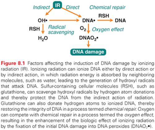
Radical scavengers normally present in cells, such as glutathione, can protect target molecules by reacting with the hydroxyl radical (see Fig. 8.1). Even after the target molecule has been hit and ionized, glutathione can contribute to cell protection by donating a hydrogen atom to the radical, allowing the unpaired electron present in the radical to pair up with the electron from the hydrogen atom. This is considered the simplest of all types of repair and is called chemical repair.3 However, if oxygen molecules are present, they will compete with scavenger molecules for the ionized molecule, and if oxygen reacts with the ionized target molecule before the hydrogen donation occurs, the damage will be solidified as a peroxide, which is not amendable to chemical repair. Instead, this lesion will require enzymatic repair for the restoration of DNA. This augmenting biologic effect of oxygen is called the oxygen effectand is considered an important factor for the effectiveness of radiation therapy.1
Damage to DNA
The direct and indirect effects of radiation induce more or less identical types of lesions in DNA. However, the density of lesions induced in a stretch of DNA is higher for high LET radiation, and this increased complexity is thought to complicate the repair of these lesions. Radiation-induced lesions consist of more than 100 chemically distinct base lesions, such as the mutagenic lesions thymine glycol and 8-hydroxyguanine.2,4,5 Furthermore, damage to the sugar moiety in the backbone of DNA and some types of base damage can result in single-strand breaks (SSB). Because the energy deposition of radiation is clustered even for low LET radiation, it is possible that two individual strand breaks are formed in close proximity on opposite strands, resulting in the formation of a double-strand break (DSB). It has been estimated that 1 Gy of ionizing radiation gives rise to about 40 DSBs, 1,000 SSBs, 1,000 base lesions, and 150 DNA-protein cross-links per cell.2 For a similarly lethal dose of UV light, about 400,000 lesions are required, demonstrating that the lesions induced by IR are much more toxic than lesions induced by UV light. It is believed that DSBs are the critical lesions that lead to cell lethality following exposure to ionizing radiation.6
Damage to Proteins
Although proteins and lipids are subject to damage following exposure to IR, the common belief is that DNA is the critical target for the biologic effects of radiation. Indeed, abrogation of DNA damage surveillance or repair processes in cells results in the enhanced induction of mutations and decreased cell survival following radiation.5 However, studies of radiation-sensitive and radiation-resistant bacteria imply that mechanisms that suppress protein damage may also play important roles in radiation resistance.7 Deinococcus radiodurans is a bacterium that can survive radiation exposures of up to 17,000 Gy, and its extreme radioresistance has been linked to high intracellular levels of manganese, which protect proteins from oxidation. The thought is that if a cell can limit protein oxidation, then its enzymes will remain active, and cellular functions such as DNA repair will be able to restore the integrity of DNA even after severe DNA damage.8 It would be interesting to explore whether the concentration of manganese can be manipulated to sensitize tumor cells to radiation therapy. Furthermore, because protein damage due to reactive oxygen species (ROS) accumulate during the aging process, could supplements of manganese turn back the clock on aging?
Cellular Responses
DNA Repair
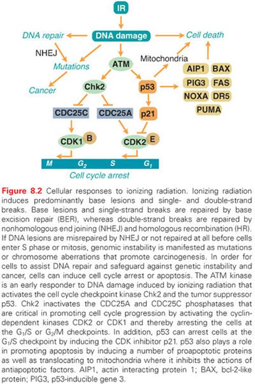
Ever since organisms started to utilize atmospheric oxygen for metabolic respiration many millions of years ago, they have been forced to deal with the cellular damage induced by ROS. Base excision repair (BER) evolved to remove many of the different types of oxidative base lesions and DNA SSBs induced by ROS. However, ROS seldom induce DSBs unless the generation of hydroxyl radicals is clustered near the DNA molecule. A more important source of intracellular generation of DSBs may instead be the process of DNA replication, and it is possible that homologous recombination (HR) repair primarily evolved to overcome DSBs sporadically induced during the replication process. The other major pathway of DSB repair is the nonhomologous end-joining (NHEJ) pathway, which is utilized by immune cells in the process of antibody generation. Although the HR pathway has high fidelity due to the utilization of homologous sister chromatids to ensure that correct DNA ends are joined, the NHEJ pathway lacks this control mechanism and therefore occasionally rejoins ends incorrectly. Thus, the NHEJ pathway may contribute to the generation of mutations following radiation (Fig. 8.2). However, NHEJ is the only mechanism available for DSB repair in postmitotic cells and cells in the G1 phase of the cell cycle because no sister chromatids are available in these cells to support HR repair.
Ataxia-Telangiectasia Mutated and Cell Cycle Checkpoints
Due to the enormous task of replicating the whole genome during the S phase and segregating the chromosomes during mitosis, proliferating cells are generally much more vulnerable to radiation than stationary cells. To prevent cells with damaged DNA from entering into these critical stages of the cell cycle, cells can activate cell cycle checkpoints (see Fig. 8.2). The major sensor of radiation-induced damage in cells is the ataxia-telangiectasia mutated (ATM) kinase, which, following activation, can phosphorylate more than 700 proteins in cells.9 Two ATM substrates, p53 and Chk2, are critical for the activation of cell cycle arrests at multiple sites in the cell cycle.10,11 The kinase p53 regulates the gene expression of specific genes such as p21, which inhibits cyclin-dependent kinase (CDK)2- and CDK4-mediated phosphorylation of the retinoblastoma protein, resulting in a block in the progression from the G1phase to the S phase of the cell cycle.12,13 The Chk2 kinase promotes checkpoint activation in G1 by targeting the cell division cycle 25 homolog A (CDC25A) phosphatase14 and, in G2/M, by targeting the CDC25C phosphatase.15 The activation of a cell cycle arrest following DNA damage provides the cell with additional time to repair the DNA before entering critical cell cycle stages, which promotes genetic stability. Loss or defects in the ATM or p53 genes result in abrogation of radiation-induced cell cycle checkpoints, which manifests itself as the highly cancer-prone human syndromes ataxia telangiectasia16 or Li-Fraumeni,17 respectively.
Radiation-Induced Cell Death
Terminally differentiated and stationary cells, such as kidney, lung, brain, muscle, and liver cells, are generally more resistant to radiation-induced killing than are cells with a high turnover rate, such as different epithelial cells, spermatogonia, and hair follicles. However, the spleen and thymus, which consist of mostly nondividing cells, are among the most radiosensitive tissues, implying that the rate of cell proliferation is not the sole determiner of the radiation sensitivity of a tissue. An important factor regulating the induction of programmed cell death (apoptosis) in tissues is the tumor suppressor p53.18 The p53 protein is activated in cells following exposure to IR by the ATM kinase (see Fig. 8.2). When activated, it regulates the expression of multiple genes that have roles in DNA repair, cell cycle arrest, and apoptosis. p53 can also localize to mitochondria following irradiation, where it triggers apoptosis through the inactivation of antiapoptotic regulatory proteins.19 Not all tissues induce the p53 response to the same degree after similar doses of IR, nor do they activate downstream pathways, such as DNA repair, cell cycle arrest, and apoptosis, in a similar way. For example, thymocytes have an intrinsic setting that favors apoptosis over cell cycle arrest following IR, whereas fibroblasts rarely induce apoptosis, but instead activate a strong and lasting cell cycle arrest.18
IR can induce cell death in tissues by many different mechanisms. Apoptosis can occur rapidly in a p53-dependent manner or later in a p53-independent manner. This later wave of radiation-induced apoptosis is often initiated by mitotic catastrophe, which occurs as a result of complications during chromosome segregation. Cell death induced by IR may in some cases be associated with autophagy, also called autophagocytosis, in which cells degrade cellular components via the lysosomal machinery. Whether autophagy is a programmed cell death or occurs in parallel with cell death is not clear. Interestingly, for some cell types, autophagy has been shown to actually protect the cells from radiation-induced death. Finally, tissue can undergo necrotic cell death following exposure to IR. Necrosis is a clinical problem following radiation therapy that can occur in normal tissues many months after treatment and can contribute to the inflammatory response.
Cancer Risks
It is clear from epidemiologic studies of radiation workers and atomic bomb and Chernobyl victims that IR can induce cancer.20 Twenty years after the atomic bomb explosions in Japan during World War II, significant increases in the incidence of thyroid cancer and leukemia were observed. However, it took almost 50 years before solid tumors appeared in the population as a result of radiation exposure from the atomic bombs.21 The incidences of solid tumors, such as breast, ovary, bladder, lung, and colon cancers, were estimated to have increased by a factor of 2 in the exposed group during this time period. The epidemiology studies following the nuclear power plant disaster in Chernobyl showed a clear increase in thyroid cancer as early as 4 years after the accident.22 Young children were the most vulnerable to radiation exposure, with 1-year-old children being 237-fold more susceptible to thyroid cancer than the control group, while 10-year-old children were found to be sixfold more susceptible to thyroid cancer. Many of the thyroid cancers that developed following the Chernobyl disaster could have been prevented if the population had not consumed locally produced milk that was contaminated with radioactive iodine.
The molecular signatures of radiation-induced tumors are complex but involve point mutations that could lead to the activation of the RAS oncogene or inactivation of the tumor suppressor gene p53. Furthermore, IR induces DNA DSBs that may be unfaithfully repaired by the NHEJ pathway, leading to chromosome rearrangements. One such rearrangement found in 50% to 90% of the thyroid cancers examined following the Chernobyl accident involved the receptor tyrosine kinase c-RET, which promotes cell growth when activated.22 Furthermore, a great majority of the thyroid cancers found in the exposed children harbored kinase fusion oncogenes affecting the mitogen-activated protein kinase (MAPK) signaling pathway.23
The correlation between high exposure to IR and cancer following the atomic bomb explosions and the Chernobyl accident is clear. What about the cancer risk following lower radiation exposures occurring in daily life? There are four theoretical risk models of radiation-induced cancer to consider. First, the linear, no threshold (LNT) model suggests that the induction of cancer is directly proportional to the dose of radiation, even at low doses of exposure. Second, the sublinear or threshold model suggests that below a certain threshold dose the risk of radiation-induced cancers is negligible. At these lower doses of radiation exposure, the DNA damage surveillance and repair mechanisms are thought to be fully capable of safeguarding the DNA to avoid the induction of mutations and cancer. Third, the supralinear or stealth model suggests that doses below a certain threshold or radiation with sufficiently low dose rates may not trigger the activation of DNA damage surveillance and repair mechanisms, resulting in suboptimal activation of cell cycle checkpoints and repair. This would be expected to lead to a higher rate of mutations and cancers than predicted by the LNT model, but may be balanced by a higher incident of cell death. Fourth, the linear-quadratic model suggest that radiation effects at low doses are due to a single track of radiation hitting multiple targets, resulting in a linear induction rate, whereas at higher doses, multiple radiation tracks hit multiple cellular targets, resulting in a quadratic induction rate.
The Biological Effects of Ionizing Radiation (BEIR) VII report, released by the Committee on Biological Effects of Ionizing Radiation of the National Academy of Sciences and commissioned by the US Environmental Protection Agency (EPA), is a review of published data regarding human health and cancer risks from exposure to low levels of IR. Although this topic is controversial and not fully settled, the BIER VII report favored the LNT model.24 Thus, the “official” view is that no level of radiation is safe; therefore, a careful consideration of risks versus benefits is necessary to ensure that the general population only receives radiation doses as low as reasonably achievable. Furthermore, the BIER VII committee concluded that the heritable effects of radiation were not evident in the published data, indicating that an individual is not likely to develop cancer due to radiation exposure of his or her parents.
The largest source of radiation exposure to the population is radon, which is a natural radioactive gas formed as a decay product of radium in the decay chain of uranium. Radon gas can accumulate to high levels in poorly ventilated basements in houses built on rock containing uranium. The major risk with radon is that some of its radioactive decay products can attach to dust particles that accumulate in the lungs, leading to a continuous exposure of the lung tissues to high LET alpha particles. Due to this radiation exposure, the EPA claims that radon is the second leading cause of lung cancer in the United States. Another important source of human exposure to IR is medical x-ray devices, and there is a growing concern about the dramatically increased use of whole body CT scans for diagnostic purposes. For a typical CT scan, a patient will receive about 100-fold more radiation than from a typical mammogram.24 It is recommended that the use of whole body CT scans for children be very restricted due to the elevated risk of developing radiation-induced cancer for this age group.
Cancer patients who receive radiation therapy are at risk of developing secondary tumors induced by the radiation therapy treatment.1 This is particularly a concern for young patients since (1) children are more prone to radiation-induced cancer, (2) children have a relatively good chance of surviving the primary cancer and would have long life expectancies so a secondary tumor would have plenty of time to develop, and (3) many childhood cancers are promoted by genetic defects in DNA damage response pathways, making these patients highly prone to the genotoxic effects of radiation and subsequent secondary cancers. The most sensitive tissues for the development of secondary cancer have been found to be bone marrow (leukemia), the thyroid, breast, and lung.1
ULTRAVIOLET LIGHT
Depending on the wavelength, UV light is categorized into UVA (320 to 400 nm), UVB (290 to 320 nm), and UVC (240 to 290 nm) radiation. Most of the UVC light emitted from the sun is absorbed by the ozone layer in the atmosphere, and, thus, living organisms are mostly exposed to UVA and UVB irradiation.
Mechanisms of Damage Induction
UVC light is more damaging to DNA than UVA and UVB because the absorption maximum of DNA is around 260 nm. UVB and UVC induce predominantly pyrimidine dimers and 6-4 photoproducts, which consist of covalent ring structures that link two adjacent pyrimidines on the same DNA strand.5 The formation of these lesions results in the bending of the DNA helix, resulting in the interference with both DNA and RNA synthesis. UVA light does not induce pyrimidine dimers or 6-4 photoproducts but can induce ROS, which in turn can form SSBs and base lesions in DNA of exposed cells.
Cellular Responses
DNA Repair
The nucleotide excision repair (NER) pathway removes pyrimidine dimers and 6-4 photoproducts from cellular DNA.5 This pathway involves proteins that recognize the DNA lesions, nucleases that excise the DNA strand that contains the lesion, a DNA polymerase that synthesizes new DNA to fill the gap, and a DNA ligase that joins the backbone in the newly synthesized strand. Genetic defects in the NER pathway result in the human syndrome xeroderma pigmentosum, with individuals more than 1,000-fold more prone to sun-induced skin cancer than normal individuals. In addition, human polymorphisms in certain NER genes are thought to predispose individuals to cancers such as lung cancer, nonmelanoma skin cancer, head and neck cancer, and bladder cancer, indicating that NER is responsible for safeguarding the genome against many types of DNA adducts in addition to UV-induced lesions.5
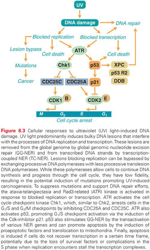
UV-induced lesions formed in the transcribed strand of active genes block the elongation of RNA polymerase II, and if a cell does not restore transcription within a certain time frame, it may undergo apoptosis (Fig. 8.3).25,26 To rapidly restore RNA synthesis and avoid cell death, NER enzymes are recruited to the sites of blocked RNA polymerase II and the lesions are removed in a process called transcription-coupled repair (TCR).27 Individuals with Cockayne syndrome (CS), trichothiodystrophy, or the UV-sensitive syndrome, are unable to utilize the TCR pathway following UV irradiation.5 Cells from these individuals do not recover RNA synthesis following UV irradiation and are therefore very prone to UV-induced apoptosis. Interestingly, despite a clear DNA repair defect, these individuals are not predisposed to UV-induced skin cancer. It is thought that the inability of CS cells to remove the toxic lesions that block transcription following UV irradiation results in the suppression of tumorigenesis by the elimination of damaged cells by apoptosis. However, while protecting against tumorigenesis, the elevated level of apoptosis in these cells leads to increased cell loss, which in turn may lead to neurologic degeneration.25,28Persistent transcription-blocking lesions in the genome have also been linked to aging.29–31
Translesion DNA Synthesis
Proliferating skin cells are very vulnerable to UV light because UV lesions block DNA replication (see Fig. 8.3). Cells that have entered the S phase and have initiated DNA synthesis have no choice but to finish replicating the whole genome or they will die. If DNA repair enzymes are not able to remove the blocking lesions from the template, the processive DNA polymerases may be exchanged for other, less processive DNA polymerases that can bypass the lesions. This is part of a “tolerance” mechanism, which allows cells to complete replication and eventually divide.5 However, the translesion DNA polymerases do not have the same fidelity as the processive DNA polymerases; thus, mutations may occur. This is thought to be a major pathway by which UV light induces mutagenesis and, subsequently, cancer (see Fig. 8.3).
ATM and Rad3-Related Mediated Cell Cycle Checkpoints
In addition to utilizing the NER and BER pathways to repair UV-induced DNA damage, proliferating cells activate cell cycle checkpoints to allow more time for repair before entering critical parts of the cell cycle, such as the S phase and mitosis. The ATM and Rad3-related (ATR) kinase is activated following UV irradiation by blocked replication or transcription (see Fig. 8.3).32 ATR phosphorylates a large number of proteins, many of which are the same as those phosphorylated by ATM after exposure to ionizing radiation.9 Two important substrates of ATR are p53 and Chk1, which are critical in promoting cell cycle arrest. When induced by ATR, p53 transactivates the gene that encodes the cell cycle inhibitor p21, leading to the arrest of cells in the G1 phase of the cell cycle, while Chk1 phosphorylates the CDK-activating phosphatases CDC25A and CDC25C, which targets them for degradation, resulting in an S-phase or G2-phase arrest (see Fig. 8.3).33
Activation of Cell Membrane Receptors
In addition to triggering cellular stress responses by inducing DNA damage, UV light can directly induce membrane receptor signaling by receptor phosphorylation. This is thought to be due to the direct UV-mediated inhibition of protein-tyrosine phosphatases that regulate the phosphorylation levels of various membrane receptors.34 In addition, membrane receptors may physically aggregate following UV irradiation, leading to the activation of signal transduction pathways that regulate cell growth35 or apoptosis.36
Cell Death
UV light effectively induces apoptosis in skin cells. The mechanism by which UV light induces cell death is not fully understood, but failure to adequately resume RNA synthesis following UV light exposure is strongly linked to apoptosis (see Fig. 8.3).25 Many potential mechanisms of how blocked transcription results in apoptosis have been suggested, such as a physical clash during the S phase between elongating replication machineries and transcription complexes stalled at UV lesions. Another possible mechanism involves the preferential loss of survival factors coded by highly unstable mRNAs.37 The induction of p53 may also contribute to UV-induced apoptosis,38 although p53 appears to protect human fibroblasts39 and keratinocytes40 from UV-induced apoptosis. Although complications induced by DNA damage may be the predominant mechanism by which cells die following UV irradiation, UV light may induce apoptosis in certain cell types by directly promoting the physical aggregation of the death receptor Fas/APO1.36
Cancer Risks
The incidence of sun-induced skin cancer, especially melanoma, is on the increase due to higher rates of sun exposure in the general population. The link between UV light exposure and skin cancer is very strong, but the role of UV light in the etiology of nonmelanoma and melanoma skin cancer differs. Although the risk of nonmelanoma cancer relates to the cumulative lifetime exposure to UV light, the risk of contracting melanoma appears to be linked to high sunlight exposure during childhood.41 What makes UV light such a potent carcinogen is that it can initiate carcinogenesis by inducing DNA lesions as well as suppressing the immune system, resulting in a greater probability that initiated cells will survive and grow into tumors.42,43
Nonmelanoma Skin Cancer
Basal cell carcinoma (BCC) and squamous cell carcinoma (SCC) are the two most common skin cancer types. BCC and SCC occur predominantly in sun-exposed areas of the skin, but there are examples of these cancers forming in nonexposed areas as well. The tumor suppressor genes p53 and p16 are frequently inactivated in BCC and SCC, while the hedgehog-signaling pathway is activated primarily by mutations to the patched gene (percutaneous transhepatic cholangiography [PTCH]). This scenario promotes proliferation without the opposition of the cell cycle inhibitors p53 and p16.
Melanoma
Melanoma arises from mutations in epidermal melanocytes and is the most dangerous form of skin cancer because it has the highest propensity to metastasize. It is formed in both sun-exposed and shielded areas of the skin; therefore, the role of UV light as the major carcinogen in melanoma has been controversial.41 Defects in the NER pathway do not seem to predispose the development of melanoma, suggesting that pyrimidine dimers or 6-4 photoproducts induced by UVB are not the initiators of melanoma carcinogenesis. Instead, ROS induced by UVA may be responsible for the development of melanoma.41 However, a study using next-generation sequencing techniques to catalog all mutations in a melanoma cell line found a mutational spectrum of the over 33,000 mutations detected that strongly indicated that pyrimidine dimers and 6-4 photoproducts are the major mutagenic lesions in melanoma, whereas a subset of mutations may be induced by ROS.44 The incidence of mutations in the p16 and ARFgenes is high, whereas p53 and RAS mutations are fairly uncommon in melanoma.
Photoimmunosuppression
Studies of transplantation of mouse skin cancers into syngeneic mice revealed that prior UVB irradiation of recipient mice promoted tumor growth, whereas transplantation into naïve nonirradiated mice led to rejection.43 These studies established that UV light has local immunosuppressing ability, and subsequent studies found that UV light preferentially depletes Langerhans cells from irradiated skin.42 Langerhans cells play an important role in the immune response by presenting antigens to the immune cells, and, thus, depletion of these cells leads to local immunosuppression. In addition to local immunosuppression, UV light has been shown to promote systemic immunosuppression.45This response is complex, but it is known that UV-induced DNA lesions in skin cells contribute to the systemic immunosuppression response.46 The secretion of the immunosuppressing cytokine interleukin (IL)-10 from irradiated keratinocytes as well as UV-induced structural alteration of the epidermal chromophore urocanic acid may mediate the long-range immunosuppressive effects of UV light.42,45
RADIOFREQUENCY AND MICROWAVE RADIATION
Radiofrequency radiation (RFR) is electromagnetic radiation in the frequency range 3 kHz to 300 MHz, whereas microwave radiation (MR) is in the frequency range between 300 MHz to 300 GHz. RFR and MR do not have sufficient energies to cause ionizations in target tissues. Rather, the radiation energy is converted into heat as the radiation energy is absorbed. Sources of radiofrequency and microwave radiation include mobile phones, radio transmitters of wireless communication, radars, medical devices, and kitchen appliances.
Mechanism of Damage Induction
Because human exposure to RFR has increased dramatically in recent years, it is important to know whether this type of radiation gives rise to genotoxic damage. Although there are many studies showing that RFR can induce ROS, leading to genetic damage in cell culture systems, other studies have generated conflicting results.47 One confounding factor when assessing the genotoxic effect of RFR, and especially MR, is the heating effect that occurs in the tissue when the radiation energy is absorbed. A recent study controlling for the potential heating effect of exposure found that RFR induces ROS and DNA damage in human spermatozoa in vitro, which is an alarming finding considering the potential hereditary implications.48 It has been suggested that MR may affect the folding of proteins in cells that promote new protein synthesis.49 Furthermore, exposure of cells to MR has been shown to lead to the phosphorylation of numerous cellular proteins largely through the activation of the p38/MAPK stress response pathway.50 However, the biologic consequences of these cellular changes are not clear. Epidemiology studies that monitored the genetic effects in individuals exposed to high levels of RF have revealed evidence of increased induction of chromosome aberrations in lymphocytes.51 However, there is a level of uncertainty in these studies about exposure levels, making it difficult to come to meaningful conclusions.
Cancer Risks
Because the population’s exposure to RFR and MR has dramatically increased in recent years, it is of great importance to assess the potential cancer risks of these types of radiation so that appropriate exposure limits can be implemented. A number of studies have focused on the potential cancer risks from mobile phone usage, and some of these studies indicate that long-term mobile phone usage may be associated with increased risks of developing brain tumors (see the following). Other epidemiologic studies of cancer incidences in populations living near radio towers or mobile phone base stations are inconclusive. Some studies have shown a connection between proximity to mobile phone base stations and increased cancer incidence,52 whereas another study found no association between exposure to RFR from mobile phone base stations and early childhood cancers.53
ELECTROMAGNETIC FIELDS
An electromagnetic field (EMF) is a physical field produced by electrically charged objects that can affect other charged objects in the field. Typical sources of EMFs are electric power lines, electrical devices, and magnetic resonance imaging (MRI) machines.
Mechanisms of Damage Induction
A low frequency EMF does not transmit energy high enough to break chemical bonds; therefore, it is not thought to directly damage DNA or proteins in cells. The data obtained from studies to assess the potential genotoxic effects of EMF do not provide a clear conclusion. Some of the results obtained in cell culture studies suggest a harmful effect of EMFs, but the concerns are that these effects may be related to heat production induced by EMFs rather than from the magnetic field itself. A recent in vitro study detected DNA strand breaks in cells exposed to EMFs, but this induction was thought to not be the result of ROS production, but rather due to indirect effects through interference with DNA replication and induction of apoptosis in a subset of cells.54 A study using an MRI found no evidence of an induced formation of DNA DSBs in cell cultures.55 EMFs have been shown to induce nongenotoxic effects in cells, such as interference with cellular signaling pathways,56 which could contribute to neurodegeneration.57
Cancer Risks
Studies with rodents have largely failed to detect an association between exposure to EMFs and cancer. This is also true for numerous epidemiology studies, with the only exception being the association between EMF exposure and childhood leukemia where children exposed to doses of 0.4 mcT or above may have about a twofold increased risk of developing leukemia.58,59 There is no strong link between EMF exposure and increased risks of contracting adult leukemia, brain tumors, or breast cancer.60,61 Furthermore, a study investigating whether EMF exposure was associated with heritable effects found no correlation between parental exposure and childhood cancer.62
Potential Cancer Risks from Mobile Phone Usage
Mobile phones emit RFR and generate EMFs. The biggest health concern with mobile phone usage is its potential role in the development of brain tumors. During mobile phone use, the brain tissue is exposed to doses, giving peak specific absorption rates (SAR) of 4 to 8 W/kg. At these intensities, the induction of DNA damage has been detected in laboratory studies.63 The current epidemiologic data are largely inconclusive on the association between mobile phone usage and brain tumor incidence. Meta-analysis studies of populations who had used mobile phones for more than 10 years concluded that mobile phone usage was associated with an elevated risk for brain tumors, such as acoustic neuroma and glioma cancer.64–66 In contrast, other large prospective studies did not observe a correlation between mobile phone usage and incidences of glioma, meningioma, or non–central nervous system (CNS) cancers.67,68 It is important to point out that, generally, it takes 30 to 40 years for brain tumors to develop, and because mobile phones have only been in general use for about 15 years, there has not been sufficient time to fully evaluate the brain cancer risks of mobile phone usage.
ASBESTOS
Asbestos is a class of naturally occurring silicate minerals that have been widely used in building materials for its heat, sound, and electrical insulating qualities. Asbestos becomes a serious health hazard if the fibers are inhaled over a long period of time, and these health effects are increased dramatically if the exposed individual is a smoker. It was first reported in 1935 that asbestos might be an occupational health hazard that could induce cancer.69,70 However, it was not until 1986 that the International Labor Organization recommended banning asbestos.71 The use of asbestos products peaked in the 1970s, yet remains a major health hazard in many places around the world today.
Mechanisms of Damage Induction
Asbestos fibers can enter cells and induce ROS, especially if they contain high levels of iron.72 In addition, ROS can be generated by “frustrated” phagocytosis, and this in turn can lead to the release of proinflammatory cytokines with subsequent inflammation of the tissue. ROS have been implicated to originate from affected mitochondria leading to induction of SSBs and base damage, such as 8-hydroxyguanine in DNA.73 Furthermore, if not successfully repaired, asbestos-induced DNA damage has been shown to result in chromosome aberrations, micronuclei formation, and increased rates of sister chromatid exchanges.74
Cellular and Tissue Responses
Asbestos-induced ROS cause base lesions and DNA strand breaks, which require base excision repair for the restoration of DNA and for minimizing mutagenesis. In addition to DNA repair, a number of cellular signaling pathways are activated by asbestos. These include the epidermal growth factor receptor (EGFR) and the MAPK pathway, leading to the activation of nuclear factor kappa B (NF-κB) and transcription factor AP-1.72,74 Activation of the NF-κB pathway leads to the induction of proinflammatory genes such as tumor necrosis factor (TNF), IL-6, IL-8, and proliferation-promoting genes such as c-Myc, leading to inflammation and increased cell proliferation. Asbestos exposure also stimulates the expression of the transforming growth factor beta (TGF-β), which, in turn, stimulates fibrogenesis in exposed tissues.74
Cancer Risks
Lung Cancer
Epidemiologic studies have found a strong link between asbestos exposure and lung cancer.74 It has been estimated that about 5% to 7% of all lung cancers are attributable to asbestos exposure, and asbestos and tobacco smoking act in synergy to induce lung cancer. Mutational spectra due to 8-hydroxyguanine lesions formed by ROS can be linked to asbestos exposure, and point mutations in the tumor suppressor genes p53 and p16/INK4A and in the KRASoncogene have been found in tumors from asbestos-exposed individuals.
Mesothelioma
After being taken up by lung tissues, asbestos fibers can translocate into the pleura, the body cavity that surrounds the lungs. The pleura are covered with a protective lining, the mesothelium, which consists of squamouslike epithelial cells. Mesothelial cells can internalize asbestos fibers, resulting in the induction of ROS and inflammatory responses, subsequently leading to the initiation and progression of malignant mesothelioma.75 Asbestos is considered one of the major causes of malignant mesothelioma, and frequent mutations are found in the p16/INK4A and NF2 genes, whereas p53 mutations are fairly rare.
NANOPARTICLES
Nanoparticles are defined as ultrafine particles of the size range 1 to 100 nm in diameter. Nanoparticle chemistry of a certain compound is different from bulk chemistry of that compound because of the high percentage of atoms at the surface of the particle. The production of nanoparticles has increased dramatically in recent years, and they are found in many industrial and consumer products such as paint, cosmetics, and sunscreens. They also have many potential medical applications, such as delivery vehicles for specific drugs to specific target tissues or tumors.
Mechanisms of DNA Damage Induction
Many of the cellular effects of nanoparticles are similar to the effects exerted by asbestos, such as the generation of ROS and inflammation.72 Nanoparticles have been shown to induce oxidative DNA damage, such as DNA strand breaks and 8-hydroxyguanine lesions both in cell culture76, 77 and in vivo.78 Nanoparticle-induced DNA lesions are manifested as histone γ-H2AX nuclear foci, chromosome deletions, and micronuclei.
Cellular Responses
Nanoparticles induce ROS either directly or indirectly, resulting in DNA lesions, such as 8-hydroxyguanine–base damage and DNA strand breaks. These lesions are repaired by the base excision repair. The phosphorylation of histone H2AX has been shown to occur following exposure of cells to nanoparticles, suggesting that the DNA lesions trigger the activation of ATM or ATR stress kinases.79 Nanoparticles have also been found to affect the immune system80 and can induce the release of the proinflammatory cytokine TNF-α from cells.
Cancer Risks
Some nanoparticles, such as titanium dioxide, which is used as pigments in paint, have been classified by the International Agency for Research on Cancer (IARC) as a group 2B carcinogen, “possible carcinogenic to humans.” However, rigorous epidemiologic data is lacking to fully evaluate the cancer-inducing potential of nanoparticles.81
REFERENCES
1. Hall E, Giaccia A. Radiobiology for the Radiologist. Philadelphia: Lippincott Williams & Wilkins; 2012.
2. Ward JF. DNA damage produced by ionizing radiation in mammalian cells: identities, mechanisms of formation, and repairability. Prog Nucleic Acid Res Mol Biol 1988;35:95–125.
3. Prutz WA. ‘Chemical repair’ in irradiated DNA solutions containing thiols and/or disulphides. Further evidence for disulphide radical anions acting as electron donors. Int J Radiat Biol 1989;56:21–33.
4. Hutchinson F. Chemical changes induced in DNA by ionizing radiation. Prog Nucleic Acid Res Mol Biol 1985;32:115–154.
5. Friedberg E, Walker G, Siede W, et al. DNA Repair and Mutagenesis. 2nd ed. Washington, D.C.: ASM Press; 2006.
6. Radford IR. The level of induced DNA double-strand breakage correlates with cell killing after X-irradiation. Int J Radiat Biol Relat Stud Phys Chem Med 1985;48:45–54.
7. Daly MJ. A new perspective on radiation resistance based on Deinococcus radiodurans. Nat Rev Microbiol 2009;7:237–245.
8. Krisko A, Radman M. Biology of extreme radiation resistance: the way of Deinococcus radiodurans. Cold Spring Harb Perspect Biol 2013;5.
9. Matsuoka S, Ballif BA, Smogorzewska A, et al. ATM and ATR substrate analysis reveals extensive protein networks responsive to DNA damage. Science 2007;316:1160–1166.
10. Kastan M, Onyekwere O, Sidransky D, et al. Participation of p53 protein in the cellular response to DNA damage. Cancer Res 1991;51:6304–6311.
11. Matsuoka S, Huang M, Elledge SJ. Linkage of ATM to cell cycle regulation by the Chk2 protein kinase. Science 1998;282:1893–1897.
12. Harper J, Adami G, Wei N, et al. The p21 cdk-interacting protein Cip1 is a potent inhibitor of G1 cyclin-dependent kinases. Cell 1993;75:805–816.
13. El-Deiry W, Tokino T, Velculescu V, et al. WAF1, a potential mediater of p53 tumor suppression. Cell 1993;75:817–825.
14. Falck J, Mailand N, Syljuasen RG, et al. The ATM-Chk2-Cdc25A checkpoint pathway guards against radioresistant DNA synthesis. Nature 2001;410:842–847.
15. Bartek J, Falck J, Lukas J. Chk2 kinase—a busy messenger [Review]. Nat Rev Mol Cell Biol 2001;2:877–886.
16. Savitsky K, Bar-Shira A, Gilad S, et al. A single ataxia telangiectasia gene with a product similar to PI-3 kinase. Science 1995;268:1749–1753.
17. Srivastava S, Zou ZQ, Pirollo K, et al. Germ-line transmission of a mutated p53 gene in a cancer-prone family with Li-Fraumeni syndrome. Nature.1990;348:747–749.
18. Gudkov AV, Komarova EA. The role of p53 in determining sensitivity to radiotherapy. Nat Rev Cancer 2003;3:117–129.
19. Mihara M, Erster S, Zaika A, et al. p53 has a direct apoptogenic role at the mitochondria. Mol Cell 2003;11:577–590.
20. Williams D, Baverstock K. Chernobyl and the future: too soon for a final diagnosis. Nature 2006;440:993–994.
21. Thompson DE, Mabuchi K, Ron E, et al. Cancer incidence in atomic bomb survivors. Part II: Solid tumors, 1958–1987. Radiat Res 1994;137:S17–67.
22. Williams D. Cancer after nuclear fallout: lessons from the Chernobyl accident. Nat Rev Cancer 2002;2:543–549.
23. Ricarte-Filho JC, Li S, Garcia-Rendueles ME, et al. Identification of kinase fusion oncogenes in post-Chernobyl radiation-induced thyroid cancers. J Clin Invest 2013;123:4935–4944.
24. National Research Council. Health Risks from Exposure to Low Levels of Ionizing Radiation: BEIR VII Phase 2. Washington, D.C.: National Academy Press; 2006.
25. Ljungman M, Zhang F. Blockage of RNA polymerase as a possible trigger for u.v. light-induced apoptosis. Oncogene.1996;13:823–831.
26. Brash DE, Wikonkal NM, Remenyik E, et al. The DNA damage signal for Mdm2 regulation, Trp53 induction, and sunburn cell formation in vivo originates from actively transcribed genes. J Invest Derm 2001;117:1234–1240.
27. Hanawalt PC, Spivak G. Transcription-coupled DNA repair: two decades of progress and surprises. Nat Rev Mol Cell Biol 2008;9:958–970.
28. Lehmann AR. DNA repair-deficient diseases, xeroderma pigmentosum, Cockayne syndrome and trichothiodystrophy. Biochimie 2003;85:1101–1111.
29. Andressoo JO, Hoeijmakers JH. Transcription-coupled repair and premature ageing. Mutat Res 2005;577:179–194.
30. de Boer J, Andressoo JO, de Wit J, et al. Premature aging in mice deficient in DNA repair and transcription. Science 2002;296:1276–1279.
31. Garinis GA, Uittenboogaard LM, Stachelscheid H, et al. Persistent transcription-blocking DNA lesions trigger somatic growth attenuation associated with longevity. Nat Cell Biol 2009;11:604–615.
32. Derheimer FA, O’Hagan HM, Krueger HM, et al. RPA and ATR link transcriptional stress to p53. Proc Natl Acad Sci U S A 2007;104:12778–12783.
33. Kastan MB, Bartek J. Cell-cycle checkpoints and cancer. Nature 2004;432:316–323.
34. Gross S, Knebel A, Tenev T, et al. Inactivation of protein-tyrosine phosphatases as mechanism of UV-induced signal transduction. J Biol Chem 1999;274:26378–26386.
35. Sachsenmaier C, Radlerpohl A, Zinck R, et al. Involvement of growth factor receptors in the mammalian UVC response. Cell 1994;78:963–972.
36. Rehemtulla A, Hamilton CA, Chinnaiyan AM, et al. Ultraviolet radiation-induced apoptosis is mediated by activation of CD-95 (Fas/APO-1). J Biol Chem 1997;272:25783–25786.
37. Ljungman M, Lane DP. Transcription guarding the genome by sensing DNA damage. Nat Rev Cancer 2004;4:727–737.
38. Ziegler A, Jonason AS, Leffell DJ, et al. Sunburn and p53 in the onset of skin cancer. Nature 1994;372:773–776.
39. McKay B, Ljungman M. Role for p53 in the recovery of transcription and protection against apoptosis induced by ultraviolet light. Neoplasia 1999;1:276–284.
40. Chaturvedi V, Sitailo LA, Qin JZ, et al. Knockdown of p53 levels in human keratinocytes accelerates Mcl-1 and Bcl-x(L) reduction thereby enhancing UV-light induced apoptosis. Oncogene 2005;24:5299–5312.
41. Maddodi N, Setaluri V. Role of UV in cutaneous melanoma. Photochem Photobiol 2008;84:528–536.
42. Murphy GM. Ultraviolet radiation and immunosuppression. Br J Dermatol 2009;161(Suppl 3):90–95.
43. Fisher MS, Kripke ML. Systemic alteration induced in mice by ultraviolet light irradiation and its relationship to ultraviolet carcinogenesis. Proc Natl Acad Sci U S A 1977;74:1688–1692.
44. Pleasance ED, Cheetham RK, Stephens PJ, et al. A comprehensive catalogue of somatic mutations from a human cancer genome. Nature 2010;463:191–196.
45. Schwarz T. Photoimmunosuppression. Photodermatol Photoimmunol Photomed 2002;18:141–145.
46. Kripke ML, Cox PA, Alas LG, et al. Pyrimidine dimers in DNA initiate systemic immunosuppression in UV-irradiated mice. Proc Natl Acad Sci U S A 1992;89:7516–7520.
47. Vijayalaxmi, Prihoda TJ. Genetic damage in mammalian somatic cells exposed to radiofrequency radiation: a meta-analysis of data from 63 publications (1990–2005). Radiat Res 2008;169:561–574.
48. De Iuliis GN, Newey RJ, King BV, et al. Mobile phone radiation induces reactive oxygen species production and DNA damage in human spermatozoa in vitro. PLoS One 2009;4:e6446.
49. Gerner C, Haudek V, Schandl U, et al. Increased protein synthesis by cells exposed to a 1,800-MHz radio-frequency mobile phone electromagnetic field, detected by proteome profiling. Int Arch Occup Environ Health 2010;83:691–702.
50. Leszczynski D, Joenvaara S, Reivinen J, et al. Non-thermal activation of the hsp27/p38MAPK stress pathway by mobile phone radiation in human endothelial cells: molecular mechanism for cancer- and blood-brain barrier-related effects. Differentiation 2002;70:120–129.
51. Verschaeve L. Genetic damage in subjects exposed to radiofrequency radiation. Mutat Res 2009;681:259–270.
52. Khurana VG, Hardell L, Everaert J, et al. Epidemiological evidence for a health risk from mobile phone base stations. Int J Occup Environ Health 2010;16:263–267.
53. Elliott P, Toledano MB, Bennett J, et al. Mobile phone base stations and early childhood cancers: case-control study. BMJ 2010;340:c3077.
54. Focke F, Schuermann D, Kuster N, et al. DNA fragmentation in human fibroblasts under extremely low frequency electromagnetic field exposure. Mutat Res 2010;683:74–83.
55. Schwenzer NF, Bantleon R, Maurer B, et al. Detection of DNA double-strand breaks using gammaH2AX after MRI exposure at 3 Tesla: an in vitro study. J Magn Reson Imaging 2007;26:1308–1314.
56. Girgert R, Hanf V, Emons G, et al. Signal transduction of the melatonin receptor MT1 is disrupted in breast cancer cells by electromagnetic fields. Bioelectromagnetics 2010;31:237–245.
57. Consales C, Merla C, Marino C, et al. Electromagnetic fields, oxidative stress, and neurodegeneration. Int J Cell Biol 2012;2012:683897.
58. Ahlbom A, Day N, Feychting M, et al. A pooled analysis of magnetic fields and childhood leukaemia. Br J Cancer 2000;83:692–698.
59. Malagoli C, Fabbi S, Teggi S, et al. Risk of hematological malignancies associated with magnetic fields exposure from power lines: a case-control study in two municipalities of northern Italy. Environ Health 2010;9:16.
60. Kheifets L, Monroe J, Vergara X, et al. Occupational electromagnetic fields and leukemia and brain cancer: an update to two meta-analyses. J Occup Environ Med 2008;50:677–688.
61. Chen C, Ma X, Zhong M, et al. Extremely low-frequency electromagnetic fields exposure and female breast cancer risk: a meta-analysis based on 24,338 cases and 60,628 controls. Breast Cancer Res Treat 2010;123:569–576.
62. Hug K, Grize L, Seidler A, et al. Parental occupational exposure to extremely low frequency magnetic fields and childhood cancer: a German case-control study. Am J Epidemiol 2010;171:27–35.
63. Hardell L, Sage C. Biological effects from electromagnetic field exposure and public exposure standards. Biomed Pharmacother 2008;62:104–109.
64. Hardell L, Carlberg M, Hansson Mild K. Mobile phone use and the risk for malignant brain tumors: a case-control study on deceased cases and controls. Neuroepidemiology 2010;35:109–114.
65. Hardell L, Carlberg M, Soderqvist F, et al. Meta-analysis of long-term mobile phone use and the association with brain tumours. Int J Oncol 2008;32:1097–1103.
66. Myung SK, Ju W, McDonnell DD, et al. Mobile phone use and risk of tumors: a meta-analysis. J Clin Oncol 2009;27:5565–5572.
67. Benson VS, Pirie K, Schuz J, et al. Mobile phone use and risk of brain neoplasms and other cancers: prospective study. Int J Epidemiol 2013;42:792–802.
68. Poulsen AH, Friis S, Johansen C, et al. Mobile phone use and the risk of skin cancer: a nationwide cohort study in Denmark. Am J Epidemiol 2013;178:190–197.
69. Lynch K, Smith W. Pulmonary asbestosis. III. Carcinoma of lung in asbestos-silicosis. Am J Cancer 1935;24:56–64.
70. Gloyne S. Two cases of squamous carcinoma of the lung occuring in asbestosis. Tubercele 1935;17:5–10.
71. LaDou J. The asbestos cancer epidemic. Environ Health Perspect 2004;112:285–290.
72. Pacurari M, Castranova V, Vallyathan V. Single- and multi-wall carbon nanotubes versus asbestos: are the carbon nanotubes a new health risk to humans? J Toxicol Environ Health A 2010;73:378–395.
73. Liu G, Cheresh P, Kamp DW. Molecular basis of asbestos-induced lung disease. Annu Rev Pathol 2013;8:161–187.
74. Nymark P, Wikman H, Hienonen-Kempas T, et al. Molecular and genetic changes in asbestos-related lung cancer. Cancer Lett 2008;265:1–15.
75. Jaurand MC, Renier A, Daubriac J. Mesothelioma: do asbestos and carbon nanotubes pose the same health risk? Part Fibre Toxicol 2009;6:16.
76. Shukla RK, Kumar A, Gurbani D, et al. TiO(2) nanoparticles induce oxidative DNA damage and apoptosis in human liver cells. Nanotoxicology 2013;7:48–60.
77. Horie M, Nishio K, Endoh S, et al. Chromium(III) oxide nanoparticles induced remarkable oxidative stress and apoptosis on culture cells. Environ Toxicol 2013;28:61–75.
78. Trouiller B, Reliene R, Westbrook A, et al. Titanium dioxide nanoparticles induce DNA damage and genetic instability in vivo in mice. Cancer Res 2009;69:8784–8789.
79. Prasad RY, Chastain PD, Nikolaishvili-Feinberg N, et al. Titanium dioxide nanoparticles activate the ATM-Chk2 DNA damage response in human dermal fibroblasts. Nanotoxicology 2013;7:1111–1119.
80. Zolnik BS, Gonzalez-Fernandez A, Sadrieh N, et al. Nanoparticles and the immune system. Endocrinology 2010;151:458–465.
81. Shi H, Magaye R, Castranova V, et al. Titanium dioxide nanoparticles: a review of current toxicological data. Part Fibre Toxicol 2013;10:15.