
Dan L. Longo
Cytokines are soluble proteins or glycoproteins that exert trophic effects on a variety of targets based on the expression of particular ligand-specific receptors on the target. All of the cytokines have not yet been identified; but at this time, more than 80 different molecules have been defined. The same cytokine can exert different effects on different cells and tissues. However, the biochemical consequences within the cell of ligand binding to its cellular receptor are similar among all the targets. A number of cytokines have been evaluated for their antitumor effects including the interferons, interleukin-1 (IL1), tumor necrosis factor, IL4, IL12, and others. The rationale for testing these agents as antitumor agents is twofold. First, many of these agents stimulate cells of the immune system, an effect that could promote the immunological killing of the tumor cells. Second, many neoplastic cells retain the cytokine receptors of their normal counterparts; thus, direct biological and potentially antitumor effects are theoretically possible.
Currently, only interferon-α (Chapter 13) and IL2 are approved for use as anticancer agents. Most other tested cytokines have either had little or no antitumor effect or were too toxic when administered systemically as a pharmacologic agent. In general, cytokines work physiologically as paracrine signals coordinating cellular responses in a localized area of release. It has been estimated that in the course of trying to develop IL2 as a therapeutic agent, we administered more of the agent to a few hundred patients than had been produced physiologically in the courses of their entire lives by every man and woman who ever lived.
INTERLEUKIN-2
Interleukin-2 (IL2) is a glycoprotein composed of 133 amino acids and has a molecular weight of 15 kD. It is structurally related to IL4, IL15, and granulocyte-macrophage colony-stimulating factor (GM-CSF). It is normally produced by stimulated T cells and NK cells and acts to promote the proliferation of activated T cells. Resting T cells do not express IL2 receptors and do not respond to the cytokine.
The IL2 receptor has three components: an α-chain, a 55-kD component, also known as CD25, that has only 13 amino acids located intracellularly and functions mainly in binding to IL2; a β-chain, a 75 kD component with a large intracellular component involved in signaling; and the common γ-chain, a 64-kD component called “common” because it is also a shared signaling component of receptors for IL4, IL7, IL9, IL15, and IL21. IL2 binds to the three-component high-affinity receptor with a Kd of 10 pmol/l; in the absence of the α-chain, IL2 binding is termed intermediate and is about 100-fold reduced. High-affinity receptors are mainly expressed on activated T cells; intermediate affinity receptors are expressed on monocytes and NK cells.
Biologic activity. IL2 stimulates the proliferation of activated T cells and promotes the secretion of cytokines from monocytes and NK cells. The main biologic consequence of IL2 stimulation is an increase in cytotoxicity in both T cells and NK cells. IL2 also has a negative regulatory effect on T cells to prevent them from overexpanding or attacking self as IL2 knockout mice have lymphadenopathy and autoimmunity.
Pharmacology. The serum half-life of IL2 after intravenous administration has an α-phase of about 13 min and a more prolonged β-phase of about 90 min. Peak serum levels vary with the dose; 6 × 10⁶ IU/m2by IV bolus produces serum levels near 2000 IU/ml. IL2 has been conjugated to polyethylene glycol to prolong its half-life (α 3 h; β 12½ h), but this form is not FDA approved. It is mainly excreted as an inactive metabolite in the urine. When 6 × 10⁶ IU/m2 IL2 is administered by continuous infusion, it reaches steady-state levels within 2 h at 123 IU/ml and levels fall rapidly after the infusion is stopped. When 6 × 10⁶ IU/m2 IL2 is administered subcutaneously, peak serum levels of 32–42 IU/ml are reached within 2–6 h.
Method of administration. Chiron IL2 (aldesleukin) is the only form of IL2 currently FDA approved. It is administered in one of three ways. High-dose IL2 is 600,000 or 720,000 IU/kg administered by IV bolus every 8 h until dose-limiting toxicity is reached or a maximum of 15 doses. Low-dose IL2 is 60,000 or 72,000 IU/kg administered by IV bolus every 8 h for 15 doses. A third regimen is for more chronic administration: 250,000 IU/kg subcutaneously daily for 5 days, then 125,000 IU/kg daily for 6 weeks. Considerable data exist on high-dose and low-dose schedules. Much less information is available on the activity of the subcutaneous regimen. Treatment is generally repeated at least once in responding patients.
Because of its life-threatening toxicities (see below), patients must be carefully screened before embarking on a course of IL2 treatment. Patients should undergo cardiac stress testing, pulmonary function tests, brain MRI, and a thorough physical examination and laboratory testing before treatment. They should have a good performance status (0.1 on ECOG scale), no active infections, and normal renal, hepatic, and thyroid function.
Clinical effects. IL2 was approved for use in metastatic renal cell cancer in 1992 and in metastatic melanoma in 1998 (1, 2). High-dose IL2 produces an overall response rate of about 19% in patients with renal cell cancer; however, 8% of patients get complete responses. Both complete and partial responses appear to be quite durable with median response durations of 8–9 years. Thus, median survival is not affected appreciably, but a subset of patients receives substantial benefit from the therapy. Unfortunately, it is not possible to distinguish in advance patients more likely to respond.
High-dose IL2 produces an overall response rate of 16% in metastatic melanoma and 6% of patients achieve complete responses, many of which are long lasting. Median response duration is about 5 years.
The role of high-dose therapy versus low-dose therapy is controversial. Many argue that response rates are the same with the two regimens. However, response durations do not seem to be as durable when low-dose IL2 is used, at least in some studies. Other groups have not seen dramatic differences in efficacy between high- and low-dose regimens, but all groups have noted dramatic differences in toxicities. The mechanism of action of IL2 against these cancers is undefined.
A novel use for IL2 has been developed as more information has emerged about T-cell subsets and their function. A subset of CD4+ T cells known as regulatory T cells (Tregs) are CD25+ and express the FoxoP3 transcription factor. These cells function to suppress T-cell mediated immune responses. Daily administration of IL2 at a dose of 106 IU/m2 is effective in some autoimmune diseases such as chronic graft-vs-host disease and hepatitis C-induced vasculitis (3).
Toxicities. The toxicities from IL2 are life-threatening and are dominated by the capillary leak syndrome (4). Intravascular fluid leaks into the extravascular space, tissues, and alveoli of the lungs. As a consequence, patients develop hypotension, edema, respiratory difficulties, confusion, tachycardia, oliguric renal failure, and electrolyte abnormalities including hypokalemia, hypomagnesemia, hypocalcemia, and hypophosphatemia. Patients may also experience nausea and vomiting, fever, chills, malaise, and thrombocytopenia. Diarrhea, abnormal liver functions, and neutropenia may occur. Patients often develop a pruritic skin rash over most of the body. Hypothyroidism may also occur. Arrhythmias are a rare complication.
Despite the severity and widespread distribution of the toxic effects of IL2, nearly all the toxicities are reversible within 24–48 h of stopping the drug.
DENILEUKIN DIFTITOX (IL2-DIPHTHERIA HYBRID TOXIN)
Mechanism of action. The fusion protein delivers a potent cellular toxin (diphtheria) to CD25-expressing malignant cells inhibiting cellular protein synthesis and leading to cell death.
Pharmacology. Following the first dose, the agent has a distribution phase half-life of 2–5 minutes and a terminal phase half-life of 70–80 minutes. The development of neutralizing antibodies enhances clearance with subsequent courses.
Administration. Because of infusion reactions, patients are usually premedicated with an antihistamine and acetaminophen before infusion. The drug is given at a dose of 9 or 18 μg/kg/day by IV infusion over 30–60 minutes on 5 consecutive days every 21 days for a total of 8 cycles. The drug is not given if serum albumin levels are less than 3 g/dl.
Toxicity. Hypersensitivity reactions (although most are controllable/preventable by slowing the rate or temporarily interrupting the infusion and treating with antihistamines, acetaminophen, and possibly glucocorticoids, they can be severe or life-threatening); respiratory (dyspnea); gastrointestinal; constitutional (flulike); vascular leak syndrome; rash; elevations of hepatic enzymes (not usually accompanied by other liver abnormalities); renal insufficiency; anemia; thrombocytopenia; hemolysis; proteinuria; and increased risk of infections. Most patients develop antibodies against the toxin/IL2, and these may impact on clearance rates that tend to be two to three times more rapid by the third course. Patients may also lose visual acuity and color vision; thus, these should be monitored during treatment.
Clinical effectiveness. Approved for treatment of persistent or recurrent cutaneous T-cell lymphomas (CTCL) expressing the CD25 antigen (5).
COLONY-STIMULATING FACTORS
The relatively disappointing antitumor efficacy of cytokines has been counterbalanced by the more effective use of a group of cytokines in supportive care of the cancer patient. The lesson learned from these development efforts is that cytokines are more effectively applied to people when they are used to influence their known physiologic targets. Thus, colony-stimulating factors are capable of increasing the production of the cells they normally regulate. However, here, too, we have learned the physiologic limitations of the hematopoietic system. Generally, when we make a patient anemic or granulocytopenic or thrombocytopenic with chemotherapy or radiation therapy, the problem is not that the physiologic response to the cytopenia is limited by poor production of the relevant colony-stimulating factor. Instead the limitation is the number of surviving marrow precursors and the obligate time period for their differentiation into end-stage cells. Thus, even when a cytokine is used to perform its physiologically relevant task, it does not act as a cure-all that erases the prior damage of disease and therapy. Nevertheless, colony-stimulating factors have made a modest contribution to more rapid recovery of blood counts after treatment.
Unfortunately, the magnitude of the effect of colony-stimulating factors has not been sufficient to influence the maximally tolerated doses of myelotoxic agents, a result that was hoped for when these agents were first introduced. However, clinical experience has defined settings in which their use can be beneficial, and guidelines for clinical use have been developed.
 GRANULOCYTE-COLONY-STIMULATING FACTOR
Granulocyte-colony-stimulating factor (G-CSF) is a 174-amino acid glycoprotein (MW 19,600) encoded by a gene on chromosome 17q11–12 that acts late in myeloid cell differentiation to promote the development of granulocytes. Not only is granulocyte production increased by G-CSF, but the generation of reactive oxygen species by granulocytes is also augmented. Over time additional functions have been uncovered, and its use is now being evaluated in cardiac disease and stroke. It may have a role in suppressing immune reactions.
G-CSF production is usually induced by inflammatory cytokines, and it is produced by fibroblasts, macrophages, and endothelial cells. The receptor for G-CSF is in the cytokine type I receptor family and signals through Janus-like kinase (JAK)/signal transducer and activator of transcription (STAT) pathways.
Biologic activity. When added to bone marrow cell cultures, G-CSF mainly stimulates the development of neutrophils, in contrast to GM-CSF, which induces neutrophil, eosinophil, basophil, monocyte, and dendritic cell development. In addition to increasing neutrophils in the marrow, G-CSF promotes the early release of these cells into the peripheral blood and promotes their ability to phagocytose and kill bacteria. Through the release of metalloproteinases, they also promote the mobilization of hematopoietic stem cells into the peripheral blood.
Pharmacology. Intravenous administration of G-CSF (filgrastim) shows an α-phase half-life of about 8 min and a β-phase half-life of about 2 h. When given subcutaneously, the half-life is 2.5–5.8 h. To prolong the half-life, a 20-kD polyethylene glycol molecule was covalently attached to the N-terminal methionine of filgrastim to produce pegfilgrastim. The half-life of subcutaneously administered pegfilgrastim is 27–47 h.
Method of administration. Filgrastim is generally administered at a dose of 5 μg/kg subcutaneously daily. When given to promote granulocyte recovery, the daily dose is continued until the neutrophil count has increased above 10,000/μl. Pegfilgrastim is usually administered only once at a dose of 100 μg/kg or a total dose of 6 mg subcutaneously. A single dose of pegfilgrastim appears comparable in efficacy to a 10–14 day course of filgrastim. For mobilization of stem cells, the usual dose of filgrastim is 10 μg/kg/day or 5–8 μg/kg twice daily.
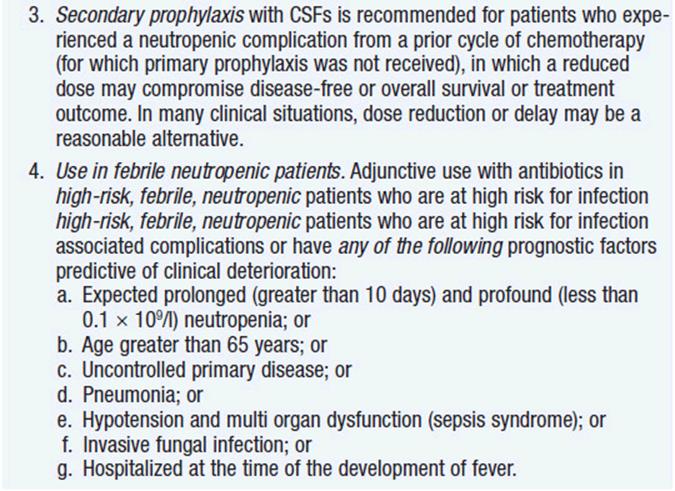
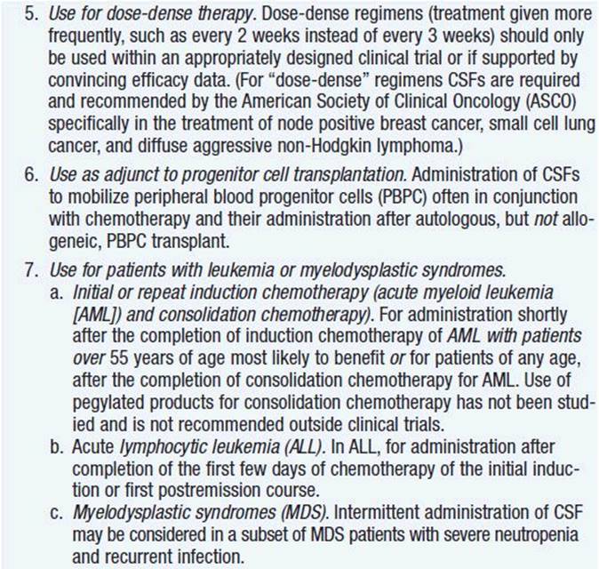
Clinical effect. Based on expert opinion and analysis of the world’s literature on G-CSF use (6, 7), guidelines have been developed to aid in decision making on who should and who should not receive G-CSF during chemotherapy (Table 14-1). In general, G-CSF is overused in clinical practice. The guidelines suggest that it be used with regimens that have a greater than 20% likelihood of inducing febrile neutropenia. Only a small fraction of frequently used regimens are in this category. Risk of developing febrile neutropenia is reduced by about 50%. In the setting of febrile neutropenia, G-CSF may speed neutrophil recovery by 2 or 3 days. However, its use has not permitted dose escalation of hemotherapy. G-CSF is extremely effective in mobilizing hematopoietic stem cells into the peripheral blood. It is so effective that bone marrow harvest has become unnecessary in the vast majority of stem cell donors. Not only are peripheral blood stem cells easier to collect from the donor, but G-CSF-mobilized cells are also more efficient at reestablishing normal hematopoiesis than bone marrow-derived cells and are associated with shorter periods of neutropenia and thrombocytopenia.
TABLE 14-1 CLINICAL INDICATIONS FOR NEUTROPHIL GROWTH FACTORS



Toxicities. The acute toxicity associated with G-CSF use is minor. A few patients may experience bone pain. In normal individuals receiving G-CSF to mobilize hematopoietic stem cells, rapid splenic enlargement is possible and rare splenic rupture has occurred. Thus, these patients need to be monitored for abdominal or shoulder pain.
More serious concerns are emerging about long-term effects. First, animal studies have shown that the amount of damage to hematopoietic stem cells by cyclic chemotherapy is increased with the use of colony-stimulating factor support to hasten recovery (8). In addition, at least three studies have reported an increase in the incidence of acute leukemia and myelodysplasia when cancer therapy was supported with G-CSF use compared to the incidence with chemotherapy alone (9–11). The precise mechanism of the G-CSF effect is unclear. Possibly, through its antiapoptotic effects, it keeps damaged cells alive that would normally die. Regardless of mechanism, the twofold increased leukemia/myelodysplasia risk is sufficient to motivate clinicians to use the agent more sparingly and only when indicated, especially when cure is the goal.
GRANULOCYTE-MACROPHAGE COLONY-STIMULATING FACTOR
Granulocyte-macrophage colony-stimulating factor (GM-CSF) is a 127-amino acid glycoprotein (MW 22 kD) encoded by a gene on chromosome 5q31 that acts early and late in myeloid cell development. The GM-CSF receptor has a unique α-chain called CSF2R and shares a β-chain with the IL3 and IL5 receptors. It stimulates the common myeloid progenitor to differentiate toward the granulocyte/monocyte progenitor rather than the erthroid/megakaryocyte progenitor, and it stimulates an increase in all the progeny of the granulocte/monocyte progenitor. It also activates granulocytes, monocytes, and macrophages and promotes the antigen-presenting function of dendritic cells. Like G-CSF, it is produced by macrophages, fibroblasts, and endothelial cells, but unlike G-CSF, GM-CSF is also produced by T cells.
Biologic activity. GM-CSF stimulates the production of all three granulocyte types, neutrophils, eosinophils, and basophils. It increases the number of peripheral blood monocytes and supports the differentiation of monocytes into professional antigen-presenting cells called dendritic cells, an activity that has stimulated its testing as a vaccine adjuvant. GM-CSF also improves target killing by antibody-dependent cellular cytotoxicity. GM-CSF is usually not detectable in the peripheral blood under normal conditions or after the induction of neutropenia. The consequences of its deletion in knockout mice were minor, only a decrease in alveolar macrophages. Thus, GM-CSF is not viewed as a major physiologic regulator of myelopoiesis. Certainly, in its absence, other cytokines are able to stand in for any essential functions it has.
Pharmacology. An intravenously administered dose of GM-CSF (sargramostim) has an α-phase of 5–20 min and a β-phase of 1.1–2.4 h. A subcutaneously administered dose has a half-life of 1.6–5.8 h. A pegylated version of GM-CSF has been generated, but the agent is not approved for use.
Method of administration. Sargramostim is generally given subcutaneously at a dose of 250 μg/m2/day for all its indications.
Clinical effect. The clinical effects of GM-CSF mimic those of G-CSF to a large degree. Unfortunately, the agents have not been compared head-to-head. However, in general, the magnitude of the beneficial effects seen with GM-CSF and G-CSF are comparable in magnitude (12). No data suggest that the use of either factor improves the response rate, response duration, or overall survival. GM-CSF has also been used as a vaccine adjuvant and appears to be capable of stimulating both antibody and cellular responses to mildly immunogenic proteins such as idiotypic determinants on immunoglobulin molecules (13).
Toxicities. GM-CSF shares the property of G-CSF to induce bone pain in some patients. In general, GM-CSF is associated with more systemic symptoms than G-CSF including more fevers, muscle aches, and fluid retention. Because of their similar effects on neutrophil counts, G-CSF is used more commonly because of the perception that it produces fewer side effects.
ERYTHROPOIETIN
Erythropoietin (EPO) is a 166-amino acid glycoprotein (MW 21 kD) encoded by a gene on chromosome 7q21 that regulates erythropoiesis. It is produced mainly in the kidney, which senses the level of tissue oxygenation. When levels fall below a certain threshold, hypoxia-inducible factor is produced and acts as a stimulus to produce more EPO. EPO is a hormone that is released by the kidney into the peripheral blood. It binds to the EPO receptor, a 66-kD single-chain molecule expressed on bone marrow erythroid progenitors.
Biologic activity. EPO acts both early and late in red cell production (14). In addition to its effects on the committed erythroid progenitor, it may also exert effects on the early multipotent progenitor cells. EPO suppresses apoptosis and improves the efficiency of red cell production. Additional studies have found that EPO is also produced in neurons and may be involved in protecting hypoxic neurons from cell death (15). Furthermore, EPO appears to exert protective effects on myocardium that has been rendered hypoxic by experimental coronary artery ligation (16). These findings led to clinical trials to evaluate the capacity of EPO to protect hypoxic brain and heart that have thus far been negative (17, 18).
Pharmacology. An intravenously administered dose of EPO in the form of epoetin has a serum half-life of 4–11 h. Subcutaneous administration leads to a more prolonged and more variable kinetics with a half-life of 9–38 h. Glycosylation can affect the pharmacokinetics greatly. Site-directed muta-genesis was performed to add two N-glycosylation sites producing the product darbepoetin. Its molecular weight is 23% greater than epoetin, but the serum half-life is prolonged about threefold. An intravenous injection has a half-life of 18–25 h; a subcutaneous injection has a half-life of 33–49 h.
Method of administration. The usual dose of epoetin in patients with cancer is 100–150 U/kg administered subcutaneously 3 times weekly. The usual dose of darbepoetin is 200 μg administered once every 2 weeks. No specific level of hemoglobin is used to trigger the intervention. Many physicians intervene when the hemoglobin level falls to 8 g/dl. In the face of cormorbid lung disease or heart disease, a threshold of 10 g/dl may be more appropriate.
Clinical effect. The patients who respond best to EPO have low levels of circulating endogenous EPO and adequate supplies of iron, B12, and folate. In the setting of renal failure, EPO has been very effective at reducing transfusion requirements and improving quality of life. However, in cancer patients, the slow response to EPO has made it difficult to show any influence on the usual efficacy endpoints of response rates, response durations, and survival. Instead, its FDA approval was based on softer quality-of-life data (19, 20). In the absence of complicating factors, a typical patient may get a 1–2 gm/dl increase in hemoglobin over 6–8 weeks of EPO administration. However, an increasing body of data suggests that EPO administration adversely affects the efficacy of concomitantly administered chemotherapy and protects the tumor from chemotherapy-induced killing. Randomized studies in patients with head and neck cancer, lung cancer, and breast cancer have demonstrated poorer response rates and shorter periods of remission in the group of patients receiving chemotherapy or chemotherapy plus radiotherapy together with EPO than in the group of patients receiving the same antitumor treatment without EPO (21). Accordingly, it appears that EPO use should be confined to the palliative care setting and should not be used in patients in whom the goal of therapy is to cure the disease.
Toxicities. EPO is relatively free of toxic symptoms. When the hemoglobin level gets as high as 12 gm/dl, EPO should be stopped because continued use in the setting of hemoglobin levels of 12 gm/dl or above can be associated with hypertension, polycythemia, and thromboembolic disease.
INTERLEUKIN-11
Interleukin-11 (IL11) is a 178-amino acid nonglycosylated protein (MW 23 kD) encoded by a gene on chromosome 19q13 that stimulates thrombopoiesis. Its receptor is a double-chain molecule with a unique a-chain and a second chain called gp130 that it shares with IL6 and leukemia inhibitory factor (LIF). It is produced by bone marrow-derived stromal cells, fibroblasts, and epithelial cells. It plays a critical role in placental and fetal development as IL11 receptor knockout mice fail to develop. IL11 appears to be involved in implantation of the embryo into the endometrium.
Biologic activity. IL11 causes the proliferation of hematopoietic stem cells and megakaryocyte precursors and promotes platelet development independent of thrombopoietin. Some evidence suggests that it may also be a growth factor for hybridomas in vitro. IL11 has also been an agent of interest in inflammatory bowel disease because of its therapeutic effects to minimize bowel inflammation probably through inhibitory effects on the production of proinflammatory cytokines, particularly by monocytes/macrophages (22, 23).
Pharmacology. Oprelvekin is administered subcutaneously and has a half-life of about 7 h.
Method of administration. Oprelvekin is administered at a dose of 50 μg/kg/day beginning the day after chemotherapy in a setting where thrombocytopenia is an expected toxicity. The agent is given daily for periods of 10–21 days until the platelet count reaches 50,000/μl. Treatment should be discontinued at least 2 days before the start of the next treatment cycle.
Clinical effect. The administration of oprelvekin to women with breast cancer who had experienced thrombocytopenia in a prior cycle reduced the requirement for platelet transfusion by about 25%. Of the 96% of women who experienced thrombocytopenia with the drugs alone, the need for platelet transfusion was noted in 70% of those who had received oprelvekin (24). A much more exciting possibility for IL11 is its application to inflammatory bowel disease where early clinical testing documented a response rate of over 40% (25).
Toxicities. Oprevelkin may produce fatigue, myalgias, arthralgias, and fluid retention with weight gain. The majority of treated patients have fluid retention. Rare patients develop atrial arrhythmias or syncope.
ELTROMBOPAG
Eltrombopag is a biphenyl hydrazine (MW 565) that interacts with the transmembrane domain of the thrombopoietin receptor (cMpl). It is orally bioavailable and stimulates platelet production. The number of megakaryocytes and their production of platelets are both enhanced by the promotion of signaling through cMpl.
Pharmacology. After oral administration, the drug achieves a peak concentration in plasma in 2–6 h. Administration with antacids or other sources of divalent cations (e.g., calcium in dairy products) decreases absorption. It is eliminated by the fecal route and metabolized by oxidation and glucuronidation. The metabolism is slower in people of Asian descent.
Method of administration. Eltrombopag is given at a starting dose of 50 mg/day on an empty stomach. If the platelet count is still less than 50K/mm3 at 2 weeks, the dose may be increased to 75 mg/day, but not higher. If no response is seen after 4 weeks of treatment, the drug should be discontinued. If the platelet count increases above 200K/mm3, but less than 400K/mm3, the dose may be reduced to 25 mg/day and the platelets rechecked in 2 weeks. If the platelet count exceeds 400K/mm3, stop the drug and monitor the platelet count twice a week until the count decreases below 200K/mm3 and then restart at a dose 25 mg lower.
Clinical effect. The majority of patients treated with eltrombopag experience an increase in their platelet count within 2 weeks. If the drug is given in support of chemotherapy, it should be stopped 2 or 3 days before the next cycle. No published randomized clinical trials demonstrate efficacy in this setting (the drug has been applied mainly to treat immune thrombocytopenia), but clinical experience has suggested that platelet nadirs from chemotherapy can be shortened.
Toxicities. Eltrombopag may cause hepatotoxicity; liver function tests should be carefully monitored. The drug is discontinued of the transaminase level exceeds three times the upper limits of normal. Eltrombopag inhibits the OATP1B1 transporter and may result in increased levels of agents that use this transporter, such as statins.
ROMIPLOSTIM
Romiplostim is an Fc peptide fusion protein (peptibody) produced in Escherichia coli; it has two identical single-chain subunits, each of which contains a human IgG1 Fc domain linked at its C-terminus to a peptide comprising two thrombopoietin receptor binding domains. The molecule has no amino acid homology with thrombopoietin, thereby reducing the risk of the development of anti-thrombopoietin neutralizing antibodies, a problem that limited the use of authentic thrombopoietin in patients.
Pharmacology. The pharmacology of the agent is variable. It is administered subcutaneously and achieves peak serum concentrations a median of 14 h later (range 7–50 h) with a half-life of 3.5 days (range 1–34 days). Serum concentrations are not correlated with dose. The agent is cleared faster when the platelet count increases because it binds to the Mpl receptor on platelets.
Method of administration. The drug is administered subcutaneously at a dose of 1 μg/kg once a week and adjusted in 1 μg/kg per week increments up to a maximum dose of 10 μg/kg per week. The drug is not given if the platelet count exceeds 400K/mm3.
Clinical effect. The drug is active in immune thrombocytopenia. Clinical trials in support of the platelet count in the setting of cancer chemotherapy are ongoing. Most patients respond to the drug with higher platelet production.
Toxicities. Chronic use has led to an increase in reticulin deposition in the marrow, but overt myelofibrosis has not been reported. When the agent is stopped, platelet counts may fall. Platelet counts may increase to a level that promotes thromboembolic complications. Decreasing responses may be due to development of neutralizing antibodies.
Other hematopoietic growth factors are being explored for clinical application, including stem cell factor and FLT-3 ligand. These agents are not currently approved for clinical use.
GROWTH FACTORS
Aside from colony-stimulating factors, most therapeutic strategies that focus on growth factors and their receptors are aimed at blocking the effects of the growth factors. However, growth factors with certain selective properties may be useful in protecting against damage from cancer treatments or in promoting tissue restoration after therapy. A prototype agent is palifermin, keratinocyte growth factor.
PALIFERMIN
Palifermin is a 140-amino acid protein (MW 16.3 kD) that differs from endogenous human keratinocyte growth factor by the removal of the first 23 N-terminal amino acids, which improves the stability of the protein. It is a member of the fibroblast growth factor family (FGF7) and binds to keratinocyte growth factor receptor, one of four receptors in the fibroblast growth factor receptor family. The receptor is expressed on epithelial cells of many tissues including the gastrointestinal tract, breast, genitourinary tract, and skin. It is not expressed on hematopoietic cells. It may have trophic effects on involuted thymi.
Biologic activity. Palifermin is produced by mesenchymal cells in response to epithelial injury. When administered to experimental animals, palifermin increases tissue thickness of the tongue, buccal mucosa, and gastrointestinal tract. When given to mice before and after chemotherapy or radiation, palifermin minimized fatalities and reduced weight loss. Palifermin is capable of enhancing the growth of epithelial-derived tumor cell lines in vitro at concentrations >10 μg/ml (generally more than a log higher than levels achieved clinically).
Pharmacology. The elimination half-life of intravenously administered palifermin is about 4.5 h. Levels do not accumulate with three consecutive daily doses. At least a threefold increase in epithelial cell proliferation was detected in healthy subjects who received 40 μg/kg/day for 3 days.
Method of administration. Palifermin is given intravenously on three consecutive days before exposure to the toxic regimen (chemotherapy, radiation therapy, or both) and on 3 consecutive days after treatment at a dose of 60 μg/kg/day. Treatment is given on 6 days.
Clinical effect. Summaries of pivotal clinical trial results are included in the FDA-approved product label (26). Among patients undergoing high-dose therapy and bone marrow transplantation, palifermin reduced duration of grade 3/4 mucositis from 9 to 3 days, reduced incidence of grade IV mucositis from 62% to 20%, and reduced requirement for pain medication by 60% (27). Furthermore, despite the concern about potential adverse effects on growth of carcinomas, palifermin has been applied to the supportive care of patients with colorectal cancer undergoing fluorouracil-based chemotherapy (28). Oral mucositis was dramatically reduced by the use of palifermin, and dose modifications were required in only 14% of the group receiving palifermin compared to 31% of placebo controls. A number of useful supportive measures can further ameliorate the unpleasant consequences of mucositis in patients undergoing cancer treatment (29).
Toxicities. The main toxic effects were grade 3 skin rashes in 3% of patients. Some patients also noted some discoloration of the tongue or mild dysesthesia. Rare patients complained of altered taste. No permanent or life-threatening toxicities were noted.
APPROACHES TO CANCER TREATMENT AND PREVENTION BASED ON ELICITING ANTIGEN-SPECIFIC IMMUNITY
A major goal of oncologists has been to find methods of activating host defenses in the effort to eliminate cancer. The awesome destructive power of the immune system is undeniable, given the consequences of its overactivity in conditions like severe rheumatoid arthritis or multiple sclerosis. We also see the antitumor effects of the immune system in graft-vs-tumor effects that are seen in patients undergoing allogeneic bone marrow transplantation. Those positive effects can be boosted and renewed in some patients with donor lymphocyte infusions. However, despite substantial efforts, not many tumor antigen-specific approaches to cancer therapy are active components of our therapeutic armamentarium. We shall briefly review some promising strategies.
INFECTIOUS DISEASE VACCINES
A number of cancers are known to be caused by infectious agents. Epstein-Barr virus causes lymphomas and nasal lymphoepitheliomas. HTLV-I causes adult T-cell leukemia. Helicobacter pylori causes gastric lymphoma and probably some gastric adenocarcinomas. The list of potential targets for vaccine development is quite large. The power of this approach is substantial. Liver cancer from hepatitis B is a major health hazard, particularly in Asia. The institution of a mandatory hepatitis B vaccination program in Taiwan in the 1990s reduced the prevalence of chronic hepatitis B infection in children by over 90% (30).
The newest vaccine that should have cancer preventive activity is the quadrivalent vaccine against the human papillomavirus (HPV) called Gardasil. The vaccine is composed of virus-like particles that express the major capsid protein L1 from four HPV types: 16 and 18 that account for about 70% of cases of cervical cancer and 6 and 11 that account for about 90% of venereal warts (31). An aggressive vaccination campaign should eliminate these types from the population. The question then is whether this would translate into fewer cases of cervical cancer and venereal warts or whether other virus types would emerge to take the place of the eliminated ones.
Additional targets for vaccine approaches to cancer prevention that would make a major impact on cancer incidence worldwide should include hepatitis C, Epstein-Barr virus, and H. pylori.
CANCER VACCINES
While cancer prevention by targeting infectious etiologic agents is a clever use of the immune system, the capacity to elicit antitumor immunity in a tumor-bearing host is a challenge we have not yet mastered. The problems are daunting. First, tumor cells are not dramatically different from normal cells; thus, finding a way to attack them uniquely is difficult. One might find a way to activate the immune system that does not distinguish between tumor cells and normal cells. Second, the tumors have undergone several adaptations to protect themselves against host immune attack. They sometimes fail to express major histocompatibility determinants, the molecules through which T cells recognize a target. They erect barriers to penetration by developing high levels of interstitial pressure. Thus, a T cell trying to get into a tumor has to navigate the various natural membrane barriers plus push against a pressure gradient that can be as high as or higher than systolic blood pressure. If the cell manages to overcome those odds, tumors can express Fas ligand, which will kill the T cell where it stands. In addition to these serious local barriers to the immune system, tumors make soluble factors that interfere with the antigen-presenting function of dendritic cells, polarize T cells to the less helpful Th2 phenotype (for making antibody) and away from the more helpful Th1 phenotype (for making cytotoxic cells), and alter the signal transduction machinery making the T cells difficult to activate. In short, efforts at activating the immune system of a tumor-bearing host are like whipping a dead horse.
Nevertheless, if we can define the barriers, we may be able to design strategies to overcome them. Many clever approaches are being tested.
Given the apparent success of allogeneic bone marrow transplantation, one idea has been to vaccinate the normal donor against the tumor and adoptively transfer an immune system that may have an even more powerful and specific antitumor effect. Anecdotal reports have been promising (32), but a systematic evaluation of the strategy is needed.
Another strategy to boost the immune response is to perform the immunization during a period of lymphopenia. Several experimental models have documented that vaccine responses are more robust in animals undergoing homeostasis-driven lymphocyte expansion after a lympholytic stimulus (33). Additional data suggest that it would be wise to selectively deplete CD4+ CD25+ regulatory T cells to boost a vaccine response.
Many investigators are focusing more on the composition of the vaccine than on the immunologic environment into which it will be introduced. Accordingly different investigators favor proteins or peptides as antigens; some use DNA that encode the antigenic determinant; some use DNA encoding both the antigen and an adjuvant molecule such as a chemokine; some pulse dendritic cells with peptides, and some augment the dendritic cells by introducing genes (e.g., GM-CSF) aimed to improve their function. In general, immunologic monitoring of such vaccinations generally shows that tumor-specific T-cell immunity is augmented; but little in the way of an antitumor effect has been seen in cancer-bearing people as a consequence of vaccination strategies.
An exception to this generalization is the work of Bendandi, first at the National Cancer Institute and later at the University of Navarre in Spain (11, 34). In one study, idiotype vaccination of patients rendered disease-free by combination chemotherapy was associated with an immune response, as expected; however, in addition, minimal residual disease detected as persistent cells bearing the t(14;18) translocation disappeared from the blood after vaccination. In a second study of follicular lymphoma patients in relapse, multiple vaccinations following conventional chemotherapy produced longer second remissions than first remissions obtained from either similar or the same chemotherapy. These data suggest that idiotype protein given with GM-CSF not only elicits idiotype-specific T cells, but also those T cells are capable of mediating antitumor effects. This is not the same as seeing a tumor mass shrink under the influence of a vaccine. However, additional evidence for an antitumor effect of the cells comes from an analysis of a relapsed patient. The idiotype of the relapsed tumor was altered; thus, the tumor appeared to have escaped the immune surveillance established by the vaccine.
These results point out an additional problem we will have to face down the line; the emergence of tumor variants that evade detection by altering the antigen that we designed our therapy to attack. The implication of this finding is that we should consider multivalent vaccines that are aimed at more than one tumor antigen, if possible.
A cancer vaccine has been approved for use in the setting of advanced prostate cancer. The vaccine is called sipuleucel T (35) and involves taking antigen-presenting cells from the patient and pulsing them with a fusion protein is composed of prostatic acid phosphatase linked to GM-CSF. These antigen pulsed antigen-presenting cells are then given back to the patient; the pheresis procedure, pulsing, and readministration occurs 3 times 2 weeks apart. Despite the fact that no tumor assessment revealed evidence of a tumor response (size of lesions, PSA level, time to progression), patients receiving the vaccine experienced a median improvement in survival of 4 months. The basis for this improvement is unclear. The study lacked appropriate controls including the administration of antigen-presenting cells pulsed only with GM-CSF.
An additional novel strategy to boost immune effects against tumors is to block the negative regulatory pathways that are designed to prevent the immune system from overreacting to any stimulus. At least two such pathways exist, the CTLA4 regulatory pathway and the PD-1 regulatory pathway. CTLA4 is a homologue of CD28 that is upregulated on activated T cells. It binds to costimulatory molecules CD80 and CD86 on dendritic cells 100 times more efficiently than the physiologic ligand CD28 and the effect of its action is to stop the interaction between the T cell and the antigen-presenting cell and turn off the immune response. Two blocking antibodies to CTLA4 are in clinical trial, ipilimumab (IgG1) and ticilimumab (IgG2). They produce a 15% response rate in metastatic malignant melanoma including complete responses and improve overall survival in metastatic melanoma by 4 months. In addition, responses are sometimes delayed and follow a period of transient tumor expansion as host T cells infiltrate the tumors (36). However, the toxicity profile suggests a breaking of self-tolerance (37). Toxicities include dermatitis, colitis, uveitis, hepatitis, hypophysitis, arthritis, nephritis, and hyperthyroidism. Additional studies are underway using these antibodies to boost vaccine responses and combine them with other targeted therapies.
A second negative regulatory pathway operates in activated T cells. A receptor on T cells called PD-1 (programmed death-1) binds to B7-H1 (also called PD-1 ligand-1 or PD-L1) or to B7-DC (PD-L2) on antigen-presenting cells to inhibit T cell proliferation and function. Antibodies to either PD-1 (nivolumab) (38) or the PD-L1 molecule (39) appear to activate antitumor immune responses and have produced responses in tumor types that are generally refractory to immune therapy, including lung cancer. These studies appear to suggest a reversal of immunologic tolerance to tumors by these agents. Studies are underway to develop combination immunotherapies and to combine these treatments with chemotherapy and targeted agents.
We have chosen not to go into more detail about the specialized studies on adoptive cellular therapies. None is ready to become treatments we need to learn how to give in the office, and the field has been associated with claims that have not withstood efforts at repetition. Suffice it to say that adoptive cellular therapy is an active area of investigation, and based on the successes of allogeneic hematopoietic stem cell transplantation, it seems likely that some adoptive therapy approach will show efficacy as we learn more about the determinants of response. One promising approach worth mentioning is the adoptive transfer of T-cell bearing chimeric antigen receptors. Recombinant receptors containing an antibody molecule and activation domains of molecules involved in T-cell signalling are introduced into T cells and administered in vivo. Responses in a few patients with acute and chronic lymphoid leukemia have been dramatic (40) and point to a novel approach to cancer treatment.
REFERENCES
1. Rosenberg SA. Interleukin 2 and the development of immunotherapy for the treatment of patients with cancer. Cancer J Sci Am. 2000; 6 (Suppl 1): S2–S7.
2. McDermott DF. Update on the application of interleukin-2 in the treatment of renal cell carcinoma. Clin Cancer Res. 2007; 13: 716s–720s.
3. Koreth J, Matsuoka K, Kim HT, et al. Interleukin-2 and regulatory T cells in graft-vs-host disease. N Engl J Med. 2011; 365: 2055–2066.
4. Schwartz RN, Stover L, Dutcher J. Managing toxicities of high-dose interleukin 2. Oncology. 2002; 16 (Suppl 13): 11–20.
5. Lansigan F, Stearns DM, Foss F. Role of denileukin diftitox in the treatment of persistent of recurrent cutaneous T-cell lymphoma. Cancer Manag Res. 2010; 2: 53–59.
6. Smith TJ, Khatcheressian J, Lyman GH, et al. 2006 update of recommendations for the use of white blood cell growth factors: an evidence-based clinical practice guideline. J Clin Oncol. 2006; 24: 3187–3205.
7. Aapro MS, Cameron DA, Pettengell R, et al. EORTC guidelines for the use of granulocyte-colony stimulating factor to reduce the incidence of chemotherapy-induced febrile neutropenia in adult patients with lymphomas and solid tumors. Eur J Cancer. 2006; 42: 2433–2453.
8. Hornung RL, Longo DL. Hematopoietic stem cell depletion by restorative growth factor regimens during repeated high-dose cyclophosphamide therapy. Blood. 1992; 80: 77–83.
9. Relling MV, Boyett JM, Blanco JG, et al. Granulocyte colony-stimulating factor and the risk of secondary myeloid malignancy after etoposide treatment. Blood. 2003; 101: 3862–3867.
10. Smith RE, Bryant J, Decillis A, et al. Acute myeloid leukemia and myelodysplastic syndrome after doxorubicin-cyclophosphamide adjuvant therapy for operable breast cancer: the National Surgical Adjuvant Breast and Bowel Project Experience. J Clin Oncol. 2003; 21: 1195–1204.
11. Hershman D, Neugut AI, Jacopson JS, et al. Acute myeloid leukemia or myelodysplastic syndrome following use of granulocyte colony-stimulating factors during breast cancer adjuvant chemotherapy. J Natl cCancer Inst. 2007; 99: 196–205.
12. Bohlius J, Reiser M, Schwarzer G, Engert A. Granulopoiesis-stimulating factors to prevent adverse effects in the treatment of malignant lymphoma. Cochrane Database Syst Rev. 2004; 1: CD003189.
13. Bendandi M, Gocke CD, Koprin CB, et al. Complete molecular remission induced by patient-specific vaccination plus granulocyte-monocyte colony-stimulating factor against lymphoma. Nat Med. 1999; 5: 1171–1177.
14. Kranz SB. Erythropoietin. Blood. 1991; 77: 419–434.
15. Brines ML, Ghezzi P, Keenan S, et al. Erythropoietin crosses the blood brain barrier to protect against experimental brain injury. Proc Natl Acad Sci USA. 2000; 97: 10526–10531.
16. Moon C, Krawczyk M, Ahn D, et al. Erythropoietin reduce myocardial infarction and left ventricular functional decline after coronary artery ligation in rats. Proc Natl Acad Sci USA. 2003; 100: 11612–11617.
17. Ehrenreich H, Weissenborn K, Prange H, et al. Recombinant human erythropoietin in the treatment of acute ischemic stroke. Stroke. 2009; 40.: e647-e656.
18. Najjar SS, Rao SV, Melloni C, et al. Intravenous erythropoietin in patients with ST-segment elevation myocardial infarction: REVEAL: a randomized controlled trial. JAMA. 2011; 305: 1863–1872.
19. Case DC Jr, Bukowski RM, Carey RW, et al. Recombinant erythropoietin therapy for anemic cancer patients on combination chemotherapy. J Natl Cancer Inst. 1993; 85: 801–806.
20. Crawford J, Cella D, Cleeland CS, et al. Relationship between changes in hemoglobin level and quality of life during chemotherapy in anemic cancer patients receiving epoetin alfa therapy. Cancer. 2002; 95: 888–895.
21. http://www.fda.gov/cder/drug/infopage/RHE/default.htm
22. Du X, Williams DA. Interleukin 11: review of molecular, cell biology and clinical use. Blood. 1997; 89: 3897–3908.
23. Williams DA. Inflammatory cytokines and mucosal injury. J Natl Cancer Inst Monogr. 2001; 29: 26–30.
24. Isaacs C, Robert NJ, Bailey FA, et al. Randomized placebo-controlled study of recombinant human interleukin-11 to prevent chemotherapy-induced thrombocytopenia in patients with breast cancer receiving dose-intensive cyclophosphamide and doxorubicin. J Clin Oncol. 1997; 15: 3368–3377.
25. Sands BE, Bank S, Sninsky CA, et al. Preliminary evaluation of safety and activity of recombinant human interleukin 11 in patients with active Crohn’s disease. Gastroenterology. 1999; 117: 58–64.
26. http://www.fda.gov/cder/foi/label/2004/125103lbl.pdf
27. Spielberger R, Stiff P, Bensinger W, et al. Palifermin for oral mucositis after intensive chemotherapy for hematologic cancers. N Engl J Med. 2004; 351: 2590–2598.
28. Rosen LS, Abdi E, Davis ID, et al. Palifermin reduces the incidence of oral mucositis in patients with metastatic colorectal cancer treated with fluorouracil-based chemotherapy. J Clin Oncol. 2006; 24: 5183–5200.
29. http://www.nci.nih.gov/cancertopics/pdq/supportivecare/oralcomplications/HealthProfessional/page5
30. Shepard CW, Simard EP, Finelli L, et al. Hepatitis B virus infection: epidemiology and vaccination. Epidemiol Rev. 2006; 28: 112.
31. Lowy DR, Schiller JT. Prophylactic human papillomavirus vaccines. J Clin Invest. 2006; 116: 1167.
32. Kwak LW, Taub DD, Duffey PL, et al. Transfer of myeloma idiotype-specific immunity from an actively immunized marrow donor. Lancet. 1995; 345: 1016.
33. Hu HM, Poehlein CH, Urba WJ, Fox BA. Development of antitumor immune responses in reconstituted lymphopenic hosts. Cancer Res. 2002.; 62: 2914.
34. Inoges S, Rodriguez-Calvillo M, Zabalegui N, et al. Clinical benefit associated with idiotypic vaccination in patients with follicular lymphoma. J Natl Cancer Inst. 2006; 98: 1292.
35. Kantoff PW, Higano CS, Shore ND, et al. Sipuleucel-T immunotherapy for castration-resistant prostate cancer. N Engl J Med. 2010; 363: 411.
36. Hodi FS, O’Day SJ, McDermott DF, et al. Improved survival with ipilimumab in patients with metastatic melanoma. N Engl J Med. 2010; 363.: 711.
37. Korman A, Yellin M, Keler T. Tumor immunotherapy: preclinical and clinical activity of anti-CTLA4 antibodies. Curr Opin Invest Drugs. 2005.; 6: 592.
38. Topalian SL, Hodi, FS, Brahmer JR, et al. Safety, activity and immune correlates of anti-PD-1 antibody in cancer. N Engl J Med. 2012; 366: 2443.
39. Brahmer JR, Tykodi SS, Chow LQ, et al. Safety and activity of anti-PD-L1 antibody in patients with advanced cancer. N Engl J Med. 2012; 366.: 2455.
40. Porter DL, Levine BL, Kalos M, et al. Chimeric antigen receptor-modified T cells in chronic lymphoid leukemia. N Engl J Med. 2011; 365: 725.