
Eyal C. Attar
INTRODUCTION
Myelodysplastic syndromes (MDS) represent premalignant entities that share many characteristics with acute myeloid leukemia (AML). These clonal hematopoietic stem cell (HSC) disorders are characterized by pancytopenia resulting from failure of normal hematopoiesis. The bone marrow shows hypercellularity, arrested maturation in one or more cellular lineages, and an increase in bone marrow myeloid precursors. Clinical symptoms result from cytopenias. Approximately one-third of patients ultimately progress to AML. Treatment involves supportive care and the use of agents capable of ameliorating cytopenias and delaying development of AML. However, hematopoietic stem cell transplantation (HCT) represents the only potentially curative treatment for MDS.
KEY FEATURES
• One or more peripheral blood cytopenias.
• Hematopoietic cell dysplasia.
• Bone marrow hypercellularity.
• Ringed sideroblasts in a subset of patients.
• Less than 20% bone marrow and peripheral blood myeloblasts.
• Abnormal cytogenetics are observed in approximately 50% of patients. Approximately 20% of patients have characteristic, interstitial deletions within the long arm of chromosome 5, which are associated with clinical response to immunomodulatory drugs (IMIDs).
• MDS may be related to prior chemotherapy and/or radiation for another medical condition (therapy-related MDS or T-MDS).
• Approximately 30% of patients with MDS develop AML.
EPIDEMIOLOGY
Approximately 15,000–30,000 new cases of MDS are diagnosed in the United States each year. MDS is three to four times more prevalent than AML and follows a more indolent course. MDS is likely underdiagnosed. MDS is one cause of anemia in the elderly.
ETIOLOGY
The exact cause of MDS is unknown in most patients. However, intrinsic defects in hematopoietic cells and extrinsic defects associated with the bone marrow microenvironment are involved in the pathogenesis of this disorder.
While most patients with MDS have spontaneously arising, de novo, disease, a portion of patients has therapy-related MDS (T-MDS). Such patients have received chemotherapy and/or radiation in the past, possibly for another malignancy or autoimmune disorder. T-MDS develops within a period of 3–10 years following chemotherapy and is associated with complex chromosomal abnormalities, often involving alterations of chromosomes 5 and/or 7. In addition, T-MDS is associated with a more aggressive clinical course and poorer prognosis in comparison to de novo MDS.
CLINICAL CHARACTERISTICS
• Median age is approximately 70 years.
• Slightly more common in males than in females.
• Prevalence is 50,000–100,000 cases in the United States.
• May be associated with prior chemotherapy, radiation, or environmental exposures to genotoxic agents.
CLASSIFICATION OF MDS
The World Health Organization (WHO) classification system includes refractory anemia (RA), RA with ringed sideroblasts (RARS), refractory cytopenia with multilineage dysplasia (RCMD), and MDS with isolated deletion of 5q for patients harboring a distinct interstitial deletion within the long arm of chromosome 5 (1). Patients with elevated bone marrow blasts of 5%–9% have RA with excess blasts I (RAEB-I) and patients with 10%–19% blasts have RAEB-II. Individuals with 20% or greater myeloblasts in the marrow or peripheral blood have AML.
PROGNOSIS
The most commonly used system to assess a patient’s prognosis is the International Prognostic Scoring System (IPSS) (2). This system assigns a score at diagnosis within each of three categories: the percentage of bone marrow blasts, cytogenetics, and the number and degrees of cytopenias (Table 25-1). The scores are added to yield the IPSS category (Low, Int-1, Int-2, and High). The median survival for patients with Low-, Int-1-, Int-2-, and High-risk disease is 5.7, 3.5, 1.2, and 0.4 years, respectively. Importantly, this system applies only to patients with de novo MDS and was not developed using information from patients with T-MDS, who uniformly have a worse prognosis.
TABLE 25-1 THE INTERNATIONAL PROGNOSTIC SCORING SYSTEM (IPSS)

Additional staging systems have emerged. The WPSS system incorporates the WHO category and whether a patient requires transfusions (3). This system may be used dynamically throughout a patient’s illness to assess prognosis. Another system stratifies patients within IPSS categories using five refined cytogenetic subgroups (4). Indeed, a revised IPSS (IPSS-R) has been developed which incorporates these five subgroups along with blast percentage, hemoglobin, platelet count, and ANC (Greenberg P., et al., American Society of Hematology Meeting, December, 2011).
Other factors affect prognosis. The presence of abnormally localized immature progenitors (ALIPS) within the bone marrow is associated with worse prognosis. These collections of immature, CD34 positive cells are displaced from their customary paratrabecular location to the central marrow space and are associated with decreased survival and increased risk of transformation to AML, even within IPSS subgroups. Dependence upon blood transfusions and elevated ferritin levels are also associated with worse prognosis.
KEY ELEMENTS OF PROGNOSIS
• IPSS scoring system (Table 25-1):
• Percentage of bone marrow blasts
• Cytogenetics
• Number and degrees of cytopenias
• Additional parameters important when assessing prognosis:
• ALIPS
• Requirement for blood product transfusions
• Therapy-related MDS
• Elevated ferritin
PATHOPHYSIOLOGY
MDS involvement of multiple hematopoietic lineages suggests the disease arises in a primitive hematopoietic cell, or HSC. However, the microenvironment, too, contributes significantly to disease pathogenesis.
 MDS IS A CLONAL STEM CELL DISORDER
The hypercellular bone marrow in MDS is clonal in origin and the clonal genetic lesion resides within a primitive HSC. MDS arises from a primitive, multipotent HSC capable of homing and engraftment.
GENOMIC INSTABILITY
Genomic instability within hematopoietic cells further indicates the presence of a cell-intrinsic defect. Clonal genetic abnormalities are observed in the bone marrow of 50% of patients with de novo MDS and 80% of patients with secondary MDS. The majority of abnormalities are nonrandom. The presence of genetic alterations, in most cases, is associated with inferior prognosis. However, one particular genetic alteration, interstitial deletion within the long arm of chromosome 5, paradoxically confers a favorable prognosis.
T-MDS is an example of the contribution of genomic instability to MDS pathogenesis. T-MDS occurs in younger patients than de novo MDS and is more often associated with chromosomal abnormalities. Karyotypic abnormalities include deletion of large portions of, or entire, chromosomes (-5, -7, 7q-, 13q-, 17p-, and -18) (5).
Clinically, T-MDS follows exposure to agents that cause DNA damage and accounts for approximately 10%–20% of MDS/AML. Affected individuals have been previously treated for lung cancer, breast cancer, childhood acute lymphoblastic leukemia, rheumatoid arthritis, and other oncologic and autoimmune disorders requiring chemotherapy and/or irradiation. T-MDS is associated with a poorer prognosis than de novo MDS, with a median survival of approximately 9 months. Injury associated with topoisomerase inhibitor chemotherapy, such as etoposide, has the earliest onset, often within 2–3 years of exposure. Typical mutations involve core binding factor on chromosomes 16 or 21 and the mixed-lineage leukemia (MLL) gene on 11q. In contrast, alkylating agents such as chlorambucil and cyclophosphamide result in a more latent T-MDS, arising 4–7 years following exposure. Alkylator-associated T-MDS is often associated with abnormalities of chromosomes 5 and/or 7. Exposure to radiation may result in delayed onset of T-MDS, even 10 years or longer following exposure. This category of injury is associated with mutations in the AML1 gene. Genotoxic insult from occupational solvents, such as benzene, is clearly associated with development of MDS/AML. Bone marrow disorders associated with stem cell defects such as paroxysmal nocturnal hemoglobinuria (PNH) and aplastic anemia (AA) may evolve into MDS.
5Q- INTERSTITIAL DELETION
Approximately 5%–20% of patients with MDS harbor an interstitial deletion within the long arm of chromosome 5, with or without additional cytogenetic abnormalities. This represents the most common single cytogenetic abnormality in MDS (6).
These patients differ from those with T-MDS, who have losses of large regions of 5q or the entire chromosome 5 and have a distinctly inferior prognosis. Instead, interstitial deletion of genomic DNA between bands q13 and q34 is prognostically favorable compared to other types of MDS. Most importantly, patients with 5q- are exquisitely responsive to the class of agents known as immunomodulators (IMIDs). Patients harboring the 5q- interstitial deletion but carrying additional cytogenetic abnormalities are also responsive to IMIDs, although they have a worse prognosis when compared to patients with isolated interstitial deletion of 5q.
Approximately half of patients with 5q- have isolated deletion of 5q- and clinical features comprising the “5q- syndrome”: RA, mild leukopenia, atypical megakaryocytes, normal or increased platelets, transfusion dependence, and extended survival with low risk of transformation to AML. 5q-syndrome is twice as common in women as men and has a median age of 68 years. The pathogenesis and clinical features of the 5q- syndrome may be related to haploinsufficiency of the RPS14 gene and to loss of miR-145, both on chromosome 5q (7, 8).
GENETIC MUTATIONS WITH NORMAL KARYOTYPE
About 50% of patients have somatic mutations in one or more of at least 18 genes. Mutations in RUNX1, TP53, and NRAS are associated with severe thrombocytopenia and a higher percentage of blast cells in the bone marrow (9). Mutations in TP53, EZH2, and ETV6 are associated with a greater than twofold increased risk of death.
ABNORMAL DIFFERENTIATION
A major clinicopathologic feature of MDS is altered differentiation on cytologic examination of the bone marrow aspirate and biopsy, which displays arrested differentiation and dysplasias affecting one or more lineages. In vitro differentiation of MDS bone marrow is diverted toward nonerythroid lineages, explaining the clinical anemia observed in these patients. MDS marrow contains lower levels of erythroid progenitors and requires severalfold higher concentrations of erythropoietin (Epo) to support in vitro erythroid colony growth. This is clinically relevant, as patients with MDS are often resistant to exogenous Epo or require higher doses to support erythropoiesis than required for other diseases.
INCREASED PROLIFERATION AND APOPTOSIS
Cell cycle analyses have demonstrated increased cellular proliferation in MDS marrow, particularly in the myeloid lineage. However, this is accompanied by an increase in cellular apoptosis. The net balance is a hypercellular bone marrow but ineffective hematopoiesis.
Proliferation and apoptosis have been specifically studied in primitive CD34+ cells from MDS. In early stages of disease such as RA and RARS, apoptosis is greatest and exceeds proliferation. In progressive stages of disease, the ratio of apoptosis to proliferation equalizes. Progression is associated with reductions in both proliferation and apoptosis (10).
MICROENVIRONMENT
Several abnormalities of the environment in which hematologic progenitors proliferate contribute to the pathogenesis of MDS. The most notable example of the potential role of the microenvironment is evidenced in a murine model where mice lacking expression of Dicer1 strictly within osteoprogenitors develop MDS and, in some case, AML (11).
Another potential contributor to MDS pathogenesis is increased secretion of pro-apoptotic cytokines by bone marrow fibroblasts and macrophages, resulting in increased hematopoietic cell apoptosis. Cells and stroma from MDS patients secrete increased levels of TNF-α, interleukin-6, and IFN-γ relative to normal controls. Agents such as IMIDs inhibit TNF-α and IL-6 secretion and reduce bone marrow angiogenesis.
T-lymphocytes are hypothesized to provide immune surveillance in MDS and undergo activation and proliferation in an attempt to eradicate the malignant clone. Indeed, use of immunosuppressive medications such as cyclosporine and eliminating activated T cells using antithymocyte globulin (ATG) can improve cytopenias, particularly in patients with hypoplastic MDS (12).
CLINICAL PRESENTATION
The clinical presentation of MDS relates to the number and degree of cytopenias. Anemia, the most common cytopenia in MDS and seen in 80%–90% of patients, may manifest as a sensation of light headedness, fatigue, chest pain, dyspnea, palpitations, and depression. Leukopenia is the second most common cytopenia in MDS, present in 50% of affected individuals. Manifestations include recurrent lung, sinus, and skin infections. Thrombocytopenia is present in 25% of patients and may result in easy bruising, epistaxis, petechiae, gastrointestinal bleeding, and hematuria. In addition, qualitative defects in hematopoietic cell function may result in clinical signs and symptoms, even in the presence of adequate blood counts. For example, neutrophil dysfunction may contribute to infection, while platelet dysfunction may result in hemorrhage even if the blood counts are within the normal range.
1. Anemia. Light headedness, fatigue, chest pain, dyspnea, palpitations, and depression.
2. Leukopenia. Lung, sinus, and skin infections.
3. Thrombocytopenia. Easy bruising, epistaxis, petechiae, gastrointestinal bleeding, and hematuria.
DIAGNOSTIC STUDIES
The initial evaluation of uni- or multilineage cytopenias begins with careful history taking. Medications, such as antibiotics and chemotherapeutic agents, are common causes. Also, infections caused by parvovirus, HBV, HCV, EBV, CMV, and HIV suppress hematopoiesis, and their presence may be discerned by history, physical examination, and laboratory studies.
When MDS is suspected, a complete blood count with differential is necessary to identify the number and severity of cytopenias. An elevated erythroid mean corpuscular volume (MCV) is often, although not always, present in MDS. Evaluation of iron levels by assessing the iron (Fe), total iron binding capacity (TIBC), and ferritin levels is necessary to assess baseline iron stores, to determine whether iron supplementation is required to enhance hematopoiesis, or identify situations of Fe overload. The B12 and folate levels should also be assessed to ensure adequate substrates for hematopoiesis. An Epo level is helpful in deciding whether exogenous erythroid growth factor administration will be likely to help patients with anemia. Levels of copper and ceruloplasmin should be checked, particularly in patients with ringed sideroblasts within the bone marrow.
A bone marrow aspiration and core biopsy is essential for the diagnosis of MDS. The percentage of bone marrow cellularity is assessed on the core biopsy, while the myeloblast percentage is calculated using the aspirate. Morphologic assessment of myeloid and erythroid dysplasia is made using the aspirate, while megakaryocyte dysplasia is most easily assessed on the core. Flow cytometry is utilized to identify monotypic populations of lymphoid and myeloid cells. Cytogenetic analysis is performed on the aspirate. If cytogenetic information cannot be obtained, FISH may be used to disclose critical genetic deletions.
Bone marrow studies may reveal alternative causes for cytopenias, such as other hematologic malignancies including AML and non-Hodgkin lymphomas, and solid tumors metastatic to the bone. Hypoplastic MDS, hypoplastic anemia, and aplastic anemia may be determined on the bone marrow core. PNH may be detected by flow cytometric assessment of the glycosylphosphatidylinositol (GPI) linked proteins CD55 and CD59.
Laboratory studies used to diagnose MDS:
• Peripheral blood
• CBC with differential, MCV
• Fe, TIBC, ferritin, copper, ceruloplasmin
• B12, folate
• Epo level
• Bone marrow aspiration and biopsy
• Morphologic analysis of dysplasia using the aspirate and core biopsy
• Quantitation of myeloblasts using the bone marrow aspirate
• Flow cytometry to identify monoclonal populations of lymphoid and myeloid cells, PNH, and T-cell large granular lymphocytes
• Cytogenetics to identify chromosomal alterations (FISH is used when cytogenetic information cannot be obtained)
THERAPIES FOR MDS
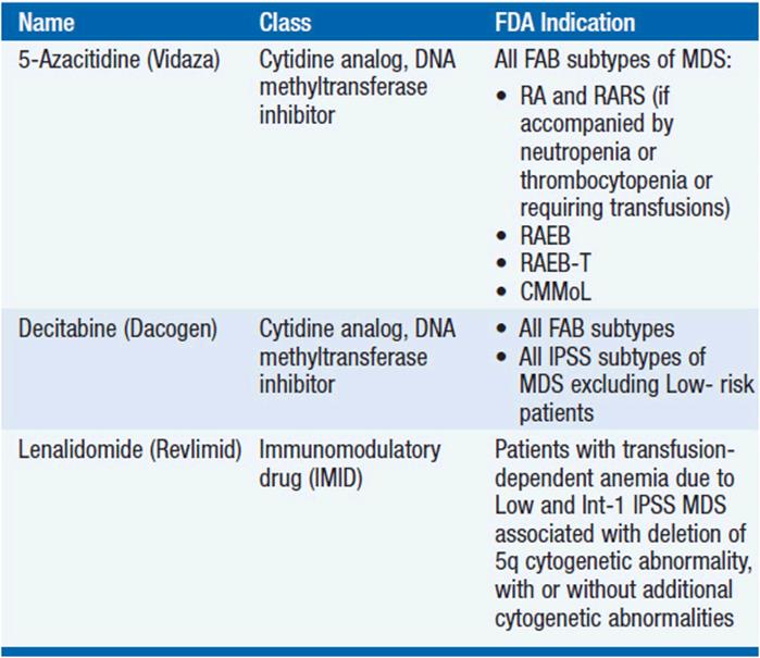
A broad range of management options exists for MDS including amelioration of hematologic deficits with blood product support and administration of growth factors, the use of novel agents aimed at restoring normal hematopoiesis and reducing the malignant clone (Table 25-2), and HCT for patients with high-risk disease for whom a donor can be identified.
TABLE 25-2 FDA-APPROVED THERAPEUTIC AGENTS FOR MDS
SUPPORTIVE CARE
The goal of supportive care is to reduce morbidity and improve quality of life (QoL). Supportive care for MDS consists of cytokine growth factors capable of stimulating myelo- and erythropoiesis, transfusion of red blood cells (RBCs) and platelets, and antibiotics. Anemia has been clearly linked to diminished QoL that can be improved by increasing the hemoglobin (HgB). Age, comorbidities, and lifestyle determine the optimal target HgB level for each patient. However, most patients derive clinical benefit by maintaining an HgB of at least 9 g/dl and HCT of at least 27%, although lower transfusion thresholds are required for patients with symptoms of anemia and with other comorbidities, such as cardiopulmonary disease.
Initial treatment of anemia may be achieved by periodic administration of erythroid growth factors such as epoetin alfa, Procrit, or darbepoetin alfa, Aranesp. Responses to epoetin alfa and darbepoetin alfa are 20%–70% depending on the patient population. Epoetin alfa may be initiated at 40,000–60,000 units weekly, while darbepoetin is given at 200–300 mcg every 1–2 weeks. However, dose adjustments will be required to achieve the desired laboratory and clinical benefit. HgB rises of 1–4 g/dl may be achieved. Both agents appear to have similar efficacy in MDS (13). There may be potential synergy achieved by adding G-CSF (filgrastim, Neupogen), which should be considered if there is no response to epoetin or darbepoetin alone after a period of 6 weeks (14).
Patients with elevated endogenous Epo levels (>500 mU/ml) and those requiring RBC transfusions are unlikely to respond to erythroid growth factors alone. Patients who fail to respond to growth factors require RBC supplementation via periodic transfusions. Individuals with advanced MDS by IPSS score are more likely to require RBC transfusions than those with lower risk disease. Leukodepleted products are recommended to decrease alloimmunization, prevent nonhemolytic febrile transfusion reactions, and reduce the transmission of cytomegalovirus (CMV). Other complications include iron and volume overload, infections, and graft-versus-host disease. Blood products should be irradiated to prevent potentially fatal graft-versus-host disease.
Infections are treated with antibiotics. Myeloid growth factors are not routinely used for prophylaxis of uninfected individuals, even if neutropenic. However, they may be added for resistant or recurrent infections, particularly in patients with an ANC <500/μL. Platelet transfusions are used to treat patients with thrombocytopenia. Prophylactic transfusions are typically used when the platelet count is <10,000/μL. However, transfusions may be required at higher levels such as 20,000 or 30,000 if accompanied by signs of bleeding or petechiae.
IRON-CHELATION THERAPY
Both increased intestinal absorption and transfusional overload account for elevated iron in MDS. While total body iron stores are normally between 3 and 5 g, tissue overload with subsequent dysfunction occurs when the total body iron load is 15–20 g. A serum ferritin of 1000 ng/ml corresponds with a total body iron load of approximately 5 g. Current guidelines are to consider iron-chelation therapy in patients who have received more than 20–25 units of packed RBCs or who have a serum ferritin exceeding approximately 2500 ng/ml. Chelation may be achieved by use of the parenteral agent deferoxamine (Desferal), or one of the two oral chelators, deferiprone (not approved in the United States) and deferasirox (Exjade). Consideration must be given toward balancing the cost and convenience of chelation therapy with the potential benefit. In general, chelation therapy should be considered in young patients with Low/Int-1 IPSS scores who have low levels of bone marrow myeloblasts and iron overload.
Although the relationship of iron-chelation therapy to overall survival has not been prospectively assessed, one retrospective study found that transfusional iron overload, defined as a ferritin level exceeding 1000 ng/ml, was associated with inferior survival in patients with RA and RARS receiving pRBC transfusions compared to patients whose ferritin levels remained below <1000 ng/ml. Retrospective analyses have demonstrated that heavily transfused patients with Low- and Int-1-risk MDS have improved survival, but prospective studies are needed (15, 16).
HYPOMETHYLATING AGENTS
Epigenetic DNA hypermethylation is common in MDS. Hypermethylation results in transcriptional silencing and may contribute to the pathogenesis of MDS by decreasing expression of tumor suppressor genes, genes involved in differentiation, and cyclin-dependent kinase inhibitors (CDKIs). DNA methyltransferase inhibitors (DMTIs) promote hypomethylation via their incorporation into DNA and inhibition of DNA methyltransferases. Like histone deacetylase inhibitors (HDACs), DMTIs have the capacity to promote cellular differentiation in vitro. Such approaches are of obvious relevance to patients with MDS, in which the bone marrow often shows hypercellularity and a block in differentiation.
The DMTI 5-azacitidine (Vidaza), a cytidine analog, was the first agent to receive approval by the FDA for the treatment of MDS. Azacitidine is approved for all FAB subtypes of MDS, including RA and RARS with neutropenia or thrombocytopenia or requiring transfusional support. Azacitidine’s activity was established in a study of 191 patients with MDS randomized to receive azacitidine versus best supportive care (17). Complete and partial responses were observed in 7% and 10% of patients, respectively, treated with azacitidine compared to none in the control arm. Overall improvement was observed in 37% of patients with azacitidine versus 5% with best supportive care. Time to leukemia or death was significantly increased from 13 to 21 months in the azacitidine group. In addition, QoL in the categories of fatigue, dyspnea, physical functioning, positive effect, and psychologic distress was improved in the treatment arm. The effect of azacitidine on improving overall survival (OS) in patients with Int-2- and High-risk MDS was established in the AZA-001 study, where 358 patients were randomized to receive azacitidine versus a conventional care regimen consisting of low-dose cytarabine, induction chemotherapy, or best supportive care alone (18). Patients treated with azacitidine had a median 9-month improvement in OS compared to the CCR arm. In addition, the complete remission (CR) rate and the hematologic response rate in patients receiving azacitidine were 17% and 49%, compared to 8% and 29% in those treated with CCR.
Decitabine (5-deoxyazacitidine, Dacogen) is a deoxycytidine analog that inhibits DNA methylation. Decitabine’s activity was established in a study of 170 patients with MDS randomized to receive either decitabine at a dose of 15 mg/m2 given intravenously over 3 h every 8 h for 3 days (at a dose of 135 mg/m2 per course) and repeated every 6 weeks, or best supportive care (19). Patients treated with decitabine achieved an overall response rate of 17%, including 9% complete responses. In comparison, no responses were seen in the supportive care group. In addition, patients treated with decitabine had a trend toward a longer median time to progression to AML or death compared with patients who received supportive care alone (all patients, 12.1 months versus 7.8 months [P = 0.16]), although improvement was statistically significant in patients with IPSS Int-2- and High-risk disease (12.0 months versus 6.8 months [P = 0.03]) and in those with de novo disease (12.6 months versus 9.4 months [P = 0.04]). In a subsequent randomized study of 233 patients with intermediate- and high-risk MDS treated with decitabine versus best supportive care, a significant survival benefit was not observed in the decitabine arm (20).
IMMUNOMODULATORS (IMIDS)
IMIDs are oral agents that suppress secretion of inflammatory cytokines but also modulate the immune response and inhibit angiogenesis. Thalidomide (Thalomid), the first member of this family to show activity in MDS, is highly teratogenic and is associated with significant side effects including neuropathies, constipation, drowsiness, and fatigue, and has been superseded by lenalidomide (Revlimid), the second agent to gain FDA approval for the treatment of MDS. Lenalidomide is approved for patients with Lowor Int-1-risk MDS who are transfusion dependent and who have a deletion in chromosome 5q. Lenalidomide has fewer side effects than thalidomide.
The efficacy of lenalidomide in MDS was initially observed in a phase II study of 43 patients. Erythroid and cytogenetic responses were most prominent in patients with the 5q- interstitial deletion. In a phase II study of 148 patients with Low- and Int-1-risk MDS who were transfusion dependent, transfusion independence was achieved in 67% of patients while an additional 9% had minor erythroid responses (21). The median duration of response exceeded 104 weeks. Also, 45% of patients had complete cytogenetic remissions, while another 28% had minor cytogenetic responses.
In a similarly designed study, 214 patients with Low- and Int-1-risk MDS who were transfusion dependent and lacked a deletion in 5q were treated with lenalidomide (22). The majority of patients (78%) had a normal karyotype. Transfusion independence was achieved in 26% of patients, while an additional 17% had minor erythroid responses. Of the 47 (22%) patients with abnormal cytogenetics, 9 (19%) achieved a cytogenetic response of which 4 were complete. The median duration of transfusion independence was 41 weeks. The most common toxicities of lenalidomide include neutropenia and thrombocytopenia, which appear to be more common in patients with the 5q- interstitial deletion than in other patients.
IMMUNE SUPPRESSION
Some patients with MDS demonstrate elevated levels of inflammatory cytokines within the blood and/or abnormal lymphoid collections within the bone marrow. These findings suggest that immune dysregulation is operative in MDS pathogenesis and raises the possibility that immune suppression may result in clinical benefit. In a study of 61 patients with primarily low-risk MDS by FAB treated with equine ATG, responses were seen in all three hematopoietic lineages (12). Of these, 21 of 61 patients became transfusion independent, with a median duration of 36 months, 10 of 21 with severe thrombocytopenia achieved sustained responses, and 6 of 11 patients with severe neutropenia had significant improvements. Other studies have shown activity of ATG in MDS, particularly in patients with RA and RARS. Variables noted to predict response to therapy include young age (<60 years), expression of the HLA-DR15 antigen, and shorter duration of RBC transfusion dependence.
ADDITIONAL AGENTS UNDER INVESTIGATION
Like DNA methyltransferases, histone deacetylases suppress gene transcription and are active in patients with MDS. Thus, the role of histone deacetylase inhibitors (HDACs) is under investigation in MDS. A trial of the orally bio-available HDAC, vorinostat (suberoylanilide hydroxamic acid, SAHA), has shown activity. The Ras/Raf kinase signaling pathway is activated in a variety of malignancies including MDS and AML. Agents capable of inhibiting this pathway include arsenic trioxide (AsO3, Trisenox), farnesyl-transferase inhibitors (FTIs), which block necessary steps required for Ras activation, and direct pathway inhibitors, such as the Raf kinase inhibitor Bay 43-9006 (Sorafenib).
The proteasome, a catalytic, multisubunit protease involved in protein degradation, is an antineoplastic target inhibited by bortezomib (Velcade). This agent, capable of suppressing levels of the master transcription factor, nuclear factor-kappa B (NF-κB), is approved for use in multiple myeloma. Elevated levels of nuclear NF-κB have been identified in primitive leukemia cells from patients with leukemia in addition to CD34+ bone marrow cells from patients with high-risk MDS, suggesting a role for the inhibitor in myeloid malignancies.
STEM CELL TRANSPLANTATION
Care of those with advanced disease presents a considerable clinical challenge. Intensive chemotherapy regimens have not provided improvements in long-term outcomes. However, HCT is an important consideration offering potential cure.
Allogeneic stem cell transplantation remains the only potential cure for MDS, although long-term survival is only approximately 30%. An estimated 10%–15% of patients are eligible for HCT, which is generally performed in patients with adequate performance status and for whom a suitable donor is identified. HCT may be performed until age 75 years using reduced-intensity conditioning approaches. A retrospective study of 836 MDS patients transplanted with stem cells from HLA-identical sibling donors revealed comparable overall survival in patients receiving either myeloablative or reduced intensity conditioning (23). Autologous HCT remains an option for individuals without suitable allogeneic candidates, although the majority of patients relapse within 2 years (24).
Optimal timing of allogeneic HCT represents a balance between maximizing chances of long-term survival while decreasing transplant-related morbidity and mortality. A decision-model analysis was conducted to determine the relationship between IPSS score and timing of HCT (25). This model concluded that for patients <60 years receiving myeloablative conditioning, early HCT increased overall survival for patients with Int-2-or High-risk disease. In contrast, HCT could be delayed for a brief period of time for those with Low- or Int-1-risk disease, provided the HCT is done before leukemic transformation. A similar analysis in adults aged 60–70 years found a life-expectancy and quality-adjusted survival benefit for early transplantation of individuals with Int-2- and High-risk disease. These strategies maximize overall survival for low-risk patients while decreasing the early morbidity and mortality of transplantation.
PRINCIPLES OF THERAPY
Treatment of MDS requires consideration of patient age, treatment preference, IPSS score, performance status, presence of antecedent hematologic disorder (AHD), and availability of an HLA-matched stem cell donor. A treatment schema is provided in Figure 25-1. Patients with secondary MDS have a worse prognosis relative to those with de novo MDS. If the patient is a candidate for intensive therapy, an allogeneic donor must be sought and preparations made for allogeneic SCT. If the patient is deemed unsuitable or an appropriate donor cannot be identified, therapies consisting of supportive care, azacitidine, decitabine, and/or clinical trial should be considered.

FIGURE 25-1 Treatment schema for patients with MDS.
For patients with Low- or Int-1-risk MDS and anemia, cytogenetic status influences the starting therapy. Initial treatment for patients with the 5q-interstitial deletion, with or without additional cytogenetic abnormalities, is with lenalidomide. If an adequate response is not achieved after a period of approximately 3 months, attention is turned toward azacitidine, decitabine, or clinical trials in addition to supportive care. For Low/Int-1-risk patients lacking the 5q- cytogenetic deletion, initial treatment is based upon the serum Epo level and transfusion status. Patients with elevated serum Epo levels and/or those dependent upon RBC transfusions are unlikely to respond to exogenous erythropoietic growth factors and, therefore, treatment commences with supportive care, azacitidine, decitabine, and/or clinical trials. Lenalidomide may be effective in improving anemia, even in patients lacking the 5q- interstitial deletion. Antilymphocyte therapy consisting of ATG may be effective in patients who are HLA-DR15 positive, who harbor trisomy 8, or who have a hypocellular MDS.
Patients with neutropenia and infections require myeloid growth factors and antibiotics. Patients with clinically significant thrombocytopenia should receive platelet transfusions and/or antifibrinolytic agents (aminocaproic acid). Azacitidine, decitabine, or clinical trials may be beneficial for individuals with thrombocytopenia and/or neutropenia.
For Int-2- and High-risk patients, HCT represents the best initial therapy for young patients with HLA-matched donors. If a donor cannot be identified or if the patient is not a candidate for HCT, azacitidine, decitabine, chemotherapy agents, and/or supportive care are reasonable choices.
REFERENCES
1. Swerdlow S, Campo E, Harris N, et al. WHO Classification of Tumours of Haematopioetic and Lymphoid Tissues. Lyon: International Agency for Research on Cancer (IARC); 2008.
2. Greenberg P, Cox C, LeBeau MM, et al. International scoring system for evaluating prognosis in myelodysplastic syndromes. Blood. 1997; 89: 2079–2088.
3. Malcovati L, Germing U, Kuendgen A, et al. Time-dependent prognostic scoring system for predicting survival and leukemic evolution in myelodysplastic syndromes. J Clin Oncol. 2007; 25: 3503–3510.
4. Schanz J, Tuchler H, Sole F, et al. New comprehensive cytogenetic scoring system for primary myelodysplastic syndromes (MDS) and oligoblastic acute myeloid leukemia after MDS derived from an international database merge. J Clin Oncol. 2012; 30: 820–829.
5. Mauritzson N, Albin M, Rylander L, et al. Pooled analysis of clinical and cytogenetic features in treatment-related and de novo adult acute myeloid leukemia and myelodysplastic syndromes based on a consecutive series of 761 patients analyzed 1976–1993 and on 5098 unselected cases reported in the literature 1974–2001. Leukemia. 2002; 16: 2366–2378.
6. Nimer SD. Clinical management of myelodysplastic syndromes with interstitial deletion of chromosome 5q. J Clin Oncol. 2006; 24: 2576–2582.
7. Ebert BL, Lee MM, Pretz JL, et al. An RNA interference model of RPS19 deficiency in Diamond-Blackfan anemia recapitulates defective hematopoiesis and rescue by dexamethasone: identification of dexamethasone-responsive genes by microarray. Blood. 2005; 105: 4620–4626.
8. Kumar MS, Narla A, Nonami A, et al. Coordinate loss of a microRNA and protein-coding gene cooperate in the pathogenesis of 5q- syndrome. Blood. 2011; 118: 4666–4673.
9. Bejar R, Stevenson K, Abdel-Wahab O, et al. Clinical effect of point mutations in myelodysplastic syndromes. N Engl J Med. 2011; 364: 2496–2506.
10. Parker JE, Mufti GJ, Rasool F, et al. The role of apoptosis, proliferation, and the Bcl-2-related proteins in the myelodysplastic syndromes and acute myeloid leukemia secondary to MDS. Blood. 2000; 96: 3932–3938.
11. Raaijmakers MH, Mukherjee S, Guo S, et al. Bone progenitor dysfunction induces myelodysplasia and secondary leukaemia. Nature. 2010; 464: 852–857.
12. Molldrem JJ, Leifer E, Bahceci E, et al. Antithymocyte globulin for treatment of the bone marrow failure associated with myelodysplastic syndromes. Ann Intern Med. 2002; 137: 156–163.
13. Mannone L, Gardin C, Quarre MC, et al. High dose darbopoetin alfa the treatment of lower risk MDS: results of a phase II study. Br J Haematol. 2006; 133: 513–519.
14. Hellstrom-Lindberg E, Gulbrandsen N, Lindberg G, et al. A validated decision model for treating the anaemia of myelodysplastic syndromes with erythropoietin + granulocyte colony-stimulating factor: significant effects on quality of life. Br J Haematol. 2003; 120: 1037–1046.
15. Rose C, Brechignac S, Vassilief D, et al. Does iron chelation therapy improve survival in regularly transfused lower risk MDS patients? A multicenter study by the GFM (Groupe Francophone des Myelodysplasies). Leuk Res. 2010; 34: 864–870.
16. Guariglia R, Martorelli MC, Villani O, et al. Positive effects on hematopoiesis in patients with myelodysplastic syndrome receiving deferasirox as oral iron chelation therapy: a brief review. Leuk Res. 2011; 35: 566–570.
17. Silverman LR, Demakos EP, Peterson BL, et al. Randomized controlled trial of azacitidine in patients with the myelodysplastic syndrome: a study of the cancer and leukemia group B. J Clin Oncol. 2002; 20: 2429–2440.
18. Fenaux P, Mufti GJ, Hellstrom-Lindberg E, et al. Efficacy of azacitidine compared with that of conventional care regimens in the treatment of higher-risk myelodysplastic syndromes: a randomised, open-label, phase III study. Lancet Oncol. 2009; 10: 223–232.
19. Kantarjian H, Issa JP, Rosenfeld CS, et al. Decitabine improves patient outcomes in myelodysplastic syndromes: results of a phase III randomized study. Cancer. 2006; 106: 1794–1803.
20. Lubbert M, Suciu S, Baila L, et al. Low-dose decitabine versus best supportive care in elderly patients with intermediate- or high-risk myelodysplastic syndrome (MDS) ineligible for intensive chemotherapy: final results of the randomized phase III study of the European Organisation for Research and Treatment of Cancer Leukemia Group and the German MDS Study Group. J Clin Oncol. 2011; 29: 1987–1996.
21. List A, Dewald G, Bennett J, et al. Lenalidomide in the myelodysplastic syndrome with chromosome 5q deletion. N Engl J Med. 2006; 355: 1456–1465.
22. Raza A, Reeves JA, Feldman EJ, et al. Phase 2 study of lenalidomide in transfusion-dependent, low-risk, and intermediate-1 risk myelodys-plastic syndromes with karyotypes other than deletion 5q. Blood. 2008; 111: 86–93.
23. Martino R, Iacobelli S, Brand R, et al. Retrospective comparison of reduced intensity conditioning and conventional high dose conditioning for allogeneic hematopoietic stem cell transplantation using HLA identical sibling donors in myelodysplastic syndromes. Blood. 2006; 108: 836–846.
24. Ducastelle S, Ades L, Gardin C, et al. Long-term follow-up of autologous stem cell transplantation after intensive chemotherapy in patients with myelodysplastic syndrome or secondary acute myeloid leukemia. Haematologica. 2006; 91: 373–376.
25. Cutler CS, Lee SJ, Greenberg P, et al. A decision analysis of allogeneic bone marrow transplantation for the myelodysplastic syndromes: delayed transplantation for low-risk myelodysplasia is associated with improved outcome. Blood. 2004; 104: 579–585.