
Jeffrey A. Barnes, Jeremy S. Abramson
INTRODUCTION
The peripheral T-cell lymphomas are a heterogeneous group of lymphoid malignancies each with a distinct clinical presentation and prognosis. The term “peripheral” denotes mature T-cell neoplasms, differentiating them from the precursor lymphoid diseases T-cell acute lymphoblastic leukemia and lymphoblastic lymphoma. Treatment regimens for peripheral T-cell lymphomas are generally derived from data extrapolated from trials of aggressive non-Hodgkin lymphoma (NHL) before the advent of CD20-directed therapies of which T-cell lymphomas represent approximately 10% of the studied population. While therapies are often overlapping, T-cell lymphomas in general have an inferior prognosis to their B-cell counterparts. Recent advances have been made, however, with development of treatments specifically directed at peripheral T-cell lymphomas.
EPIDEMIOLOGY
In 2012, there were an estimated 70,130 new cases of NHL in the United States with approximately 18,940 deaths (1). T-cell lymphoma is an uncommon subtype of NHL representing just 12% of all cases (2). The T-cell lymphomas have been subdivided by the most recent WHO classification scheme into predominantly leukemic, nodal, and extranodal variants (Table 35-1) (3). The International Peripheral T-cell Lymphoma project determined the relative frequencies and geographic variation of T-cell lymphoma of subtypes with peripheral T-cell lymphoma-not otherwise specified (PTCL-NOS) being the most common, followed by angioimmunoblastic T-cell lymphoma (AITL), anaplastic large-cell lymphoma(ALCL), and adult T-cell leukemia/lymphoma (ATLL) (Figure 35-1) (4). Notably, there are significant geographic differences in the incidence of these entities with PTCL-NOS being the most common in North America and Europe, whereas extranodal nasal NK T-cell lymphoma (ENKTL) and ATLL are more common in Asia due to the seroprevalence of the EBV virus and HTLV-1 viruses, respectively (Table 35-2).
TABLE 35-1 T/NK-CELL NEOPLASMS
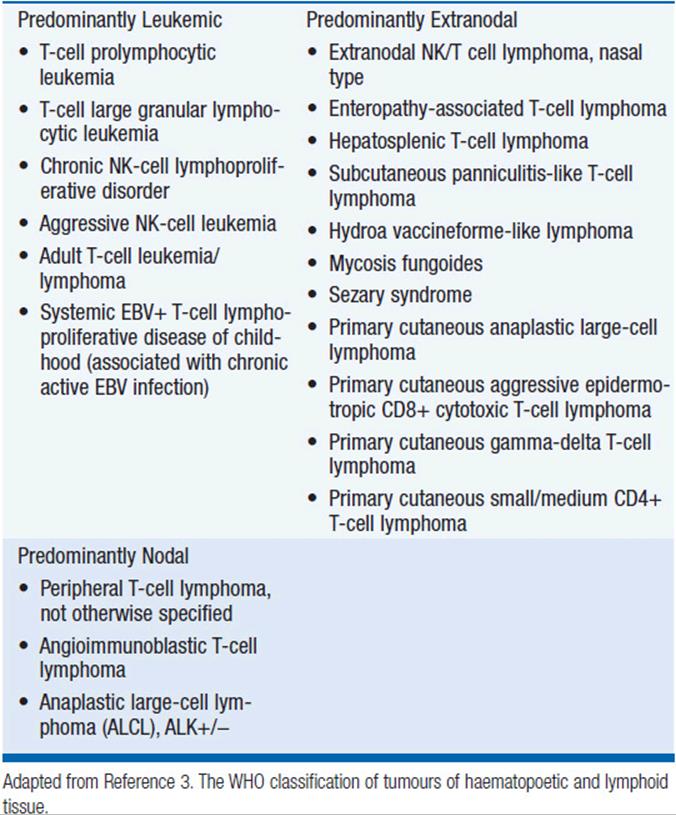

Figure 35-1 International T-cell Lymphoma Study: frequency of subtypes. (From Reference 4.)
TABLE 35-2 MAJOR LYMPHOMA SUBTYPES BY GEOGRAPHIC REGION

PERIPHERAL T-CELL LYMPHOMA NOS
 DIAGNOSIS
PTCL-NOS is the most common subtype of T-cell lymphoma in the United States representing approximately 35% of all T-cell lymphomas (4%–5% of all lymphomas). PTCL-NOS presents at a median age of 61 years with a male-to-female ratio of approximately 2:1 (5). Typically patients present with advanced stage disease (69%), elevated LDH (49%), B symptoms (35%), and extranodal disease (56%) (6). The diagnosis is optimally made based on excisional lymph node biopsies with final needle aspirates normally inadequate. PTCL can be a heterogeneous disease by morphology with most biopsies demonstrating medium-sized or large cells with irregular, pleomorphic, hyperchromatic, or vesicular nuclei with prominent nucleoli and many mitotic figures (3). Immunophenotypic features include CD4 expression more common than CD8 expression with frequent antigen loss of CD5 and CD7, antigens normally expressed on T cells. The T-cell receptor beta chain is usually expressed allowing differentiation from gamma/delta T-cell lymphomas and NK cell lymphomas. Similar to other lymphomas, PTCL-NOS is staged according to the Ann Arbor staging system (Table 35-3) (37). Initial evaluation should include:
TABLE 35-3 ANN ARBOR STAGING SYSTEM

• Physical examination with attention paid to nodal areas, hepatosplenomegaly, and performance status.
• Laboratory studies including complete blood count with differential, LDH, uric acid, calcium, and liver function tests.
• Bone marrow biopsy in select cases (of prognostic importance)
• Imaging studies including CT scans of the chest, abdomen, and pelvis. The majority of PTCL-NOS will also be PET avid, and so PET/CT may be obtained.
• Evaluation of cardiac function including ejection fraction for those patients receiving an anthracycline.
PROGNOSIS
The 5-year overall survival for PTCL-NOS is approximately 32%. The prognostic index for PTCL-NOS (PIT) was developed to risk stratify patients based on four adverse risk factors including age greater than 60, performance status of 2 or higher, elevated LDH, and bone marrow involvement. Patients in group 1 with no adverse factors, group 2 with one factor, group 3 with two factors, and group 4 with three or four factors have a 5-year overall survival of 62%, 52.9%, 33%, and 18% respectively (Figure 35-2) (7).
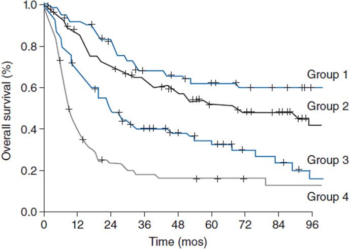
Figure 35-2 Overall survival according to the prognostic index for PTCL-NOS (PIT). (From Reference 7.)
TREATMENT
Peripheral T-cell lymphomas are traditionally treated like other aggressive NHLs with combination chemotherapy including CHOP-like regimens (cyclophosphamide, doxorubicin, vincristine, and prednisone). The complete remission rate for patients receiving an anthracycline-containing regimen such as CHOP is approximately 56% compared to 70%–90% for patients with diffuse large B-cell lymphoma receiving R-CHOP (6). The German high-grade NHL study group retrospectively analyzed 343 patients (70 with PTCL-NOS) comparing 6–8 cycles of CHOP to CHOP plus etoposide as well as 14- versus 21-day treatment intervals. While no difference was seen in CHOP 14 versus CHOP 21, there was a statistically significant improvement in 3-year event-free survival favoring inclusion of etoposide (75.4%) compared to CHOP alone (51.0%) (8).
Given the poor outcomes with standard chemotherapy, consolidation with high-dose chemotherapy and autologous stem-cell transplant (ASCT) has been explored. Retrospective studies have shown an improvement in 5-year overall survival to 68% compared to the predicted 32% (9); however, such analyses are confounded by selection bias of the most favorable patients and inclusion of patients with ALK+ ALCL who have a decidedly more favorable prognosis compared to PTCL-NOS. A prospective trial of up-front ASCT excluding patients with ALK+ ALCL showed a 3-year overall survival of 48% (10). The 3-year overall survival rate was 71% for the two-thirds of patients with chemosensitive disease enrolled on the trial that underwent ASCT versus 11% for the patient who did not undergo ASCT. There are also retrospective data for allogeneic stem-cell transplant for patients with refractory peripheral T-cell lymphomas including PTCL-NOS with 2-year overall survival to 55% but with nonrelapse mortality of 22% (11).
Several novel agents have activity in PTCL-NOS (Table 35-4). The histone deactylase inhibitor romidepsin is approved in the United States for patients with PTCL-NOS having received one prior therapy based on a phase II trial showing a response rate of 25% but with a complete response rate of 15% and a median duration of response of 17 months (12). The main toxicities of romidepsin include cytopenias, GI toxicity, and the potential for arrhythmias. Pralatrexate, a novel antifolate agent, is also approved for patients with relapsed or refractory PTCL-NOS with an overall response rate of 29% and complete response rate of 11% with main toxicities including cytopenias and mucositis (38). Gemcitabine, a nucleoside analog, also has significant activity with overall response rates of approximately 50% (13). There are several other agents that have shown promise in small clinical trials including brentuximab vedotin (for patients with CD30+ disease) (39), alemtuzumab (40), denileukin diftitox (41), and bortezomib (42). Both alemtuzumab and denileukin diftitox have been combined with CHOP-like chemotherapy but early trials were limited due to increased infectious complications. Given the poor predictive outcomes for both up-front and salvage therapy, participation in clinical trials both at diagnosis and relapse are encouraged.
TABLE 35-4 NOVEL AGENTS FOR PERIPHERAL T-CELL LYMPHOMAS

ANAPLASTIC LARGE CELL LYMPHOMA
DIAGNOSIS
Anaplastic large-cell lymphoma (ALCL) represents approximately 12% of the peripheral T-cell lymphomas seen in the United States each year or approximately 2% of all non-Hodgkin lymphomas (4). ALCL can be further subclassified as expressing or lacking expression of the protein anaplastic lymphoma kinase (ALK). ALK-positive tumors have a translocation of chromosome 2 and chromosome 5 with resultant fusion of ALK on chromosome 2 with the nucleophosmin gene on chromosome 5 to produce t(2;5), and tend to present in the first three decades of life with a median age of 34 years. ALK-negative patients present at a median age of 58 (14). Both ALK+ and ALK– ALCL often present at advanced stage (58%–65%) with elevated LDH (37%–46%), extranodal disease ~20%, and B symptoms (60%). As with PTCL-NOS, the diagnosis is made on excisional lymph node biopsies showing effacement of lymph node architecture with large anaplastic cells often with reniform nuclei and are positive for CD30, CD25, and epithelial membrane antigen (EMA). The staging and pretreatment evaluations are similar to those as listed above for PTCL-NOS.
PROGNOSIS
ALCL has an improved prognosis compared to PTCL-NOS, but with a distinct difference between the ALK+ and ALK– variants. ALK+ ALCL has a 5-year overall survival of 70% compared to 49% for ALK– ALCL (Figure 35-3) (14). The IPI score typically used for DLBCL is able to effectively risk stratify patients with ALCL regardless of the ALK status.

Figure 35-3 Overall survival according to ALK status in ALCL. (From Reference 14.)
TREATMENT
Patients with ALCL are treated with anthracycline-containing CHOP-like regimens similar to PTCL-NOS based on extrapolation of data from trials aggressive NHLs including both B- and T-cell lymphomas. As above with PTCL-NOS, retrospective studies evaluating the addition of etoposide to a CHOP-like regimen have shown improvement in both the 3-year event-free survival and overall survival for ALK+ ALCL (75.8% and 89.8%) and ALK– ALCL (45.7% and 62.1%) (8).
Given the excellent prognosis of ALK+ ALCL patients treated with CHOP-like chemotherapy, upfront consolidation with high-dose chemotherapy and autologous stem-cell transplant is not recommended. Patients with ALK– ALCL are much more likely to relapse and go onto salvage therapies including autologous stem-cell transplant resulting in long-term remissions of only 30%–40%. ALCL is uniformly CD30 positive making the CD-30 directed antibody-drug conjugate brentuximab vedotin an appealing agent. A phase II trial of 58 patients (26% of whom had received prior autologous SCT and 62% of whom were refractory to front-line treatment) demonstrated an overall response rate of 86%, complete response rate of 57% with 97% of patients having observed reductions in tumor size (Figure 35-4) (15). The median duration of response was 12.6 months with an estimated 12-month overall survival rate of 70%. Given brentuximab vedotin’s remarkable single agent activity in relapsed and refractory disease, studies are ongoing with this agent in combination with chemotherapy and earlier in the treatment paradigm. The ALK inhibitor crizotinib (recently approved for non-small cell lung cancer harboring ALK mutations) has been tested in four refractory ALK+ ALCL patients all of whom responded (16); however, its role in this otherwise favorable disease subset is unclear at this time as the majority of patients with ALK+ ALCL will be cured with existing standard therapies.
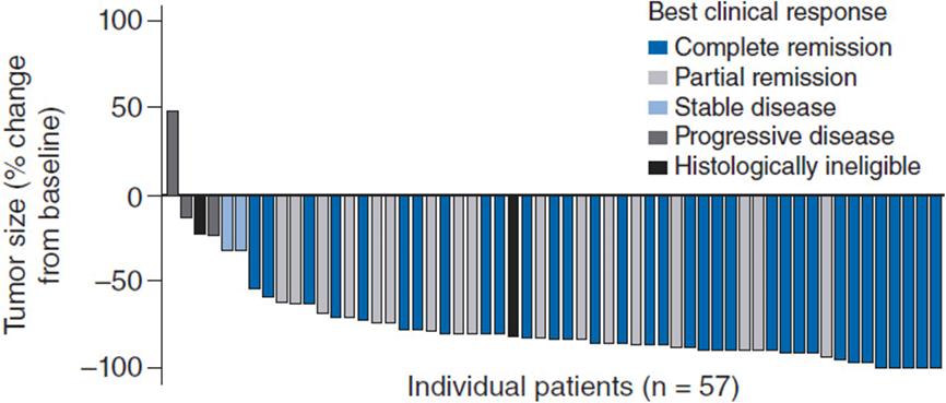
Figure 35-4 Maximum tumor reduction in patients with relapsed refractory ALCL after brentuximab vedotin. (From Reference 15.)
ANGIOIMMUNOBLASTIC T-CELL LYMPHOMA
DIAGNOSIS
Angioimmunoblastic T-cell lymphoma (AITL) represents approximately 19% of all the T-cell lymphoma seen in the United States every year or approximately 1%–2% of all NHLs (4). A majority of patients with AITL present with the relatively abrupt onset of systemic symptoms including fevers, a pruritic skin rash, nonbulky adenopathy, and organomegaly; a unique clinical presentation compared to other non-Hodgkin lymphomas. The median age of onset is 65 years with a slight male predominance (17). The majority of patients present with advanced stage and extranodal disease is present in approximately one-third of patients. As the historical name of angioimmunoblastic lymphadenopathy with dysproteinemia (AILD) suggests, 30% of patients will present with polyclonal hypergammaglobulinemia, or other protein abnormalities including small amounts of paraprotein or positive tests for autoantibodies. Given the constellation of rapid onset fevers, rashes, and nonbulky adenopathy, patients with AITL are often sent for rheumatologic or infectious disease evaluation before their lymphoma diagnosis, leading to a delay in therapy. Positive tests for rheumatoid factor, antinuclear antibody, Lyme, and other conditions contribute to initially incorrect diagnoses. These patients are frequently started on glucocorticoids confounding the diagnosis of lymphoma with a median time to diagnosis over 6 months from the onset of symptoms. Patients may also present with recurrent infections related to immune deficiency secondary to both the neoplastic process and their immunosuppressive treatments. Ten to twenty percent of patients will have concurrent autoimmune cytopenias, immune-mediated thrombocytopenia (ITP), and/or autoimmune hemolytic anemia (AIHA). As with PTCL-NOS and ALCL, the diagnosis is made on excisional lymph node biopsies demonstrating effacement of lymph node architecture with a paracortical polymorphous infiltrate of small- to medium-sized CD4-positive T cells and proliferation of arborizing high endothelial venules. There is also often an associated expansion of EBV-positive B immunoblasts that are polyclonal. The staging and pretreatment evaluations are similar to those as listed above for PTCL-NOS with the addition of testing for circulating paraproteins and autoimmune cytopenias.
PROGNOSIS
AITL has a poor prognosis with a 5-year overall survival of 32% and 5-year failure-free survival of 18% (4). The prognostic index score is less valuable in this histology since the majority of patients present with high-risk scores.
TREATMENT
As with PTCL-NOS and ALCL, AITL is customarily treated with CHOP-like regimens based on extrapolation from aggressive NHL trials with a complete remission rate for those receiving an anthracycline-containing regimen of approximately 60%. No survival differences have been observed in patients treated with combination chemotherapy that included an anthracycline compared to those receiving an anthracycline-sparing regimen, but definitive conclusions cannot be reached based on this retrospective data (17). Given the poor outcome in the majority of patients treated with standard chemotherapy, autologous stem-cell transplant in first and second remission has been retrospectively evaluated. In 146 patients with a median follow-up of 31 months, there was an estimated 4-year overall survival of 59%, which is improved compared to historical controls (18). As with all retrospective studies, these findings must be interpreted with attention to biases including selecting for patients that are younger with fewer comorbidities making them eligible for transplant and therefore likely to do better than historical controls regardless of the transplant procedure.
EXTRANODAL NK/T-CELL LYMPHOMA, NASAL TYPE
DIAGNOSIS
Extranodal NK/T-cell lymphoma, nasal type (ENKTL) is an uncommon T-cell lymphoma in North America and Europe representing around 5% of cases but with a significantly increased incidence in Asia where it represents approximately 22% of new T-cell lymphomas and 9% of all lymphomas (4). Patients present at a median age of 43 years with a slight male predominance and symptoms of nasal obstruction due to the presence of a mass lesion in the upper aerodigestive tract. The majority of patients present with localized disease (76%), with a good performance status, normal LDH (63%), and lack of B symptoms (35%) (19). Diagnostic biopsies show frequent ulceration of the mucosal surfaces by a diffuse lymphomatous infiltrate positive for CD2, CD3, and CD56, and will often demonstrate an angiocentric and angiodestructive growth pattern.
PROGNOSIS
ENKTL nasal type has an improved outcome compared to other PTCLs, owing to the more likely presentation at limited stage disease. The estimated 5-year overall survival is about 49%, but is improved in patients with localized disease who have a 5-year overall survival of approximately 76%. Specific prognostic models for ENKTL have been proposed with adverse risk factors including the presence of B symptoms, advanced stage, elevated LDH, and lymph node involvement demonstrating 5-year overall survival of 81%, 64%, 34%, and 7%, for those with 0, 1, 2, and 3 or more risk factors, respectively. (Figure 35-5) (19). Local invasion through bone or soft tissues also negatively affects prognosis.
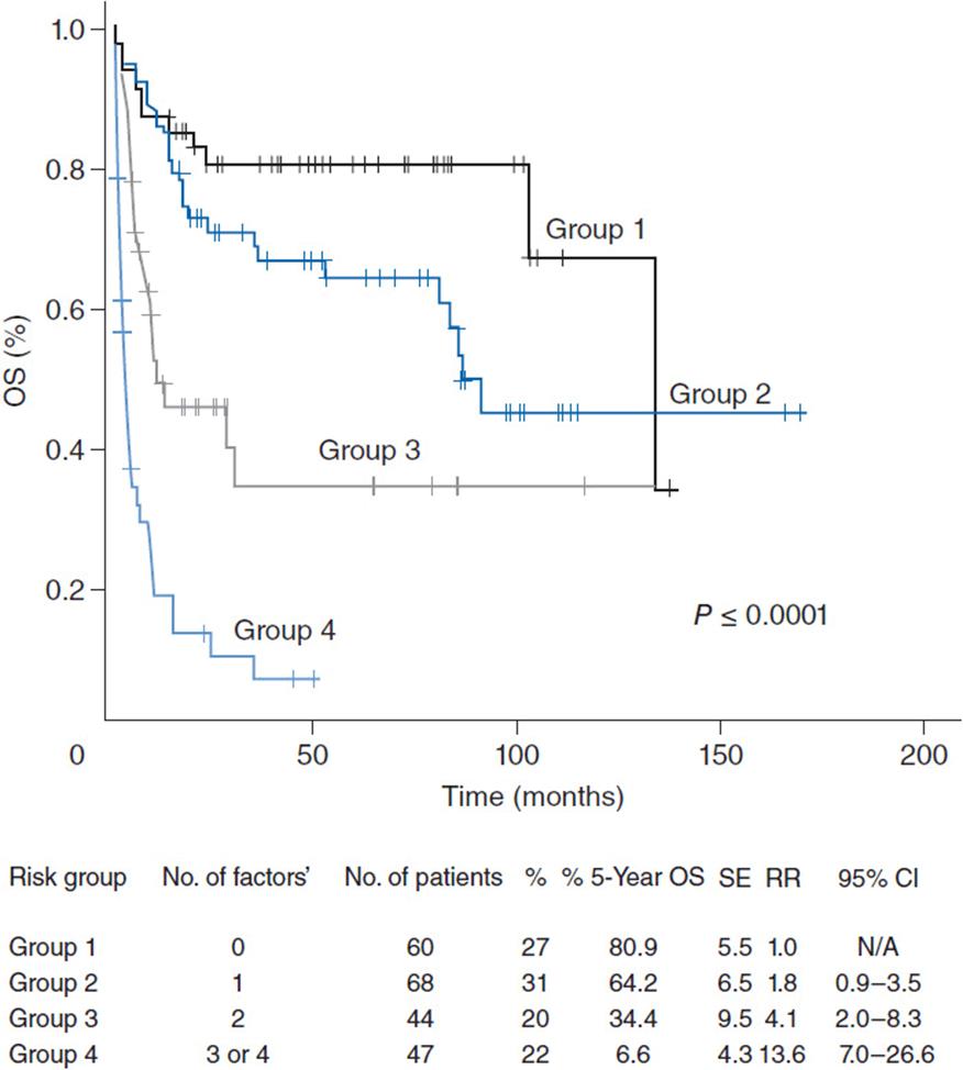
Figure 35-5 Overall survival for patients with extranodal NK/T-cell lymphoma. (From Reference 19.)
TREATMENT
ENKTL nasal type relies heavily upon the use of radiation therapy for curative intent. With doses ranging from 40 to 65Gy (higher than typical radiation doses for other lymphomas), complete remission rates are approximately 87% for radiation therapy alone with a 5-year overall survival of 71% for patients with stage IE and IIE disease (20). Small prospective trials of combined modality therapy with radiation therapy given either with concurrent cisplatin followed by VIPD (etoposide, ifosfamide, cisplatin, and dexamethasone) or DeVIC (dexamethasone, etoposide, ifosfamide, and carboplatin) have shown modestly increased overall survival but with increased toxicity (21, 22). Patients with advanced stage disease are not candidates for curative radiotherapy, and asparaginase-based regimens may perform better than other chemotherapy programs in this disease. The steroid, methotrexate, ifosfamide, L-asparaginase, and etoposide (SMILE) regimen produces an overall response rate of 79% in patients with stage IV disease, with a 1-year overall survival of 55%, though at the cost of significant toxicity (23). The AspaMetDex regimen (L-asparaginase, methotrexate and dexamethasone) is less toxic than SMILE and has shown an encouraging complete remission rate of 61% in patients with refractory disease with median overall survival of 1 year, suggesting that further testing is warranted of L-asparaginase in the up-front treatment of ENKTL (24).
ADULT T-CELL LEUKEMIA/LYMPHOMA
DIAGNOSIS
Adult T-cell leukemia/lymphoma (ATLL) is a rare entity in North America and Europe but has a significantly increased incidence in Asia and in the Caribbean where it is directly linked to the prevalence of human T-cell leukemia virus 1 (HTLV-1) infection. There is often a long latency with most affected individuals exposed to the virus early in life. HTLV-1 is transmitted in breast milk and by exposure to peripheral blood or blood products. There are four clinical variants of ATLL that affect the clinical presentation and prognosis including acute, lymphomatous, chronic, and smoldering types. The acute type is most common and presents with disseminated leukemic and bone marrow involvement, lymphocytosis, skin rash, and generalized lymphadenopathy. Hypercalcemia at presentation is quite common, as are pulmonary infiltrates. The differential diagnosis of the lung infiltrates include leukemic infiltration and opportunistic infection given the uniform immunosuppressive state associated with the disease. The lymphomatous type, as the name implies, presents with adenopathy and/or extranodal masses, but without significant involvement of peripheral blood and bone marrow. The chronic type presents with a lymphocytosis but with mild adenopathy and follows a more indolent course. Finally the smoldering type presents without peripheral blood or marrow involvement and with frequent cutaneous involvement. The median age of onset is 62 years with the majority of patients presenting with stage IV disease (73%) (25). While a broad spectrum of cytologic features may be present, the characteristic finding in the peripheral blood is of CD4+, CD8–, CD25+ T cells with the characteristic flower-shaped nucleus with many nuclear convolutions and lobules (Figure 35-6). Unlike the other T-cell lymphomas, the diagnosis often can be made on peripheral blood with correlation with HTLV-I serologies. Unlike other leukemias, bone marrow involvement is often patchy. ATLL is staged according to the Ann Arbor system, with a similar workup as for other T-cell lymphomas, but with staging of the blood with flow cytometry and attention to the risk of hypercalcemia, opportunistic infection, and occasionally spontaneous tumor lysis syndrome.

Figure 35-6 Peripheral blood smear from patient with acute ATLL. (Photo courtesy of Dr. Judith Ferry, Massachusetts General Hospital Department of Pathology.)
PROGNOSIS
The prognosis for ATLL is dependent on the subtype with the smoldering and chronic variants having significantly better survival compared to the lymphomatous and acute subtypes. The IPI score can be used to stratify patients with the lymphomatous subtype but is not very helpful in the other variants (25). For patients with the acute subtype, the ATL prognostic index is based on five factors including advanced Ann Arbor stage, performance status greater than one, serum albumin less than 3.5 g/dL, age greater than 70, and levels of soluble IL-2 receptors <20,000 U/ml and can separate patients into low- (scores 0–2), intermediate- (scores 3–4), and high-risk groups (score 5) with a 2-year overall survival rates of 37%, 17%, and 2%, respectively (26).
TREATMENT
The smoldering and chronic variants of ATLL can follow an indolent course and patients may be followed with observation alone until the disease become symptomatic. The aggressive variants of ATLL including the acute and lymphomatous types carry a poor prognosis when treated with CHOP-like regimens. Given that ATLL presents in a leukemic phase with a significant burden of disease and a high incidence of preexisting hypercalcemia, close attention must be paid to development of treatment-related tumor lysis syndrome. Overall response rates to initial therapy are approximately 70% but the duration of response is frequently short (25). Given the poor prognosis with standard therapies, allogeneic stem-cell transplantation should be considered in eligible ATLL patients with available donors given the evidence for a graft versus leukemia effect and signal of efficacy in small series.
Consideration of novel agents and early enrollment in clinical trials is warranted. Novel treatment approaches including targeting the HTLV-I virus itself may offer additional benefit compared to chemotherapy. Small studies examining the use of antiviral therapy with zidovudine plus interferon produced high response rates approaching 90% but a short median event-free survival of approximately 7 months (27). The addition of zidovudine and interferon to chemotherapy improved overall response rates from 49% to 81% although no difference was seen in progression-free or overall survival (28). For patients with relapsed or refractory disease, treatment with denileukin diftitox, alemtuzumab, pralatrexate, and histone deacetylase inhibitors may be considered based on the PTCL-NOS data, but response rates and duration of response are quite limited based on small amounts of data.
UNCOMMON SUBTYPES
CUTANEOUS T-CELL LYMPHOMAS
Mycosis Fungoides, Sézary Syndrome
Cutaneous lymphomas represent less than 2% of all NHLs, 75% of which are cutaneous T-cell lymphomas (CTCL). Mycosis fungoides is the most common type of CTCL, often presenting in older adults with a male-to-female ratio of 2:1. Patients often present with a prolonged indolent course with scaling skin plaques/lesions that wax and wane. Most patients present with limited stage disease confined to the skin alone and are treated with skin directed therapies including topical glucocorticoids, topical nitrogen mustard, topical retinoids, and UV light therapy usually enhanced by psoralen sensitization. For disease that is unresponsive to skin-directed therapies, the oral retinoid bexarotene and low-dose methotrexate can be used. The prognosis for limited stage patients is excellent with a median overall survival in excess of 10 years. Patients with advanced stage disease including disseminated cutaneous disease or visceral involvement have dramatically inferior prognosis. Multiagent chemotherapy, such as CHOP-like regimens, have overall response rates of 60%–80% but significant infectious risks and very brief duration of response; therefore, single-agent approaches are more often employed. Liposomal doxorubicin, gemcitabine, and methotrexate all have significant activity and can be used with palliative intent.
Sézary syndrome (SS) accounts for 5% of all cutaneous T-cell lymphomas, and is defined as the presence of a circulating CTCL count of 1000 cells per microliter of peripheral blood. The disease is often accompanied by widespread skin disease that is called erythroderma. SS is an aggressive disease with an overall survival rate at 5 years of less than 10%. Several targeted agents have been approved for CTCL and but few have been evaluated in patients with Sézary syndrome. Denileukin diftitox has response rates in CTCL of up to 50% with a 10% complete response rate (29). The histone deacetylase inhibitors vorinostat and romidepsin are effective in CTCL with responses seen in up to one-third of patients but no complete responses have been seen (30, 31). The CD52-directed monoclonal antibody alemtuzumab is highly active in patients with SS, where low-dose therapy (10- or 15-mg delivered subcutaneously three times a week) was found to have an overall response rate of 86% and a complete response rate of 21% in one small study (32).
Primary Cutaneous CD30-Positive T-Cell Lymphoproliferative Disorders
Primary cutaneous CD30-positive T-cell lymphoproliferative disorders are the second most common type of CTCL behind mycosis fungoides (33). This group of diseases includes lymphomatoid papulosis (LyP) and primary cutaneous anaplastic large-cell lymphoma. LyP is a chronic, recurrent, self-healing papulonodular skin eruption with histologic features of a CD30-positive CTCL. LyP generally occurs in adults and can be differentiated from primary cutaneous ALCL by the presence of skin lesions at different stages of development that resolve without intervention. LyP in general does not require therapy although in patients with truly persistent painful disease, low-dose methotrexate can be used as suppressive therapy. Primary cutaneous ALCL shares similar histologic features with LyP demonstrating nonepidermotropic infiltrates of large CD30-positive tumor cells (34). Unlike systemic ALCL, most cutaneous ALCLs do not express ALK. While primary cutaneous ALCL exists on a spectrum with LyP, the lesions in ALCL do not spontaneously remit. The major differential diagnosis of primary cutaneous ALCL is systemic ALCL with secondary skin involvement. Patients should be carefully examined for lymphadenopathy or organ involvement. Isolated lesions can be treated with local radiation therapy (electron beam) or surgical excision with chemotherapy rarely indicated. Primary cutaneous ALCL has an excellent prognosis; estimated 10-year survival exceeds 90%.
Subcutaneous Panniculitis-Like T-Cell Lymphoma/Primary Cutaneous Gamma-Delta T-Cell Lymphoma (SPTCL)
SPTCL is a rare T-cell lymphoma representing less than 1% of the T-cell lymphomas seen each year. It is a disease with a female predominance with a median age of 36 years with 20% of patients presenting before age 20 years (35). Historically, it has been divided in two groups based on T-cell receptor expression. Those with alpha/beta T-cell receptors carry a more favorable prognosis and usually present without skin findings and rarely have involvement of other nonsubcutaneous sites. Those with the gamma/delta T-cell receptor have an unfavorable prognosis and may present with ulcerated skin lesions. Patients often will have an associated autoimmune disorder such as SLE or juvenile RA, and presentation with an associated hemophagocytic syndrome may be seen in >20% of cases.
T-CELL LARGE GRANULAR LYMPHOCYTIC LEUKEMIA
T-cell large granular lymphocytic leukemia (T-LGL) is a rare lymphoproliferative disorder characterized by a population of clonal LGL cells between 2 and 20 K/ul in the peripheral blood without an identified cause (3). Reactive T-cell LGLs can be seen in response to viral infections, posttransplant T-cell expansions, and connective tissue diseases and should be differentiated from a malignant process. LGL is a rare disease occurring primarily in older adults with a strong association with rheumatoid arthritis. Patients usually present with recurrent infections secondary to neutropenia. Other cytopenias may also be seen including anemia and thrombocytopenia. Up to 14% of patients may present with pancytopenia making the distinction from aplastic anemia difficult. Splenomegaly is usually present, but is not massive as may be seen in other splenic predominant disease such as splenic marginal zone lymphoma or hairy cell leukemia.
Evaluation of the peripheral blood smear typically shows large lymphocytes with moderate to abundant cytoplasm and prominent azurophilic granules. The bone marrow often shows a normocellular or hypocellular marrow. The LGL infiltrate can be variable and difficult to visualize. When present there is usually an interstitial/intrasinusoidal infiltrate of CD3+, CD8+, TCR-alpha/beta+ cytotoxic T cells. Abnormally, decreased or lost expression of CD5 and/or CD7 is common. Most are positive for CD57 and CD16 as well as perforin and granzyme B, typical of cytoxic T cells. TCR PCR for gene rearrangements can be used to confirm clonality, though false positives do occur and should be interpreted in the context of the entire clinicopathologic picture.
The prognosis for LGL is excellent with most cases taking an indolent course. Indications for treatment are cytopenias, particularly neutropenia. Several agents can be employed including low-dose weekly methotrexate with or without prednisone, daily oral cyclosporine or cyclophosphamide, and less commonly chemotherapy agents such as pentostatin.
ENTEROPATHY-ASSOCIATED T-CELL LYMPHOMA
Enteropathy associated T-cell lymphoma (EATL) is a rare peripheral T-cell lymphoma representing less than 5% of cases. This disease occurs exclusively in patients with underlying celiac disease, but the celiac disease and lymphoma may be diagnosed concurrently at the time the patient presents with a lymphoma complication. Patients with celiac disease are recommended to maintain a gluten-free diet, which reduces the risk of developing this lymphoma. Patients typically present with abdominal pain, GI bleeding, and occasionally bowel perforation. Patients may have a poor performance status and hypoalbuminemia at diagnosis due to protein wasting. Abdominal masses show ulcerating lesions that invade the wall of the intestine with medium to large lymphoid cells with vesicular nuclei, prominent nucleoli, and moderate pale cytoplasm. Prognosis is quite poor with a 5-year overall survival of 29% (4). As with other T-cell lymphomas patients are often treated with CHOP-like regimens. A novel regimen of ifosfamide, etoposide, epirubicin, and methotrexate followed by autologous stem-cell transplant generated 5-year progression-free and overall survival of 52% and 60%, respectively. These numbers are greatly improved over historical controls; however, this is a small study (36).
HEPATOSPLENIC T-CELL LYMPHOMA
Hepatosplenic T-cell lymphoma (HSL) is an extremely rare form of T-cell lymphoma representing less than 2% of all cases of T-cell lymphoma (4). The median age of onset is 34 years with a male predominance. There is preferential involvement of the vascular sinusoids of the liver, spleen, and bone marrow, but adenopathy is generally not present. Patients frequently present with B symptoms organomegaly, and cytopenias. This disease may occur in patients with immune suppression, most commonly after a solid-organ transplantation when it occurs at median of 10 years posttransplant. All patients may initially respond to multiagent chemotherapy, but durations of remission are brief and the 5-year overall survival approaches 0% (4).
CONCLUSIONS
The peripheral T-cell lymphomas are mature T-cell neoplasms each with distinct clinical presentations. Attention to pattern recognition of their unique clinical presentations and close collaboration with expert hematopathologists are needed to make accurate diagnoses. Treatment regimens are extrapolated from trials of aggressive lymphomas of which T-cell lymphomas represent an approximate 10% of the study populations. Aside from ALK+ ALCL, prognosis is generally poor and early consideration for participation in clinical trials is warranted to identify more effective therapies for these uncommon and aggressive diseases.
REFERENCES
1. American Cancer Society. Cancer facts and figures 2012. 2012.
2. Anderson JR, Armitage JO, Weisenburger DD, for the Non-Hodgkin’s Lymphoma Classification Project. Epidemiology of the non-Hodgkin’s lymphomas: distributions of the major subtypes differ by geographic locations. Ann Oncol. 1998; 9: 717–720.
3. Swerdlow S, Campo E, Harris NL, et al (eds). WHO classification of tumours of haematopoetic and lymphoid tissue. Lyon, IARC, 2008.
4. Vose J, Armitage J, Weisenburger D, International T-Cell Lymphoma Project. International peripheral T-cell and natural killer/T-cell lymphoma study: pathology findings and clinical outcomes. J Clin Oncol. 2008; 26: 4124–4130.
5. Rüdiger T, Weisenburger DD, Anderson JR, et al. Peripheral T-cell lymphoma (excluding anaplastic large-cell lymphoma): results from the non-Hodgkin’s lymphoma classification project. Ann Oncol. 2002; 13: 140–149.
6. Weisenburger DD, Savage KJ, Harris NL, et al. Peripheral T-cell lymphoma, not otherwise specified: a report of 340 cases from the international peripheral T-cell lymphoma project. Blood. 2011; 117: 3402–3408.
7. Gallamini A, Stelitano C, Calvi R, et al. Peripheral T-cell lymphoma unspecified (PTCL-U): a new prognostic model from a retrospective multicentric clinical study. Blood. 2004; 103: 2474–2479.
8. Schmitz N, Trumper L, Ziepert M, et al. Treatment and prognosis of mature T-cell and NK-cell lymphoma: an analysis of patients with T-cell lymphoma treated in studies of the german high-grade non-Hodgkin lymphoma study group. Blood. 2010; 116: 3418–3425.
9. Rodriguez J, Conde E, Gutierrez A, et al. The results of consolidation with autologous stem-cell transplantation in patients with peripheral T-cell lymphoma (PTCL) in first complete remission: the spanish lymphoma and autologous transplantation group experience. Ann Oncol. 2007; 18: 652–657.
10. Reimer P, Rudiger T, Geissinger E, et al. Autologous stem-cell transplantation as first-line therapy in peripheral T-cell lymphomas: results of a prospective multicenter study. J Clin Oncol. 2009; 27: 106–113.
11. Delioukina M, Zain J, Palmer JM, et al. Reduced-intensity allogeneic hematopoietic cell transplantation using fludarabine-melphalan conditioning for treatment of mature T-cell lymphomas. Bone Marrow Transplant. 2012; 47: 65–72.
12. Coiffier B, Pro B, Prince HM, et al. Results from a pivotal, open-label, phase II study of romidepsin in relapsed or refractory peripheral T-cell lymphoma after prior systemic therapy. J Clin Oncol. 2012; 30: 631–636.
13. Zinzani PL, Venturini F, Stefoni V, et al. Gemcitabine as single agent in pretreated T-cell lymphoma patients: evaluation of the long-term outcome. Ann Oncol. 2010; 21: 860–863.
14. Savage KJ, Harris NL, Vose JM, et al. ALK- anaplastic large-cell lymphoma is clinically and immunophenotypically different from both ALK+ ALCL and peripheral T-cell lymphoma, not otherwise specified: report from the international peripheral T-cell lymphoma project. Blood. 2008; 111: 5496–5504.
15. Pro B, Advani R, Brice P, et al. Brentuximab vedotin (SGN-35) in patients with relapsed or refractory systemic anaplastic large-cell lymphoma: results of a phase II study. J Clin Oncol. 2012; 30: 2190–2196.
16. Pogliani EM, Dilda I, Villa F, et al. High response rate to crizotinib in advanced, chemoresistant ALK+ lymphoma patients. ASCO Meeting Abstracts. 2011; 29(15_Suppl): e18507.
17. Federico M, Rudiger T, Bellei M, et al. Clinicopathologic characteristics of angioimmunoblastic T-cell lymphoma: analysis of the international peripheral T-cell lymphoma project. J Clin Oncol. 2013; 31: 240–246.
18. Kyriakou C, Canals C, Goldstone A, et al. High-dose therapy and autologous stem-cell transplantation in angioimmunoblastic lymphoma: complete remission at transplantation is the major determinant of outcome-lymphoma working party of the European group for blood and marrow transplantation. J Clin Oncol. 2008; 26: 218–224.
19. Lee J, Suh C, Park YH, et al. Extranodal natural killer T-cell lymphoma, nasal-type: a prognostic model from a retrospective multicenter study. J Clin Oncol. 2006; 24: 612–618.
20. Li Y, Yao B, Jin J, et al. Radiotherapy as primary treatment for stage IE and IIE nasal natural killer/T-cell lymphoma. J Clin Oncol. 2006; 24: 181–189.
21. Yamaguchi M, Tobinai K, Oguchi M, et al. Phase I/II study of concurrent chemoradiotherapy for localized nasal natural killer/T-cell lymphoma: Japan clinical oncology group study JCOG0211. J Clin Oncol. 2009; 27: 5594–5600.
22. Kim SJ, Kim K, Kim BS, et al. Phase II trial of concurrent radiation and weekly cisplatin followed by VIPD chemotherapy in newly diagnosed, stage IE to IIE, nasal, extranodal NK/T-cell lymphoma: consortium for improving survival of lymphoma study. J Clin Oncol. 2009; 27: 6027–6032.
23. Yamaguchi M, Kwong YL, Kim WS, et al. Phase II study of SMILE chemotherapy for newly diagnosed stage IV, relapsed, or refractory extranodal natural killer (NK)/T-cell lymphoma, nasal type: the NK-cell tumor study group study. J Clin Oncol. 2011; 29: 4410–4416.
24. Jaccard A, Gachard N, Marin B, et al. Efficacy of L-asparaginase with methotrexate and dexamethasone (AspaMetDex regimen) in patients with refractory or relapsing extranodal NK/T-cell lymphoma, a phase 2 study. Blood. 2011; 117: 1834–1839.
25. Suzumiya J, Ohshima K, Tamura K, et al. The international prognostic index predicts outcome in aggressive adult T-cell leukemia/lymphoma: analysis of 126 patients from the international peripheral T-cell lymphoma project. Ann Oncol. 2009; 20: 715–721.
26. Katsuya H, Yamanaka T, Ishitsuka K, et al. Prognostic index for acute-and lymphoma-type adult T-cell Leukemia/Lymphoma. J Clin Oncol. 2012; 30: 1635–1640.
27. Hermine O, Allard I, Levy V, et al. A prospective phase II clinical trial with the use of zidovudine and interferon-alpha in the acute and lymphoma forms of adult T-cell leukemia/lymphoma. Hematol J. 2002; 3: 276–282.
28. Hodson A, Crichton S, Montoto S, et al. Use of zidovudine and interferon alfa with chemotherapy improves survival in both acute and lymphoma subtypes of adult T-cell leukemia/lymphoma. J Clin Oncol. 2011; 29: 4696–4701.
29. Prince HM, Duvic M, Martin A, et al. Phase III placebo-controlled trial of denileukin diftitox for patients with cutaneous T-cell lymphoma. J Clin Oncol. 2010; 28: 1870–1877.
30. Piekarz RL, Frye R, Turner M, et al. Phase II multi-institutional trial of the histone deacetylase inhibitor romidepsin as monotherapy for patients with cutaneous T-cell lymphoma. J Clin Oncol. 2009; 27: 5410–5417.
31. Duvic M, Olsen EA, Breneman D, et al. Evaluation of the long-term tolerability and clinical benefit of vorinostat in patients with advanced cutaneous T-cell lymphoma. Clin Lymphoma Myeloma. 2009; 9: 412–416.
32. Bernengo MG, Quaglino P, Comessatti A, et al. Low-dose intermittent alemtuzumab in the treatment of Sezary syndrome: clinical and immunologic findings in 14 patients. Haematologica. 2007; 92: 784–794.
33. Willemze R, Jaffe ES, Burg G, et al. WHO-EORTC classification for cutaneous lymphomas. Blood. 2005 May 15; 105: 3768–3785.
34. Bekkenk MW, Geelen FA, van Voorst Vader PC, et al. Primary and secondary cutaneous CD30(+) lymphoproliferative disorders: a report from the dutch cutaneous lymphoma group on the long-term follow-up data of 219 patients and guidelines for diagnosis and treatment. Blood. 2000 Jun 15; 95: 3653–3661.
35. Willemze R, Jansen PM, Cerroni L, et al. Subcutaneous panniculitis-like T-cell lymphoma: definition, classification, and prognostic factors: an EORTC cutaneous lymphoma group study of 83 cases. Blood. 2008; 111: 838–845.
36. Sieniawski M, Angamuthu N, Boyd K, et al. Evaluation of enteropathy-associated T-cell lymphoma comparing standard therapies with a novel regimen including autologous stem cell transplantation. Blood. 2010; 115: 3664–3670.
37. Lister TA, Crowther D, Sutcliffe SB, et al. Report of a committee convened to discuss the evaluation and staging of patients with hodgkin’s disease: Cotswolds meeting. J Clin Oncol. 1989; 7: 1630–1636.
38. O’Connor OA, Pro B, Pinter-Brown L, et al. Pralatrexate in patients with relapsed or refractory peripheral T-cell lymphoma: results from the pivotal PROPEL study. J Clin Oncol. 2011; 29: 1182–1189.
39. Pro B, Advani R, Brice P, et al. Brentuximab vedotin (SGN-35) in patients with relapsed or refractory systemic anaplastic large-cell lymphoma: results of a phase II study. J Clin Oncol. 2012; 30: 2190–2196.
40. Zinzani P, Alinari L, Tani M, et al. Preliminary observations of a phase II study of reduced-dose alemtuzumab treatment in patients with pre-treated T-cell lymphoma. Haematologica. 2005; 90: 702–703.
41. Dang NH, Pro B, Hagemeister FB, et al. Phase II trial of denileukin diftitox for relapsed/refractory T-cell non-hodgkin lymphoma. Br J Haematol. 2007; 136: 439–447.
42. Zinzani PL, Musuraca G, Tani M, et al. Phase II trial of proteasome inhibitor bortezomib in patients with relapsed or refractory cutaneous T-cell lymphoma. J Clin Oncol. 2007; 25: 4293–4297.