
David P. Ryan
EPIDEMIOLOGY
 STATISTICS
In the United States, 102,480 new cases of colon cancer were expected in 2013 (men 50,090; women 52,390) (1), and 40,340 new cases of rectal cancer were expected in 2013 (men 23,590; women 16,750). Colorectal cancer is the second leading cause of cancer-related death in the United States with 50,830 deaths annually. Age is a major risk factor in developing colon cancer. The lifetime risk of developing colorectal cancer is approximately 5% with the vast majority of cancers occurring after age 50 years. The overall incidence has been falling perhaps due to screening.
EPIDEMIOLOGIC ASSOCIATIONS
The vast majority of colorectal cancers are sporadic and not familial. Epidemiologic studies demonstrate an increased risk of colorectal cancer with the following conditions/characteristics:
• Family history of colorectal cancer is associated with an increased risk of developing colorectal cancer. If one first-degree family member had colorectal cancer, the risk increases 1.7-fold
• Western/urbanized societies
• Diet high in red or processed meat
• Increased bowel anaerobic flora
• Diabetes mellitus/insulin resistance: the risk of colon cancer may be 30% higher in diabetics compared with nondiabetics
• Inflammatory bowel disease. Increased incidence is seen with both Crohn’s disease and ulcerative colitis and is associated with the severity, extent, and duration of disease affecting the colon. The risk of colon cancer in ulcerative colitis is approximately 10% at 10-year duration, 20% at 20-year duration, and >35% at 30-year duration. Total colectomy eliminates the risk of colon cancer
• Cigarette smoking
• Alcohol consumption
• Ureterosigmoidostomy
• Streptococcus bovis bacteremia
• Prior pelvic radiation
INHERITED SYNDROMES
Fewer than 10% of colon cancers are known to be associated with an inherited predisposition to colon cancer. The most common inherited syndromes are FAP and HNPCC. The MYH gene mutations are associated with an inherited predisposition to colon cancer as well (2).
Familial Adenomatous Polyposis (FAP)
Most cases of FAP are due to mutations in the APC gene on chromosome 5q21. These mutations are inherited in an autosomal dominant fashion.
APC is a tumor suppressor gene whose product interacts with critical cell proliferation genes in part by its interaction with transcription factor, beta catenin.
FAP is associated with hundreds to thousands of polyps throughout the colon. Fewer polyps and a later onset of colorectal cancer characterize an attenuated form of FAP. The use of COX-2 inhibitors can result in regression of some polyps.
By age 10 years, 15% of carriers will have adenomas; by age 20 years, 75% will have adenomas; and by age 30 years more than 90% will have adenomas. Screening of first-degree relatives should be done by age 10 years. Treatment is a total proctocolectomy.
FAP accounts for <1% of colon cancers and is associated with congenital hypertrophy of the retinal pigment, desmoid tumors (Gardner’s syndrome), and brain tumors (Turcot’s syndrome).
Hereditary Nonpolyposis Colon Cancer (HNPCC)
HNPCC is due to mutation in mismatch repair genes (e.g., MLH1, MSH2), which leads to microsatellite instability and errors in DNA replication. Inherited in an autosomal dominant fashion, HNPCC may account for up to 6% of all colon cancers. The median age for development of colon cancer is less than 50 years. Right-sided tumors are much more common than left-sided tumors. HNPCC is associated with endometrial cancer, ovarian cancer, upper gastrointestinal cancers, and transitional cell cancers of the renal pelvis/ureter.
An individual is likely to belong to a family with HNPCC and require genetic testing if: (1) three or more relatives have had colon cancer (or another cancer associated with HNPCC such as uterine, small bowel, urethral, or renal pelvic cancer) and at least one of the relatives is a first-degree relative, (2) two or more generations of the family have colon cancer, or (3) one or more relatives were diagnosed with colon cancer before age 50 years. Screening should begin by age 21 years in affected patients and be done at least every 5 years thereafter.
These criteria for identifying HNPCC are referred to as the Amsterdam II Criteria. The Bethesda criteria modify the Amsterdam II Criteria to include in the evaluation those patients who have had family members with adenomatous colonic polyps in addition to colon cancers.
MYH
MYH is a base excision repair gene located on the short arm of chromosome 1. Homozygous mutations in MYH have been associated with a syndrome manifesting itself as multiple colonic polyps and colorectal cancer. It is inherited in an autosomal recessive fashion. MYH mutations are thought to account for less than 1% of colorectal cancers.
PRIMARY PREVENTION AND SCREENING
PREVENTION STRATEGIES
Nonsteroidal anti-inflammatory drugs (NSAIDs), calcium, folate, and estrogens prevent the development of polyps, but there is no clear prevention of cancer. Their role is unknown in the patient who is getting adequately screened (3).
Antioxidants do not prevent colon cancer and conflicting data exist for the preventive ability of calcium, vitamin D, and statins. Diets high in fiber do not prevent colon cancer and diets high in red/processed meat and low in fish have been associated with an increased risk of colon cancer. However, physical activity may have a protective effect.
SCREENING
Most colorectal cancers arise from adenomatous polyps. The progression of adenomatous polyps from small polyps, to larger polyps, to dyspastic polyps, and finally to cancer occurs over at least a 10-year period. Villous adenomas have a higher rate of progression to colon cancer than tubular or hyperplastic polyps. This progression is due to a series of acquired mutations as outlined by the work of Bert Vogelstein. It is generally felt that only 1% of polyps will progress through this sequence to form a cancer. This progression is due to a series of acquired mutations and is often referred to as the Vogelgram model after Bert Vogelstein, who initially described these events (Figure 47-1). The purpose of screening is to detect polyps before they turn into cancer. Guidelines for screening take into account the effectiveness, sensitivity, specificity, cost, and morbidity of the test (4).

FIGURE 47-1 Vogelgram model of colon carcinogenesis.
Approximately 3%–6% of Americans undergoing colonoscopic screening in their 50s will have a colon cancer, dysplastic polyp, or villous adenoma. Over the last 20 years, there has been a movement away from barium enema as the screening tool of choice toward endoscopic screening. The preferred method (i.e., flexible sigmoidoscopy vs. colonoscopy) is controversial, but it is clear that colonoscopy is a more effective means of detecting any polyps in the colon. Any diagnosis of a polyp on sigmoidoscopy should prompt a full colonoscopy examination.
For patients without a family history of colon cancer, the United States Multisociety Task Force recommends screening patients beginning at age 50 years with annual fecal occult blood test as well as sigmoidoscopy every 5 years.
Alternatively, the task force recommended screening colonoscopy beginning at age 50 years and repeated every 10 years in those patients without any colonic pathology.
For patients with two or more affected first-degree relatives or any first-degree relative with colon cancer under the age of 60 years, screening should begin by age 40 years or at least 10 years younger than the age at which the affected family member was diagnosed.
Some experts recommend colonoscopy rather than FOBT and sigmoidoscopy on the basis of the following facts: (1) the combination of FOBT and sigmoidoscopy has a sensitivity of 75%; that is, it will miss 25% of lesions, (2) approximately 2% of asymptomatic adults at age 50 years will have a precancerous or cancerous proximal colonic lesion and have a totally normal sigmoidoscopy (5, 6). However, recent studies suggest that colonoscopy may not be as protective against death from right-sided colon cancer as it is for left-sided colon cancer (7).
PRESENTATION AND STAGING
SIGNS AND SYMPTOMS
Approximately 50% of colon cancers are found in the right side of the colon that is different than 50 years ago when the majority of tumors were found on the left side of the colon. The reason for this change is not known. The presenting symptoms depend on the location of the tumor. Obstruction, perforation, change in stool character, and hematochezia are more common with left-sided tumors. Fe-deficiency anemia is more common with right-sided tumors.
STAGING OF COLON CANCER
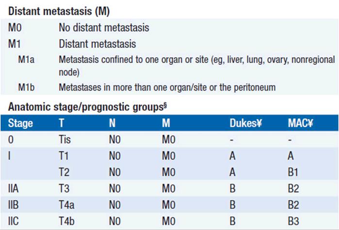
The process of staging a colon cancer is based on the American Joint Committee on Cancer (AJCC) TNM system and replaces the previous Duke’s and Astler-Collier’s systems (Table 47-1).
TABLE 47-1 COLORECTAL CARCINOMA STAGING SYSTEM OF THE AMERICAN JOINT COMMITTEE ON CANCER, 7TH EDITION



TREATMENT
SURGICAL MANAGEMENT
At presentation, the initial evaluation should consist of routine chemistries and a complete blood count. An elevated carcinoembryonic antigen (CEA) preoperatively is associated with a poor prognosis. The routine use of imaging is controversial. It is reasonable to obtain CT scans of the chest, abdomen, and pelvis to evaluate for the presence of metastatic disease.
For patients with stage I, II, or III colon cancer, surgical resection of the colon cancer is the mainstay of therapy. Open colectomy or laparoscopic colectomy is equally effective. For patients with stage IV colon cancer who are not considered candidates for cure, resection of the primary lesion can be based upon the symptoms of the patient. In the asymptomatic patient, surgical resection of the primary tumor is not necessary and can be deferred until the patient experiences local symptoms. Some patients will die of metastatic disease without ever experiencing symptoms from the primary tumor.
STAGE 1
Surgical resection cures >90% of patients with stage 1 colon cancer. Adjuvant therapy is not recommended. Patients should undergo surveillance colonoscopy 1 year after the diagnosis and then again in 3–5 years. Patients with more than two first-degree relatives with colon cancer, a first-degree relative with colon cancer under the age of 50 years, or who are under 50 years themselves, should undergo evaluation in a genetic/high-risk clinic.
STAGE 2
Surgical resection cures approximately 80% of patients with stage 2 colon cancer. The use of adjuvant chemotherapy is controversial and is currently not recommended by the American Society of Clinical Oncology. Randomized studies have not shown a statistically significant benefit for the use of adjuvant chemotherapy in patients with stage 2 colon cancer. However, many experts advocate for the use of adjuvant chemotherapy in high-risk patients, because these patients carry a greater than 20% risk of dying from recurrent disease. Patients with stage 2 colon cancer who are considered high risk carry the following features:
• T4 disease
• Presentation with perforation or obstruction
• Inadequate nodal evaluation; the American College of Pathology recommends that at least 12 regional lymph nodes be examined for the presence of nodal metastases
• Poorly differentiated tumors
Patients with microsatellite unstable (or MSI-high) tumors have a favorable prognosis that may supersede any poor risk feature. Many experts argue against treating patients with MSI-high tumors with adjuvant chemotherapy
For type of adjuvant chemotherapy see stage 3 section.
STAGE 3
Surgical resection cures approximately half of patients with stage 3 colon cancer. Patients with N1 disease can expect a cure rate with surgery alone of approximately 60%–70%. Patients with N2 disease can expect a cure rate of 30% with surgery alone. Adjuvant chemotherapy is recommended for all patients with stage 3 colon cancer at improved overall survival.
Standard treatment can consist of 6 months of 5-fluorouracil (5-FU) and leucovorin. Six months of capecitabine, an oral fluoropyrimidine, has equivalent efficacy to intravenous 5-FU and leucovorin. Recently, the addition of oxaliplatin to intravenous 5-FU and leucovorin has been associated with an improved disease-free survival compared with 5-FU and leucovorin for patients with stage 2 and 3 colon cancer. Subset analysis of patients with stage 2 disease did not reveal a statistically significant advantage in disease-free survival or overall survival for patients receiving FOLFOX compared with 5-FU and leucovorin. Subset analyses in patients with stage 3 disease demonstrated a significant improvement in disease-free and overall survival.
STAGE 4
All patients with isolated liver or lung metastases should be evaluated by a surgical specialist for consideration of resection of the metastases. Approximately 30% of patients undergoing complete resection of isolated liver or lung metastases will be cured (8).
For patients in whom a curative resection cannot be done, the median survival is approximately 6–8 months without chemotherapy and 2 years with chemotherapy. Approximately 10%–20% of patients who undergo aggressive chemotherapy will live for 5 years.
Until 1997, 5-FU was the only active chemotherapy. Studies demonstrated that the addition of folinic acid (leucovorin) to 5-FU improved the response rates and time to tumor progression. Since 1997, irinotecan, oxaliplatin, bevacizumab, and cetuximab have been approved for use in patients with metastatic colon cancer. The major randomized studies are presented in Table 47-2.
TABLE 47-2 MAJOR PHASE 3 STUDIES IN STAGE 4 COLON CANCER

First-line chemotherapy for patients with metastatic disease of consists of either FOLFOX (5-FU, leucovorin, oxaliplatin) or FOLFIRI (5-FU, leucovorin, irinotecan) with bevacizumab. Second-line chemotherapy typically consists of either an irinotecan-based regimen if FOLFOX was used as the first-line regimen or an oxaliplatin-based regimen if FOLFIRI was used as the first-line regimen. The addition of bevacizumab in second-line chemotherapy regimens when bevacizumab was a component in first-line regimens is associated with an improvement in overall survival. The addition of aflibercept to FOLFIRI in second-line treatment of patients with metastatic colorectal cancer is associated with an improved overall survival benefit compared with FOLFIRI alone. Cetuximab and panitumumab are approved for use either alone or in combination with chemotherapy for patients with metastatic colorectal cancer. Cetuximab and panitumumab are not effective in kras mutant colorectal cancers and their use is restricted to patients who have kras wild-type colorectal cancers. The combination of cetuximab or panitumumab with bevacizumab containing chemotherapy regimens is associated with worse survival outcomes. Capecitabine is often substituted for 5-FU and leucovorin in the FOLFOX regimens. The use of regorafenib in the chemotherapy-refractory setting improves overall survival by 1.4 months compared with the use of a placebo.
REFERENCES
1. Jemal A, Siegel R, Ward E, et al. Cancer statistics, 2013. CA Cancer J Clin. 2013; 63: 11–30.
2. Lynch HT, de la Chapelle A. Hereditary colorectal cancer. N Engl J Med. 2003; 348: 919–932.
3. Janne PA, Mayer RJ. Chemoprevention of colorectal cancer. N Engl J Med. 2000; 342: 1960–1968.
4. Winawer S, Fletcher R, Rex D, et al. Colorectal cancer screening and surveillance: clinical guidelines and rationale-update based on new evidence. Gastroenterology. 2003; 124: 544–560.
5. Imperiale TF, Wagner DR, Lin CY, et al. Risk of advanced proximal neoplasms in asymptomatic adults according to the distal colorectal findings. N Engl J Med. 2000; 343: 169–174.
6. Lieberman DA, Weiss DG, Bond JH, et al. Use of colonoscopy to screen asymptomatic adults for colorectal cancer. Veterans Affairs Cooperative Study Group 380. N Engl J Med. 2000; 343: 162–168.
7. Baxter NN, Warren JL, Barrett MJ, et al. Association between colonoscopy and colorectal cancer mortality in a US cohort according to site of cancer and colonoscopist specialty. J Clin Oncol. 2012; 30: 2664–2669.
8. Fong Y, Fortner J, Sun RL, Brennan MF, Blumgart LH. Clinical score for predicting recurrence after hepatic resection for metastatic color-ectal cancer: analysis of 1001 consecutive cases. Ann Surg. 1999; 230: 309–318; discussion 18–21.
9. Saltz LB, Cox JV, Blanke C, et al. Irinotecan plus fluorouracil and leucovorin for metastatic colorectal cancer. Irinotecan Study Group. N Engl J Med. 2000; 343: 905–914.
10. Goldberg RM, Sargent DJ, Morton RF, et al. A randomized controlled trial of fluorouracil plus leucovorin, irinotecan, and oxaliplatin combinations in patients with previously untreated metastatic colorectal cancer. J Clin Oncol. 2004; 22: 23–30.
11. Tournigand C, Andre T, Achille E, et al. FOLFIRI followed by FOLFOX6 or the reverse sequence in advanced colorectal cancer: a randomized GERCOR study. J Clin Oncol. 2004; 22: 229–237.
12. Hurwitz H, Fehrenbacher L, Novotny W, et al. Bevacizumab plus irinotecan, fluorouracil, and leucovorin for metastatic colorectal cancer. N Engl J Med. 2004; 350: 2335–2342.