
Jorg Dietrich
INTRODUCTION
Paraneoplastic neurologic syndromes are heterogenous disorders that can occur in the setting of various types of cancer (1). Paraneoplastic neurologic disorders are commonly caused by immune-mediated mechanisms triggered by an underlying tumor and have to be distinguished from neurological symptoms related to direct tumor invasion, infection, vasculopathy, ischemia, metabolic disturbances, or treatment-related toxicities.
Abnormal antibody or T-cell-mediated responses can target any part of the central, peripheral or autonomic nervous system to cause a diverse range of neurological symptoms. In general, the incidence of paraneoplastic syndromes is less than 1% in the general cancer population, but may be more frequently seen in specific types of cancer, such as small cell lung cancer (SCLC), cancers of the ovary and breast, and thymoma.
The diagnosis of a paraneoplastic neurologic syndrome is primarily clinical. Classical paraneoplastic syndromes (Table 64-1) may develop before the diagnosis of cancer, in a patient with known cancer, or in a patient considered to be in cancer remission. While an extensive search for an underlying tumor is warranted in classical paraneoplastic syndromes, such as paraneoplastic cerebellar degeneration (PCD) or Lambert-Eaton myasthenic syndrome, it may be noteworthy that several neurologic syndromes designated as “paraneoplastic” may also be seen in non-neoplastic autoimmune diseases.
TABLE 64-1 CLASSICAL PARANEOPLASTIC SYNDROMES OF THE NERVOUS SYSTEM

Since paraneoplastic syndromes often herald the diagnosis of cancer, patients may be diagnosed and treated early in the course of the disease. Earlier cancer diagnosis may correlate with a higher likelihood of cure and remission of neurologic symptoms. It remains controversial, however, whether a better prognosis in patients with paraneoplastic syndromes is related to the immune control of the underlying cancer.
PATHOGENESIS
While the detailed pathomechanism of most paraneoplastic syndromes remains elusive, production of antibodies directed against antigens expressed by both tumors and nervous system tissues have been described as a key mechanism in some distinct types of paraneoplastic syndromes (Table 64-2). Antibodies can be directed against cytoplasmic antigens (e.g., Purkinje cells and anterior horn cells) or cell surface proteins (e.g., voltage gated potassium and calcium channels). Immune mechanisms may involve both humoral and T-cell-mediated responses, although the exact contribution of cellular and antibody-mediated mechanisms to the disease manifestation remains controversial (2–4). Abnormal antibodies can frequently be detected in serum and cerebrospinal fluid (CSF); however, a direct pathogenic effect of certain antibodies on nervous system tissue or the neuromuscular junction has only been demonstrated in a subset of paraneoplastic disorders. Importantly, some antibodies can also be detected in the absence of a clinical paraneoplastic syndrome (5) (Table 64-3).
TABLE 64-2 WELL-CHARACTERIZED PARANEOPLASTIC ANTIBODIES DIRECTED TO NON-SURFACE ANTIGENS, ASSOCIATION WITH NEUROLOGIC SYNDROMES AND UNDERLYING TUMORS

TABLE 64-3 PARTIALLY CHARACTERIZED ANTIBODIES THAT CAN OCCUR WITH AND WITHOUT CANCER ASSOCIATION

DIAGNOSTIC AND THERAPEUTIC CONSIDERATIONS
The diagnosis of a paraneoplastic neurologic syndrome can be challenging and, in classical syndromes, is usually based on clinical features of the neurologic condition, such as in cerebellar degeneration and Lambert-Easton myasthenic syndrome (1, 6, 7). Neurological symptoms commonly develop in the early stages of cancer and in two-thirds of patients before a cancer diagnosis has been established (1). Therefore, early recognition and treatment of a paraneoplastic neurologic disorder along with treatment of the underlying cancer are critical to reduce neurologic morbidity and to improve overall patient survival. Paraneoplastic neurologic syndromes usually have an acute or subacute onset, but the diagnosis may remain challenging if no typical antibodies are found in serum or CSF (7). As most syndromes and malignancies are associated with more than one specific antibody, screening for a panel of paraneoplastic antibodies may increase the yield to establish a diagnosis (7, 8). In patients with CNS manifestation, antibody screening in CSF is usually recommended, as antibody titers in CSF are generally higher than in serum (9). CSF analysis typically reveals an inflammatory pattern with mild pleocytosis, elevated protein, and evidence of oligoclonal bands. Positron emission tomography (PET) in combination with computed tomography (CT) is helpful in the search for an underlying malignancy (10, 11). Mammography and pelvic ultrasound may be indicated in women with suspected paraneoplastic neurologic syndromes, given the frequent association with breast and gynecologic cancers. Neuroimaging of the brain with magnetic resonance imaging (MRI) may be unremarkable but is abnormal in the majority of patients with encephalo-myelitis and limbic encephalitis (1). In cases in which no underlying tumor can be identified but a high clinical suspicion for a classic paranepolastic syndrome exists, or in patients tested positive for paraneoplastic antibodies in the setting of a less classic form of a paraneoplastic neurologic syndrome, repeat cancer screening studies are recommended every 6 months (7). An underlying malignancy usually can be identified within the first 4 years from onset of neurological symptoms (7).
The most important management goals in patients with paraneoplastic neurologic syndromes are identification and treatment of an underlying cancer. In general, antibody-mediated neurologic disorders affecting the peripheral nervous system will have a higher likelihood of response to treatment and symptom improvement. In these patients, immunomodulatory strategies with intravenous immunoglobulins, plasmapheresis, or immunosuppressants are commonly effective. While most paraneoplastic syndromes affecting the central nervous system generally respond poorly to immunomodulatory therapies, patients with opsoclonus-myoclonus syndrome (12) or limbic encephalitis and presence of anti-NMDAR, anti-AMPAR, or anti-Ma antibodies (13, 14) may show a remarkable benefit from treatment. In patients not actively treated with chemotherapy for an underlying cancer and with progressive neurological decline, more aggressive immunosuppression may be considered, including the use of cyclo-phosphamide, tacrolimus, azathioprine, and cyclosporine.
SYNDROMES OF THE CENTRAL NERVOUS SYSTEM
 PARANEOPLASTIC CEREBELLAR DEGENERATION
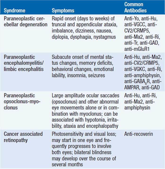
Paraneoplastic cerebellar degeneration (PCD) is a classical paraneoplastic neurologic syndrome. Symptoms commonly precede tumor diagnosis by months or occasionally years and are characterized by rapidly progressive cerebellar dysfunction. The development of gait dysfunction, appendicular ataxia, and speech dysfunction may develop and progress over the course of days to weeks, unlike in primary degenerative cerebellar disorders, where symptoms develop over months to years. Brain stem and cerebellar dysfunction, including oculomotor symptoms with diplopia, oscillopsia, nystagmus, nausea, and dysphagia are usually irreversible (15, 16). Emotional lability and cognitive deficits may occur, underlining the importance of the cerebellum in mood and cognitive function (17). In addition, patients may develop pyramidal tract signs, posterior column signs, and polyneuropathy, suggesting a more widespread involvement of the nervous system in the pathology of the disease process. The majority of patients develop severe gait difficulties, inability to write and to swallow (18).
PCD is frequently associated with anti-Yo antibodies (Purkinje-cell antibody-1; PCA-1; also termed cerebellar degeneration related-2; cdr2), directed against Purkinje cells of the cerebellum, and usually occurs in patients with breast or ovarian cancer (16, 19). Because presence of anti-Yo antibodies is so frequently associated with underlying breast or ovarian cancer, careful and repeated cancer screening is mandated. Notably, more than two-thirds of patients with anti-Yo-associated cerebellar degeneration do not have a cancer diagnosis at the onset of neurologic symptoms. PCD has also been described in the presence of anti-Hu antibodies and anti-CV2/CRMP5 antibodies and SCLC (20), anti-Tr antibodies and Hodgkin lymphoma (21, 22), or anti-Ri antibodies and breast cancer (23). Interestingly, PCD has also been reported in patients with non-Hodgkin lymphoma and lung cancer without detectable anti-neuronal antibodies (24), and in SCLC patients with evidence of P/Q calcium channel antibodies (25).
As seen in other paraneoplastic neurologic syndromes, CSF analysis may reveal lymphocytic pleocytosis, elevated protein, and oligoclonal bands. At later disease stages neuroimaging with CT or MRI may identify loss of cerebellar architecture and eventually global cerebellar atrophy.
Treatment with plasmapheresis or intravenous immunoglobulins do usually not alter the progressive course of the disease (26), although clinical improvement has been seen in some cases (18, 27), with treatment of the underlying tumor (28, 29), or in some cases of PCD and underlying anti-Tr and anti-Ri antibodies (22, 30).
PARANEOPLASTIC ENCEPHALOMYELITIS
Paraneoplastic encephalomyelitis (PEM) may affect multiple sites and levels of the nervous system, including the temporal and frontal lobes, midbrain, pons, cerebellum, spinal cord, dorsal root ganglia, and autonomic nervous system. Patients may therefore present with multiple neurological dysfunctions depending on the site of nervous system involvement. Symptoms usually develop over weeks to months. Involvement of the limbic system is characterized by short-term memory deficits, mood changes, emotional lability, and seizures. Brain stem involvement is classically associated with oculomotor dysfunction, nystagmus, and other cranial nerve palsies, whereas movement disorders are seen with midbrain involvement. Symptoms of autonomic dysfunction, such as cardiac arrhythmias, hypotension, and impaired gastrointestinal motility, can be identified in about 25% of patients. PEM typically occurs in the setting of lung cancer, particularly SCLC with presence of anti-Hu (ANNA-1) antibodies (20). Although less common, other antibodies have been linked to PEM, including anti-Ri antibodies (ANNA-2) in breast and gynecologic cancers (31), anti-CV2/CRMP5 antibodies in SCLC and thymoma (32), anti-amphiphysin antibodies in SCLC (33), anti-Ma2 antibodies in germ cell tumors and other cancers (34–36), and anti-NMDAR antibodies in ovarian teratomas (37, 38).
Diagnosis of PEM is usually clinical and supported by the presence of paraneoplastic antibodies. Neuroimaging studies with MRI frequently reveal T2/Flair signal hyperintensities of affected nervous system sites with and without gadolinium enhancement. In general, PEM is poorly responsive to treatment, although disease stabilization or mild symptom improvement has been reported with combined tumor treatment and immunotherapies, including intravenous immunoglobulins, plasmapheresis, or cyclophosphamide (38, 39).
PARANEOPLASTIC LIMBIC ENCEPHALITIS
Paraneoplastic limbic encephalitis (PLE) is clinically associated with short-term memory deficits, mood disturbances, emotional lability, sleep alterations, behavioral abnormalities, and seizures (40). Symptoms may stabilize or improve over time; however, anterograde amnesia and cognitive deficits frequently persist. PLE typically occurs in the setting of SCLC with presence of anti-Hu antibodies, but can also be seen in presence of other antibodies, such as anti-Ma2, anti-NMDAR, anti-CV2/CRMP5, anti-amphiphysin, and anti-Ri (35, 38, 40–42). Notably, the clinical and radiographic picture of limbic encephalitis can also be seen in non-neoplastic autoimmune disorders characterized by presence of anti-VGKC (43, 44) and anti-AMPAR antibodies (14).
The diagnosis of PLE commonly precedes tumor diagnosis. Although less frequently, tumors other than SCLC have been associated with PLE, such as Hodgkin lymphoma, ovarian cancer, teratomas, testicular cancer, breast cancer, and thymomas.
Pathological findings may reveal perivascular and interstitial lymphocytic infiltrates and microglia activation in limbic structures, including hippocampus, amygdala, cingulate gyrus, and hypothalamus. There may be some overlap with the clinical picture of PEM, and neuropathology may not be restricted to limbic structures.
CSF findings usually demonstrate mild pleocytosis, protein elevation, and oligoclonal bands. Neuroimaging with MRI may infrequently demonstrate unilateral or bilateral T2/Flair signal abnormalities in mesial temporal lobes with or without gadolinium enhancement. Some patients develop progressive temporal lobe atrophy as a delayed consequence of encephalitis. Hypermetabolism in various brain regions may be detectable on PET (45). EEG studies frequently show temporal lobe dysfunction with or without epileptiform activity.
Treatment includes immunotherapy with glucocorticoids, intravenous immunoglobulins, and plasmapheresis. Successful treatment has been described in patients with presence of antibodies directed against surface antigens (41).
PARANEOPLASTIC OPSOCLONUS-MYOCLONUS
Opsoclonus refers to spontaneous, arrhythmic, large-amplitude, and multidirectional eye movements. Myoclonus describes involuntary muscle jerks in extremities and trunk, and usually occurs along with hypotonia, gait difficulties, and irritability. Paraneoplastic opsoclonus-myoclonus syndrome (POM) occurs in 2% of children with neuroblastoma, and nearly 50% of children with POM have underlying neuroblastoma. Since neurologic symptoms frequently precede tumor diagnosis, a search for an underlying malignancy is warranted in all children with POM (46). In adults, POM is associated with SCLC in the majority of cases, but can also be seen with other malignancies. The most commonly identified antibodies include anti-Hu, anti-Ri, anti-Yo, anti-Ma2, and anti-amphiphysin (12, 47).
In women with anti-Ri, opsoclonus also occurs as a separate clinical entity characterized by imbalance, ataxia, and movement disorders, suggesting a more widespread brain stem and midbrain involvement. Symptoms develop within weeks to months. Breast cancer and, to some degree, SCLC are by far the most common tumors in which anti-Ri syndromes develop. Adult patients with opsoclonus without evidence of anti-Ri antibodies usually develop myoclonus, and the underlying cause is commonly SCLC. Gait difficulties, ataxia, encephalopathy, lethargy, and coma may develop.
POM may respond to treatment of the underlying tumor or immuno-therapies, including use of steroids, intravenous immunoglobulins, and plasmapheresis. Rituximab has been successfully used in some cases (48).
CANCER-ASSOCIATED RETINOPATHY
Cancer-associated retinopathy (CAR) is characterized by acute to subacute onset of photosensitivity and visual loss (49). Symptoms can present monocular, but commonly progress to involve both eyes (50). Exam findings include poor visual acuity, retinal artery narrowing, and scotomata. Blindness eventually develops over the course of months. CAR is associated with antibodies targeting recoverin, a calcium-binding protein on photoreceptors. Most commonly SCLC is the underlying tumor, and visual symptoms usually precede the diagnosis of cancer. Association with other tumors has been reported, including breast, gynecologic, pancreatic, prostate, bladder, colon, non-small cell lung cancer (NSCLC), and lymphoma (18). A distinct entity occurs in patients with metastatic melanoma (melanoma associated retinopathy), in which antibodies reacting with bipolar cells of the retina can be identified. Visual symptoms usually develop in the setting of known metastatic disease, sometimes months to years after initial tumor diagnosis (51).
STIFF PERSON SYNDROME
Stiff person syndrome (SPS), also known as stiff man syndrome, is characterized by fluctuating axial stiffness and muscle rigidity that can result in significant spine deformity. Extremities and facial muscles can be involved (52). Loud noise, sudden movement, and emotional upset often exacerbate painful muscle spasms. The remaining neurological exam is usually unremarkable. Notably, symptoms disappear in sleep or under deep anesthesia. Electrophysiologic studies with EMG show continuous motor unit activity in affected muscle groups at rest. Anti-amphiphysin antibodies can be identified in SPS, and both SCLC and breast cancer have been linked to this syndrome. However, SPS more frequently manifests as an autoimmune disorder in the absence of a tumor diagnosis (53). In these patients anti-GAD antibodies are more commonly identified. Treatment strategies include GABA-enhancing agents such as benzodiazepines, gabapentin, and baclofen that are beneficial in limiting painful muscle spasms. Symptoms may improve with treatment of the underlying tumor and with the use of intravenous immunoglobulins (54).
SYNDROMES OF THE PERIPHERAL NERVOUS SYSTEM
DORSAL ROOT GANGLIONITIS
Dorsal root ganglionitis, also referred to as subacute sensory neuronopathy, causes impairment of all sensory modalities, including pain, temperature, touch, and proprioception. Most common symptoms are numbness, paresthesias, and pain involving face, trunk, and extremities that develop over the course of days to weeks. Isolated cranial nerves can be affected. Neurological exam demonstrates loss of deep tendon reflexes and vibration sense. Patients may develop gait difficulties due to sensory impairment.
Dorsal root ganglionitis is nearly pathognomonic for paraneoplastic disease and can occur in combination with multifocal PEM (20). SCLC is the most frequent underlying cancer, but this syndrome may also be seen in lymphoma and NSCLC (55). No specific antibodies have been described with this condition; however, the same antibodies that are present in PEM (e.g., anti-Hu and anti-CV2/CRMP5) can be detected in dorsal root ganglionitis. Notably, the differential diagnosis includes symptoms related to Sjogren’s syndrome, in which ganglion cell damage also occurs. Unfortunately, most patients with paraneoplastic dorsal root ganglionitis and underlying SCLC do not show meaningful neurologic improvement despite aggressive antitumor and immunotherapy.
SENSORY AND SENSORIMOTOR POLYNEUROPATHY
Subacute and chronic peripheral neuropathies may occur for a variety of reasons in cancer patients, such as a treatment complication of chemotherapy, and as a consequence of metabolic disorders. The clinical distinction from paraneoplastic polyneuropathies can be challenging but should be considered in patients without known underlying malignancy (56). A wide variety of neuropathies may result from diverse paraneoplastic antibodies (e.g., anti-MAG, anti-Hu, anti-CV2/CRMP5). The exact pathogenic mechanisms have not been identified in most syndromes, but antibody reaction with peripheral nerve fibers may play a role, resulting in inflammation and possibly secondary demyelination. As a consequence, symptoms are usually complex and may impact both motor and sensory function.
Exam findings may demonstrate gait disorders, plexopathies, and clinical pictures that resemble Guillain-Barre syndrome, chronic inflammatory demyelinating polyneuropathy (CIPD), and anterior horn cell disease. Hodgkin’s disease is commonly identified as the underlying cancer. Chronic peripheral neuropathy has also been described in patients with SCLC, melanoma, lymphoma, and plasma cell disorders (55, 57).
The neuropathy seen in POEMS syndrome (P = polyneuropathy, O = organomegaly, E = endocrinopathy, M = elevated IgM protein, and S = skin changes) usually occurs in the setting of myeloma (58). Unlike other paraneoplastic syndromes, the involved antibody is produced by the underlying malignancy and not against it. Amyloidosis associated with hematological malignancies or paraproteinemias may complicate the clinical picture.
Response to therapy is usually unsatisfactory, but may include treatment of the underlying tumor, immunotherapies, radiation to bony lesions in myeloma, and chemotherapy (55). Symptom improvement has been reported in patients with POEMS following peripheral blood stem cell transplantation (59).
AUTONOMIC NEUROPATHY
Paraneoplastic syndromes associated with cancer can affect both the sympathetic and parasympathetic autonomic nervous system. Associated clinical symptoms include blood pressure dysregulation and postural hypotension, bowel and bladder dysfunction, arrhythmias, xerostomia, pupillary abnormalities, impotence and erectile dysfunction, diaphoresis, and anhidrosis. Autonomic dysfunction can occur as an isolated syndrome or in combination with encephalomyelitis, sensory neuronopathy, or sensorimotor neuropathy. This syndrome is commonly seen in patients with SCLC and presence of anti-Hu antibodies, but has also been described with other underlying cancers, including Hodgkin lymphoma, thymoma, and carcinoid tumors (18, 60).
Treatment of the underlying cancer may stabilize or improve symptoms. In addition, immunotherapies, plasmapheresis, and rituximab have been successfully used in some patients (61, 62).
SYNDROMES OF THE NEUROMUSCULAR JUNCTION AND MUSCLE
LAMBERT-EATON MYASTHENIC SYNDROME
Lambert-Eaton myastenic syndrome (LEMS) is one of the most common paraneoplastic syndromes. SCLC is most frequently identified as underlying cancer (63). LEMS frequently develops before the cancer diagnosis and should raise a high suspicion for an underlying cancer. Development of LEMS after cancer therapy should prompt investigation for tumor recurrence. Antibodies directed against the P/Qvoltage-gated calcium channels (VGCC) (64) interfere with acetylcholine release at the neuromuscular junction, resulting in predominant proximal muscle weakness, mostly affecting the lower extremities with associated gait difficulties. Other symptoms include xerostomia, generalized fatigue, and autonomic dysfunction (65). The neurologic exam reveals diminished deep tendon reflexes that typically improve with repetitive muscle activity (66). The diagnosis is further supported by EMG studies that show low muscle amplitudes at rest with diminished response at low rate and elevated response at high rates.
Treatment of the underlying cancer in combination with immunotherapies, such as intravenous immunoglobulins and plasmapheresis, may result in symptom improvement. Immunosuppression with azathioprine has been applied in long-term management. In addition, the use of 3,4-diaminopyridine, which facilitates acetylcholine release at the neuromuscular junction, has been associated with clinical benefit (67).
NEUROMYOTONIA
Neuromyotonia is the consequence of peripheral nerve hyperexcitability and is characterized by (often painful) muscle cramps, delayed relaxation, fasciculations, and weakness. The autonomic nervous system may also be affected and some patients demonstrate cognitive deficits as well as behavioral and personality changes (Morvan’s syndrome). This syndrome occurs in the setting of an underlying cancer or as an autoimmune condition unrelated to malignancy. Antibodies directed against CASPR2 (previously attributed to voltage-gated potassium channels) can be identified in this syndrome (68, 69). Other antibodies such as anti-Hu and anti-amphiphysin have also been identified in this syndrome. Among the cancers commonly underlying this syndrome are thymoma, SCLC, and Hodgkin’s disease. The treatment of choice is immunosuppression with plasmapheresis or intravenous immunoglobulins (70). Some patients have demonstrated symptom improvement with anticonvulsants and muscle relaxants.
NECROTIZING MYOPATHY, POLYMYOSITIS, AND DERMATOMYOSITIS
Cancer-associated necrotizing myopathy typically starts as painful proximal weakness and rapidly progresses to generalized weakness. Respiratory muscles can be involved. Laboratory studies usually reveal elevated creatinine kinase (CK), and muscle biopsy demonstrates necrotic muscle fibers without significant inflammation. Among the cancers linked to this syndrome are SCLC, breast, prostate, and gastrointestinal malignancies.
In contrast to necrotizing myopathy, biopsy findings in polymyositis and dermatomyositis show significant inflammation. Polymyositis is associated with an elevated cancer risk of up to 20% (18), including non-Hodgkin lymphoma, lung, ovarian, and bladder cancer (71). Dermatomyositis is associated with a higher frequency of malignancies when compared to polymyositis, typically ranging from 20% to 25% (72) and symptom onset should prompt a thorough investigation for an underlying malignancy (7). Patients with inflammatory myopathies typically develop proximal and symmetric muscle weakness over several weeks to months. Patients with dermatomyositis may also demonstrate skin manifestations (73), including an erythematous V-shaped rash over chest and shoulders, a heliotrope upper eyelid rash, and scaling erythema on the extensor surfaces of the extremities (“Grotton’s sign”). The most common cancers associated with dermatomyositis are lymphomas, lung, pancreatic, gastrointestinal, and gynecologic malignancies (71, 74). Treatment of inflammatory myopathies consists of treatment of the underlying tumor and immunosuppression with intravenous immunoglobulins, glucocorticoids, azathioprine, cyclosporine, and methotrexate (73, 75, 76).
SUMMARY
Paraneoplastic syndromes are heterogenous disorders with a low incidence in the general cancer population. As many paraneoplastic syndromes occur prior to the diagnosis of cancer, recognition of distinct paraneoplastic syndromes is essential to initiate thorough screening for malignancy. Both humoral and T-cell-mediated immune mechanisms play a role in the pathogenesis of paraneoplastic syndromes. Most important treatment goal is the identification and treatment of the underlying cancer in order to slow or to reverse neurological impairment. While long-term remission of neurological symptoms is infrequently achieved, early recognition of paraneoplastic syndromes is crucial. In general, antibody-mediated disorders affecting the peripheral nervous system have a higher likelihood of response to treatment. Immunomodulatory therapies have been frequently employed, including plasmapheresis, intravenous immunoglobulins, and glucocorticoids. In addition, more aggressive immunosuppressive strategies have proven beneficial in some circumstances, including treatment with rituximab, azathioprine, cyclosporine, and cyclophosphamide.
REFERENCES
1. Darnell RB, Posner JB. Paraneoplastic Syndromes. Oxford University Press, New York, NY, 2011.
2. Albert ML, Austin LM, Darnell RB. Detection and treatment of activated T cells in the cerebrospinal fluid of patients with paraneoplastic cerebellar degeneration. Ann Neurol. 2000; 47: 9–17.
3. Benyahia B, et al. Cell-mediated autoimmunity in paraneoplastic neurological syndromes with anti-Hu antibodies. Ann Neurol. 1999; 45: 162–167.
4. Tanaka M, Tanaka K, Shinozawa K, et al. Cytotoxic T cells react with recombinant Yo protein from a patient with paraneoplastic cerebellar degeneration and anti-Yo antibody. J Neurol Sci. 1998; 161: 88–90.
5. Graus F, Saiz A, Dalmau J. Antibodies and neuronal autoimmune disorders of the CNS. J Neurol. 2010; 257: 509–517.
6. Graus F, et al. Recommended diagnostic criteria for paraneoplastic neurological syndromes. J Neurol Neurosurg Psychiatry. 2004; 75: 1135–1140.
7. Titulaer MJ, et al. Screening for tumours in paraneoplastic syndromes: report of an EFNS task force. Eur J Neurol. 2011; 18: 19–e3.
8. Monstad SE, Knudsen A, Salvesen HB, et al. Onconeural antibodies in sera from patients with various types of tumours. Cancer Immunol Immunother. 2009; 58: 1795–1800.
9. Rosenfeld MR, Dalmau J. Update on paraneoplastic neurologic disorders. The Oncologist. 2010; 15: 603–617.
10. Linke R, Schroeder M, Helmberger T, et al. Antibody-positive paraneo-plastic neurologic syndromes: value of CT and PET for tumor diagnosis. Neurology. 2004; 63: 282–286.
11. Younes-Mhenni S, et al. FDG-PET improves tumour detection in patients with paraneoplastic neurological syndromes. Brain. 2004; 127: 2331–2338.
12. Bataller L, Graus F, Saiz A, et al. Clinical outcome in adult onset idiopathic or paraneoplastic opsoclonus-myoclonus. Brain. 2001; 124: 437–443.
13. Dalmau J, et al. Anti-NMDA-receptor encephalitis: case series and analysis of the effects of antibodies. Lancet Neurol. 2008; 7: 1091–1098.
14. Lai M, et al. AMPA receptor antibodies in limbic encephalitis alter synaptic receptor location. Ann Neurol. 2009; 65: 424–434.
15. Peterson K, Rosenblum MK, Kotanides H, et al. Paraneoplastic cerebellar degeneration. I. A clinical analysis of 55 anti-Yo antibody-positive patients. Neurology. 1992; 42: 1931–1937.
16. Shams’ili S, et al. Paraneoplastic cerebellar degeneration associated with antineuronal antibodies: analysis of 50 patients. Brain. 2003; 126: 1409–1418.
17. Schmahmann JD, Weilburg JB, Sherman JC. The neuropsychiatry of the cerebellum—insights from the clinic. Cerebellum. 2007; 6: 254–267.
18. Darnell RB, Posner JB. Paraneoplastic syndromes affecting the nervous system. Semin Oncol. 2006; 33: 270–298.
19. Rojas-Marcos I, et al. Spectrum of paraneoplastic neurologic disorders in women with breast and gynecologic cancer. Medicine (Baltimore). 2003; 82: 216–223.
20. Graus F, et al. Anti-Hu-associated paraneoplastic encephalomyelitis: analysis of 200 patients. Brain. 2001; 124: 1138–1148.
21. Bernal F, et al. Anti-Tr antibodies as markers of paraneoplastic cerebellar degeneration and Hodgkin’s disease. Neurology. 2003; 60: 230–234.
22. Graus F, et al. Immunological characterization of a neuronal antibody (anti-Tr) associated with paraneoplastic cerebellar degeneration and Hodgkin’s disease. J Neuroimmunol. 1997; 74: 55–61.
23. Sutton IJ, Barnett MH, Watson JD, et al. Paraneoplastic brainstem encephalitis and anti-Ri antibodies. J Neurol. 2002; 249: 1597–1598.
24. Sabater L, et al. Protein kinase Cgamma autoimmunity in paraneoplastic cerebellar degeneration and non-small-cell lung cancer. J Neurol Neurosurg Psychiatry. 2006; 77: 1359–1362.
25. Graus F, et al. P/Q type calcium-channel antibodies in paraneoplastic cerebellar degeneration with lung cancer. Neurology. 2002; 59: 764–766.
26. Rojas I, et al. Long-term clinical outcome of paraneoplastic cerebellar degeneration and anti-Yo antibodies. Neurology. 2000; 55: 713–715.
27. Phuphanich S, Brock C. Neurologic improvement after high-dose intravenous immunoglobulin therapy in patients with paraneoplastic cerebellar degeneration associated with anti-Purkinje cell antibody. J Neuro Oncol. 2007; 81: 67–69.
28. Blaes F, et al. Intravenous immunoglobulins in the therapy of paraneoplastic neurological disorders. J Neurol. 1999; 246: 299–303.
29. David YB, et al. Autoimmune paraneoplastic cerebellar degeneration in ovarian carcinoma patients treated with plasmapheresis and immunoglobulin. A case report. Cancer. 1996; 78: 2153–2156.
30. Dropcho EJ, Kline LB, Riser J. Antineuronal (anti-Ri) antibodies in a patient with steroid-responsive opsoclonus-myoclonus. Neurology. 1993; 43: 207–211.
31. Pittock SJ, Lucchinetti CF, Lennon VA. Anti-neuronal nuclear autoantibody type 2: paraneoplastic accompaniments. Ann Neurol. 2003; 53: 580–587.
32. Honnorat J, et al. Onco-neural antibodies and tumour type determine survival and neurological symptoms in paraneoplastic neurological syndromes with Hu or CV2/CRMP5 antibodies. J Neurol Neurosurg Psychiatry. 2009; 80: 412–416.
33. Dropcho EJ. Antiamphiphysin antibodies with small-cell lung carcinoma and paraneoplastic encephalomyelitis. Ann Neurol. 1996; 39: 659–667.
34. Rosenfeld MR, Eichen JG, Wade DF, et al. Molecular and clinical diversity in paraneoplastic immunity to Ma proteins. Ann Neurol. 2001; 50: 339–348.
35. Dalmau J, et al. Clinical analysis of anti-Ma2-associated encephalitis. Brain. 2004; 127: 1831–1844.
36. Mathew RM, et al. Orchiectomy for suspected microscopic tumor in patients with anti-Ma2-associated encephalitis. Neurology. 2007; 68: 900–905.
37. Vitaliani R, et al. Paraneoplastic encephalitis, psychiatric symptoms, and hypoventilation in ovarian teratoma. Ann Neurol. 2005; 58: 594–604.
38. Dalmau J, et al. Paraneoplastic anti-N-methyl-D-aspartate receptor encephalitis associated with ovarian teratoma. Ann Neurol. 2007; 61: 25–36.
39. Vernino S, O’Neill BP, Marks RS, et al. Immunomodulatory treatment trial for paraneoplastic neurological disorders. Neuro Oncol. 2004; 6: 55–62.
40. Gultekin SH, et al. Paraneoplastic limbic encephalitis: neurological symptoms, immunological findings and tumour association in 50 patients. Brain. 2000; 123: 1481–1494.
41. Bataller L, et al. Autoimmune limbic encephalitis in 39 patients: immu-nophenotypes and outcomes. J Neurol Neurosurg Psychiatry. 2007; 78: 381–385.
42. Alamowitch S, et al. Limbic encephalitis and small cell lung cancer. Clinical and immunological features. Brain. 1997; 120: 923–928.
43. Pozo-Rosich P, Clover L, Saiz A, et al. Voltage-gated potassium channel antibodies in limbic encephalitis. Ann Neurol. 2003; 54: 530–533.
44. Buckley C, et al. Potassium channel antibodies in two patients with reversible limbic encephalitis. Ann Neurol. 2001; 50: 73–78.
45. Ances BM, et al. Treatment-responsive limbic encephalitis identified by neuropil antibodies: MRI and PET correlates. Brain. 2005; 128: 1764–1777.
46. Pranzatelli MR. The immunopharmacology of the opsoclonus-myoclonus syndrome. Clin Neuropharmacol. 1996; 19: 1–47.
47. Luque FA, et al. Anti-Ri: an antibody associated with paraneoplastic opsoclonus and breast cancer. Ann Neurol. 1991; 29: 241–251.
48. Pranzatelli MR, Tate ED, Travelstead AL, et al. Immunologic and clinical responses to rituximab in a child with opsoclonus-myoclonus syndrome. Pediatrics. 2005; 115: e115–119.
49. Ko MW, Dalmau J, Galetta SL. Neuro-ophthalmologic manifestations of paraneoplastic syndromes. J Neuro Ophthalmol. 2008; 28: 58–68.
50. Keltner JL, Thirkill CE. Cancer-associated retinopathy vs recoverin-associated retinopathy. Am J Ophthalmol. 1998; 126: 296–302.
51. Lei B, Bush RA, Milam AH, et al. Human melanoma-associated retinopathy (MAR) antibodies alter the retinal ON-response of the monkey ERG in vivo. Invest Ophthalmol Vis Sci. 2000; 41: 262–266.
52. Silverman IE. Paraneoplastic stiff limb syndrome. J Neurol Neurosurg Psychiatry. 1999; 67: 126–127.
53. Brown P, Marsden CD. The stiff man and stiff man plus syndromes. J Neurol. 1999; 246: 648–652.
54. Dalakas MC, et al. High-dose intravenous immune globulin for stiff-person syndrome. N Engl J Med. 2001; 345: 1870–1876.
55. Rudnicki SA, Dalmau J. Paraneoplastic syndromes of the spinal cord, nerve, and muscle. Muscle Nerve. 2000; 23: 1800–1818.
56. Antoine JC, et al. Carcinoma associated paraneoplastic peripheral neuropathies in patients with and without anti-onconeural antibodies. J Neurol Neurosurg Psychiatry. 1999; 67: 7–14.
57. Rudnicki SA, Dalmau J. Paraneoplastic syndromes of the peripheral nerves. Curr Opin Neurol. 2005; 18: 598–603.
58. Silberman J, Lonial S. Review of peripheral neuropathy in plasma cell disorders. Hematol Oncol. 2008; 26: 55–65.
59. Kuwabara S, et al. Neurologic improvement after peripheral blood stem cell transplantation in POEMS syndrome. Neurology. 2008; 71: 1691–1695.
60. Vernino S, Adamski J, Kryzer TJ, et al. Neuronal nicotinic ACh receptor antibody in subacute autonomic neuropathy and cancer-related syndromes. Neurology. 1998; 50: 1806–1813.
61. Vernino S, Sandroni P, Singer W, et al. Invited article: autonomic ganglia: target and novel therapeutic tool. Neurology. 2008; 70: 1926–1932.
62. Iodice V, et al. Efficacy of immunotherapy in seropositive and seronegative putative autoimmune autonomic ganglionopathy. Neurology. 2009; 72: 2002–2008.
63. Titulaer MJ, et al. Screening for small-cell lung cancer: a follow-up study of patients with Lambert-Eaton myasthenic syndrome. J Clin Oncol. 2008; 26: 4276–4281.
64. Motomura M, et al. Incidence of serum anti-P/O-type and anti-N-type calcium channel autoantibodies in the Lambert-Eaton myasthenic syndrome. J Neurol Sci. 1997; 147: 35–42.
65. O’Suilleabhain P, Low PA, Lennon VA. Autonomic dysfunction in the Lambert-Eaton myasthenic syndrome: serologic and clinical correlates. Neurology. 1998; 50: 88–93.
66. Titulaer MJ, et al. The Lambert-Eaton myasthenic syndrome 1988-2008: a clinical picture in 97 patients. J Neuroimmunol. 2008; 201–202, 153–158.
67. Wirtz PW. et al. Efficacy of 3,4-diaminopyridine and pyridostigmine in the treatment of Lambert-Eaton myasthenic syndrome: a randomized, double-blind, placebo-controlled, crossover study. Clin Pharmacol Ther. 2009; 86: 44–48.
68. Newsom-Davis J, et al. Autoimmune disorders of neuronal potassium channels. Ann N Y Acad Sci. 2003; 998: 202–210.
69. Lai M, et al. Investigation of LGI1 as the antigen in limbic encephalitis previously attributed to potassium channels: a case series. Lancet Neurol. 2010; 9: 776–785.
70. van den Berg JS, van Engelen BG, Boerman RH, et al. Acquired neuromyotonia: superiority of plasma exchange over high-dose intravenous human immunoglobulin. J Neurol. 1999; 246, 623–625.
71. Hill CL, et al. Frequency of specific cancer types in dermatomyositis and polymyositis: a population-based study. Lancet. 2001; 357: 96–100.
72. Callen JP, Wortmann RL. Dermatomyositis. Clin Dermatol. 2006; 24: 363–373.
73. Mastaglia FL, Garlepp MJ, Phillips BA, et al. Inflammatory myopathies: clinical, diagnostic and therapeutic aspects. Muscle Nerve. 2003; 27: 407–425.
74. Callen JP. Relation between dermatomyositis and polymyositis and cancer. Lancet. 2001; 357, 85–86.
75. Dalakas MC. The role of high-dose immune globulin intravenous in the treatment of dermatomyositis. Int Immunopharmacol. 2006; 6: 550–556.
76. Feist E, Dorner T, Sorensen H, et al. Longlasting remissions after treatment with rituximab for autoimmune myositis. J Rheumatol. 2008; 35: 1230–1232.