
Anita Kumar
Definition of Hematopoiesis: The formation of blood cells
Sites of Hematopoiesis
• Bone marrow (BM) is the principal site of hematopoiesis in adults, predominantly in pelvis, vertebrae, ribs, sternum, & skull
• During fetal development, sites include yolk sac → liver → spleen → BM
Stem Cell Niche
• In the BM, hematopoietic stem cells (HSCs) develop in a complex microenvironment of stromal cells (fibroblasts, smooth muscle cells, endothelial cells, adipocytes, osteoblasts, osteoclast, & MΦ) & extracellular matrix
• Unique sites w/in the marrow, termed niches, favor HSC self-renewal vs. differentiation
Bone Marrow Morphology
• BM comprises hematopoietic tissue islands & adipose cells surrounded by vascular sinuses
• In nl adults, the ratio of fat cells to hematopoietic elements is 1:1
• Thin-walled sinusoids are lined by endothelial cells, & differentiated blood cells enter the circulation by transcellular migration through endothelial cells
Hierarchical Map of Hematopoietic Development
• HSCs: 2 cardinal features (1) pluripotency (can give rise to all mature blood cell types) & (2) self-renewal capacity
• HSCs give rise to all formed elements of blood – RBCs, granulocytes, monocytes, plt, & lymphocytes
• HSCs give rise to two kinds of multipotent cells, the common lymphocyte progenitors (CLPs) & common myeloid progenitors (CMPs)
• CMPs give rise to committed progenitors called colony forming units (CFUs) that develop into specific mature cells
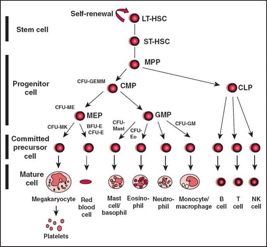
Figure 28-1 Cantor, A. B. et al. ASH-SAP 2010;2010:331–372.
• CLP = common lymphoid progenitor, CFU = colony forming unit, CMP = common myeloid progenitor, GMP = granulocyte-MΦ progenitor, LT-HSC = long-term repopulating hematopoietic stem cell, MEP = megakaryocyte-erythroid progenitor, MPP = multipotential progenitor cell, NK = natural killer, ST-HSC = short-term repopulating hematopoietic stem cell
Regulation of Blood Cell Differentiation and Production
• Various factors modulate BM response to physiologic needs & production of mature blood cells
• Cytokines (IL-1, IL-2, IL-3, IL-5, IL-6, & IL-7) & chemokines
• Hematopoietic growth factors: Epo, GM-CSF, G-CSF, & Tpo act on myeloid progenitors
Bone Marrow Examination:
• Sites: Preferred site is the posterior iliac spine. Alternate sites include anterior iliac crest or sternum.
• BM aspiration: Advance needle through cortex into medullary space. Aspirate marrow using syringe & spread on glass slides. Additional cell suspensions used for cytogenetics (karyotype & FISH), flow cytometry, & molecular studies (eg, FLT-3 or JAK-2 Mt). Use aspirate to assess morphology of hematopoietic cells.
• BM bx: Use longer needle (such as Jamshidi needle) to cut a cylinder of bone from the medullary space. W/bx, assess BM architecture, cellularity, iron stores, cell lineage.
• Risks: Hemorrhage, infxn
Interpretation of Aspirate and Biopsy:
• Cellularity: 100 – pt age = nl % cellularity.
• Myeloid: Erythroid ratio: Nl range 1.2:1–5:1 → assess on bx or aspirate. >5:1 ratio – physiologic stress (infxn), <1.2:1 ratio – erythroid response to anemia (hemolysis).
• Megakaryocytes: Assess number & appearance. Nl cells are large, multilobulated, irregularly lobed nuclei.
• Myeloid maturation (see below), erythroid maturation (see below). Excess blasts or differentiation arrest suggestive of leukemia
• Dysplasia: Nuclear & cytoplasmic blebs & dysmorphic nuclei.
Erythroid dysplasia: Multinucleated erythroid precursors, nuclear-cytoplasmic dyssynchrony, RS
Myeloid dysplasia: Hypersegmented neutrophils, hyposegmented neutrophils (Pseudo-Pelger–Huet), hypogranular neutrophils, Auer rods, dimorphic granules (basophilic & eosinophilic granules in eosinophils)
Megakaryocyte dysplasia: Hyper-/hypolobulated megakaryocytes, micromegakaryocytes (smaller in size)
• Lymphocytes & plasma cells: Benign lymphoid population, more T cells than B cells. Abnl, such as a monotonous B-cell lymphocyte population, is suggestive of lymphoma. Plasma cells <2% of BM cells in nl adult, ↑ % in plasma cell dyscrasia
• Iron stores: Can be ↑ (hemochromatosis) or ↓ (IDA)
• Other cells: Marrow infiltration by tumor or fibrosis (“myelophthisis”)
Hematology Maturation
• Erythroid maturation: The nucleus becomes progressively smaller and nuclear chromatin more condensed. The cytoplasm gradually loses the bluish color from RNA, w/c is replaced by the pink-staining Hb.
Stages of maturation:
Proerythroblast, basophilic erythroblast, polychromatophilic erythroblast, orthochromatic erythroblast (normoblast), polychromatic erythrocyte (reticulocyte), erythrocyte
• Myeloid maturation: Granulocytes are the precursors & mature forms of leukocytes w/neutrophilic, eosinophilic, or basophilic granules in their cytoplasm in mature forms
Stages of maturation:
Myeloblast, promyelocyte, myelocyte, metamyelocyte, band cell, mature cell (basophil, neutrophil, eosinophil)