
Patrick W. Burke and Gerald A. Soff
General Overview (ASH-SAP 2010;1:133)
• Definition: RBC-accelerated destruction → ↓ RBC lifespan (Nl ∼ 120 d).
RBC intrinsic vs. extrinsic disorder. Intravascular vs. extravascular (most hemolysis in-between on spectrum & manifest s/s of both.
Extravascular destruction 2° to mϕ of RE system in spleen >> liver.
Inherited vs. acquired. Compensated vs. decompensated.
• Sxs: General anemia Sxs: ↓ CaO2 → ↓ DO2 → fatigue, DOE, CP, & CNS Sxs.
• Signs: Gen- pale, jaundice, ↓ Hb (decompensated), ↑ indirect bili., ↑ LDH, ↑ retic (RI > 2 & ↑ absolute number if nl BM response), ↓ haptoglobin. ↑ urobilinogen.
Extravascular- splenomegaly, pigmented cholelithiasis.
Intravascular- ↑↑ LDH. ↓↓ haptoglobin. ↑↑ indirect bili. ↑ plasma Hb → ↓ NO (scavenged by free Hb) → esophageal spasm & vasoconstriction → nonhealing ulcers (esp lower ext). ↑ hemoglobinuria. ↑ hemosiderinuria.
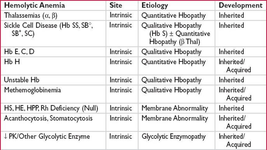
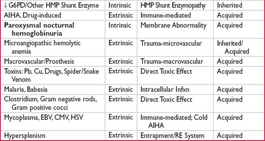
Germline Hemoglobinopathies (Hbopathy): See “Hemoglobinopathies” Chapter.
Acquired a thal a/w MDS: Clonal del α gene cluster/inactivating somatic Mt: ATRX (Blood 2005;105:443) → B4 (Hb H) ∼5–40% Hb H. Phenotypic variance.
G6PD Deficiency (ASH-SAP 2010;1:133; Lancet 2008;371:64)
G6PD: X-linked gene. > 300 variants → ↓ G6PD in males. Lyonization pattern determines female phenotypic variance.
Commonest human enzyme defect. HMP Shunt 1st enzyme: ↓ G6PD → ↓ NADPH → ↓ renewal reduced glutathione →↑ RBC/Hb oxidant stress susceptibility → extravascular + ↑↑ intravascular hemolysis w/↑↑ oxidant + ↓↓ G6PD. DAT–. Smear: Eccentrocytes, bite cells, Heinz bodies.
Oxidant challenges: DKA, hepatic injury, drugs, infxn, naphthalene, & “favism.”
Polymorphisms (Class I–V). Variants’ G6PD levels: Near nl → ↓↓↓↓. G6PD B = nl.
• G6PD A: 10–15% African-American males. Unstable enzyme → ↓ activity in aged RBCs, not retics. Vital oxidants may be continued b/c ↑↑ retics.
• G6PD Mediterranean: B variant. Both retics/mature RBCs affected → ↑↑↑ sev. course. Must stop oxidant agent/process.
• Other Variants: ↓ G6PD activity + marked instability/↓ production → mature
RBC & retics affected. ↑↑ severity. Seen in Asia/Mediterranean area.
False negative/false – G6PD enzyme level during acute hemolysis b/c ↑↑ retics compensation. Screen before starting oxidant drug or mos postacute hemolytic episode.
Variants determined w/restriction enzyme analysis/electrophoresis pattern.
Other RBC Enzymopathies (ASH-SAP 2010;1:133)
Other HMP Shunt: Rare-phosphogluconate dehydrogenase; glutathione reductase.
Glycolysis: ↓ PK > 80% hemolytic anemias a/w glycolysis. ↑ N. & E. Europeans. Nl osmotic fragility/MCV. Dx w/quant. RBC enzyme analysis.
Tx depends on severity. Consider folate supplementation & splenectomy.
Wilson disease → ↑ Cu inhibits hexokinase → acquired hexokinase deficiency.
Nucleotide Metabolism AbNl: ↓ Pyrimidine-5′-NT (Pb tox) & ↓ ADA.
RBC Membrane Abnormalities (Br J Haematol 1999;104:2)
Wide clinical spectrum a/w AbNl membrane protein: ↓ or dysfunction → RBC AbNl shape/deformability → extravascular hemolysis. Splenomegaly/cholelithiasis.
• Hereditary spherocytosis (HS) ↑ in N. Europeans: ∼1/2000. Variable penetrance. 75% autosomal dominant. Etiologies: 30–45% ↓ ankyrin & spectrin, 30% ↓ spectrin, 20% band 3 Mt. ↓ Protein 4.2 as well.
AbNl protein → unstable membrane → ↓ bilayer/↓surface area → spherocyte.
Range: Asymptomatic → Symptomatic sev. A/w aplastic/megaloblastoid/
hyperhemolytic crises.
Dx: Spherocytes, DAT–, ↑ MCHC, + osmotic fragility test, EMA-binding test.
Tx: Consider folic acid in ↑ hemolysis. Definitive Tx: Splenectomy if ↑↑ severity.
• Hereditary elliptocytosis (HE) and Hereditary pyropoikilocytosis (HPP):
Defect spectrin dimerization/spectrin-protein 4.1 interaction → weakened cytoskeleton → deformation under shear stress → ↓ membrane fidelity.
• Acanthocytosis: Liver disease → nonesterified cholesterol in RBC membrane → AbNl shape → splenic remodeling. A/w Zieve syndrome.
Abetalipoproteinemia: AbNl RBC membrane lipid structure.
McLeod Phenotype: No Kx protein (X-linked) → ↓ RBC Kell Ag.
• Stomatocytosis: Inherited: AbNl RBC cation permeability. Acquired: CA,
Alcoholism, & hepatobiliary disease. ↑ Thrombosis postsplenectomy.
• Rh Deficiency/Null: No or ↓↓↓ Rh (RhCE, RhD, & Rh50). Autosomal recessive → Δ cation xport/RBC dehydration → spherocytes/stomatocytes.
Autoimmune Hemolytic Anemia (AIHA) (Blood 2010;116:1831; ASH-SAP 2010;1:133)
AutoAbs characterized by temperature of optimal RBC binding. 1° (idiopathic) vs. 2°.
• Warm AIHA: 80–90% adult AIHA cases. IgG bind RBCs optimally at 37°C. ± C′ fixation. No agglutination. IgG-coated RBCs → removed by splenic mφ w/Fc receptors. 2° Warm AIHA a/w: HL, NHL, CTD (SLE), solid tumor (ovary), chronic inflammation, (UC) & drugs (α-methyldopa).
Tx: 1°: 1st-line steroids. 2nd-line splenectomy/rituximab. 3rd-line cyclophosphamide,
AZA, MMF, CsA, danazol, alemtuzumab. ↓↓ Efficacy w/IVIg. 2°: SLE: 1st–line steroids. 2nd-line rituximab. ↓↓ Efficacy w/splenectomy. CLL: Steroids, R-CVP, R-CD, chlorambucil, rituximab, CsA, splenectomy, alemtuzumab. NHL: Tx malignancy. splenectomy in splenic MZL. Drugs: Stop agent; steroids.
• Cold AutoAb: Mostly IgM optimally binding RBCs <37°C. A/w agglutination.
RBC destruction mediated by C′ C3b fixation → removal by splenic mφ/hepatic Kupffer cells. + Intravascular lysis w/terminal C′.
Cold Agglutinin: A/w B-cell LPD (monoclonal IgM against Carbohydrate I or i Ags). Cold agglutinin w/2° Cold AIHA (IgM): Mycoplasma & EBV.
Cold (Hemolysin) AIHA: 1°: Paroxysmal cold hemoglobinuria (IgG). 2°: Donath–Landsteiner Hemolytic Anemia (1/3 pediatric AIHA; a/w viral infxn; anti-P system IgG). Congenital/3° syphilis cold AIHA (IgG).
Tx: General-avoid cold; rituximab. No efficacy w/steroids/IVIg. Plasmapheresis acute ↓ IgM. Tx LPD. Donath–Landsteiner: Nl self-limited. ?Eculizumab; ?bortezomib.
• Mixed AIHA: IgM + IgG (↑severity) or IgA + IgG. 1° or 2° a/w CTD (esp SLE).
Dx: Usu DAT+. Coombs level ≠ hemolysis level necessarily. ELISA ↑ Sn.
DAT: IgG: Warm AIHA, hapten-mediated/drug adsorption or true autoAb. C3d: Warm AIHA w/↓IgG bound, cold agglutinin, paroxysmal cold hemoglobinuria or ternary/immune complex. IgG + C3d:Warm AIHA or true autoAb.
Xfusion is complex → autoAbs mask alloAbs (potentially sev.) → work w/blood bank to find compatible units. Obtain Hx of prior pregnancy/abortion/xfusion.
Drug-induced Hemolytic Anemia (ASH-SAP 2010;1:133)
True autoAb formation: Several drugs (α-methyldopa) → IgG autoAb formation → hemolysis w/o requiring drug presence. Hapten-mediated/Drug Adsorption: Drug binds RBC membrane proteins (PCNs) → IgG against drug epitope while drug coats RBC → hemolysis. Ternary/Immune Complex: IgG/IgM Ab against complex of drug/metabolite + RBC membrane protein → hemolysis.
PNH See “Anemias of Underproduction” Chapter.
Traumatic Hemolysis: MAHA and Macrovascular Hemolysis
MAHA: HUS, TTP, DIC, HELLP, AFLP, Kasabach–Merritt Syndrome\, Foot Strike.
Typical HUS (N Engl J Med 2009;361:1676): MAHA, ↓ plats & ARF. ↑↑ a/w children <5 y & diarrhea prodrome. E.coli O157:H7 or other Shiga toxin strain. Less commonly a/w other infxns (pneumococcus). Tx: Supportive care.
Atypical HUS: MAHA, ↓ plats & ARF. 10% of HUS. No diarrhea prodrome. ↑ Activation alternative C′ pathway. Poorer prognosis. Tx: Consider plasma exchange, immunosuppressive Rx, eculizumab (anti-C5 mAb).
TTP (Blood 2010;116:4060): Pentad-fever, MAHA, ↓ plats, ARF, neurodeficits. Suspect if MAHA & ↓ plats. Inherited vs. acquired & 1° (AutoAb ↑ clearance/inhibit) vs. 2° ↓ ADAMTS13. Not all TTP a/w ↓ ADAMTS13. 2° causes: Drugs (quinine, calcineurin inhibitors, thienopyridines, GEM, & MMC), SLE, HIV, post-HSCT & disseminated malignancy. Emergent plasma exchange: OS 10% → > 80%. Adjunct Tx: Steroids, rituximab, & immunosuppressives.
Macrovascular: Cardiac valve/vascular prosthesis, aneurysms, IABP, malignant HTN, scleroderma renal crisis.